
CBC Interpretations Dr Duaa Hiasat In this hour
CBC --- Interpretations Dr. Duaa Hiasat
In this hour we will review: Definition of anemia Signs and symptoms Cause Possible complication Utility of the CBC and reticulocyte count And give you a simple plan to: Categorize anemias into 3 morphologic groups Develop a differential diagnosis Decide on further laboratory evaluation Quiz questions
• Component of the CBC: • Red Blood Cells (RBCs) • Hematocrit (Hct) • Hemoglobin (Hgb) • Mean Corpuscular Volume (MCV) • Mean Corpuscular Hemoglobin Concentration (MCHC) - Red cell distribution width (RDW) • White Blood Cells (WBCs) • Platelet
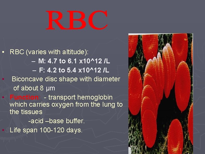
• RBC (varies with altitude): – M: 4. 7 to 6. 1 x 10^12 /L – F: 4. 2 to 5. 4 x 10^12 /L • Biconcave disc shape with diameter of about 8 µm • Function: - transport hemoglobin which carries oxygen from the lung to the tissues -acid –base buffer. • Life span 100 -120 days.
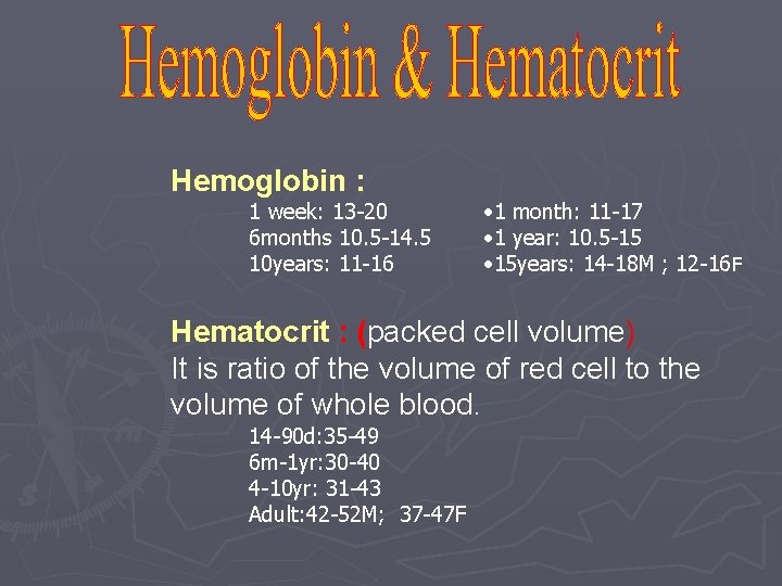
Hemoglobin : 1 week: 13 -20 6 months 10. 5 -14. 5 10 years: 11 -16 • 1 month: 11 -17 • 1 year: 10. 5 -15 • 15 years: 14 -18 M ; 12 -16 F Hematocrit : (packed cell volume) It is ratio of the volume of red cell to the volume of whole blood. 14 -90 d: 35 -49 6 m-1 yr: 30 -40 4 -10 yr: 31 -43 Adult: 42 -52 M; 37 -47 F
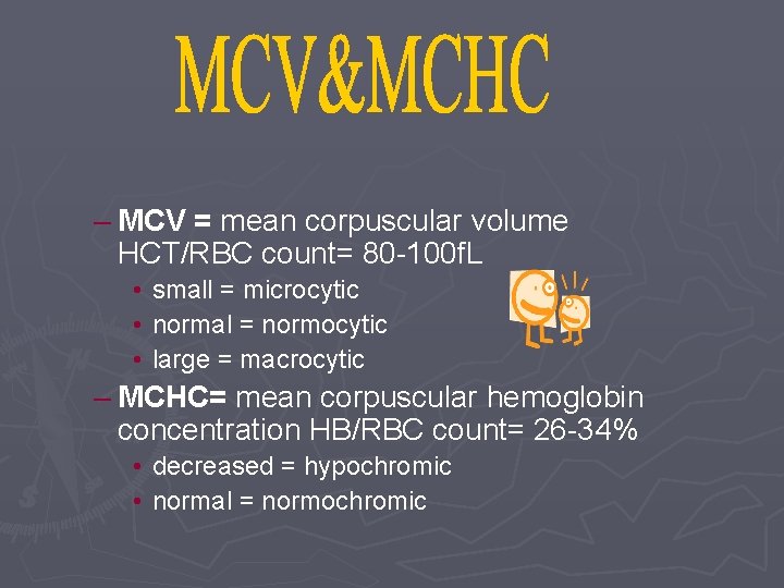
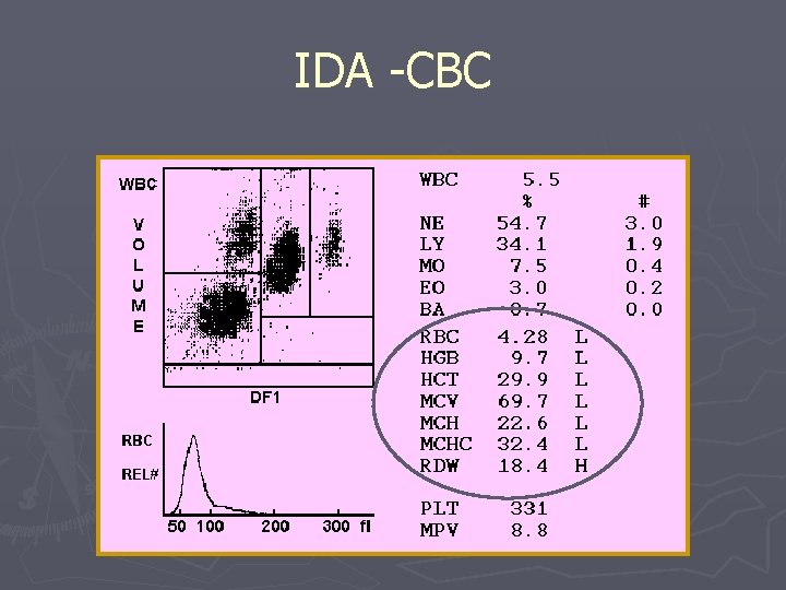
– MCV = mean corpuscular volume HCT/RBC count= 80 -100 f. L • small = microcytic • normal = normocytic • large = macrocytic – MCHC= mean corpuscular hemoglobin concentration HB/RBC count= 26 -34% • decreased = hypochromic • normal = normochromic
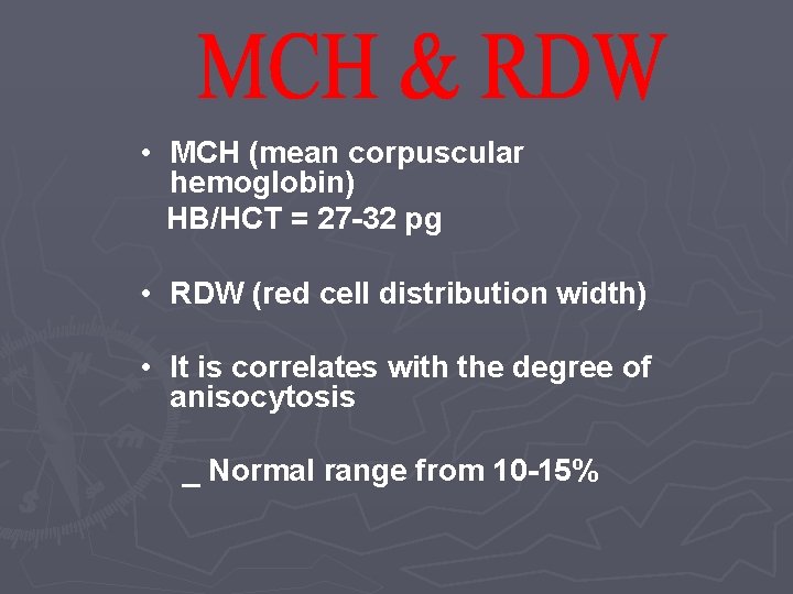
• MCH (mean corpuscular hemoglobin) HB/HCT = 27 -32 pg • RDW (red cell distribution width) • It is correlates with the degree of anisocytosis _ Normal range from 10 -15%
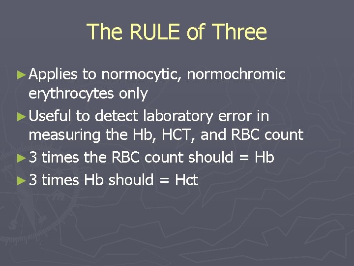
The RULE of Three ► Applies to normocytic, normochromic erythrocytes only ► Useful to detect laboratory error in measuring the Hb, HCT, and RBC count ► 3 times the RBC count should = Hb ► 3 times Hb should = Hct
• This important value is needed in the evaluation of any anemia. • Normal range 1 -2% Must correct for degree of anemia Observed retic count (Actual HCT ÷ Normal HCT) = Corrected retic count • Retic count goes up with – Hemolytic anemia • Retic goes down with – Nutritional deficiencies _ Diseases of the bone marrow itself
Definition of Anaemia Anemia is a level of Red cells, Hemoglobin, or Hematocrit that is below the lower limit of normal for age and sex. Anemia is not a disease, it is a sign of disease As such, critical for all practitioners to know how to evaluate / determine its cause / treat Mechanisms: § Increased loss (external) - hemorrhage § Increased destruction (internal) - hemolysis § Decreased production (trouble in the bone marrow)
So What’s the Plan? 1. 2. 3. 4. 5. 6. 7. 8. Hx and examination CBC- define anemia Look at red cell indices Categorize the anemia based on RBC size Reticulocyte count - marrow response Note any RBC anisopoikilocytosis Consider the differential diagnosis Plan your laboratory evaluation
Symptoms ► Can minor or vague ► Dizziness ► Weakness and fatigue ► intermittent claudication ► Poor concentration ► Symptoms of heart failure ► Dyspnea on exertion ► Impaired infant development ► Reduced school ► Pica performance ► Behavioral disturbances in children ► Palpitation and angina
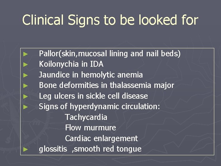
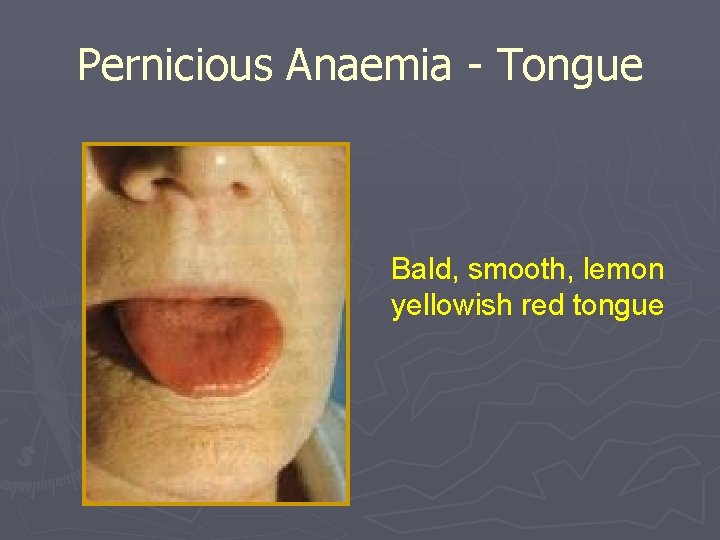
Clinical Signs to be looked for Pallor(skin, mucosal lining and nail beds) ► Koilonychia in IDA ► Jaundice in hemolytic anemia ► Bone deformities in thalassemia major ► Leg ulcers in sickle cell disease ► Signs of hyperdynamic circulation: Tachycardia Flow murmure Cardiac enlargement ► glossitis , smooth red tongue ►
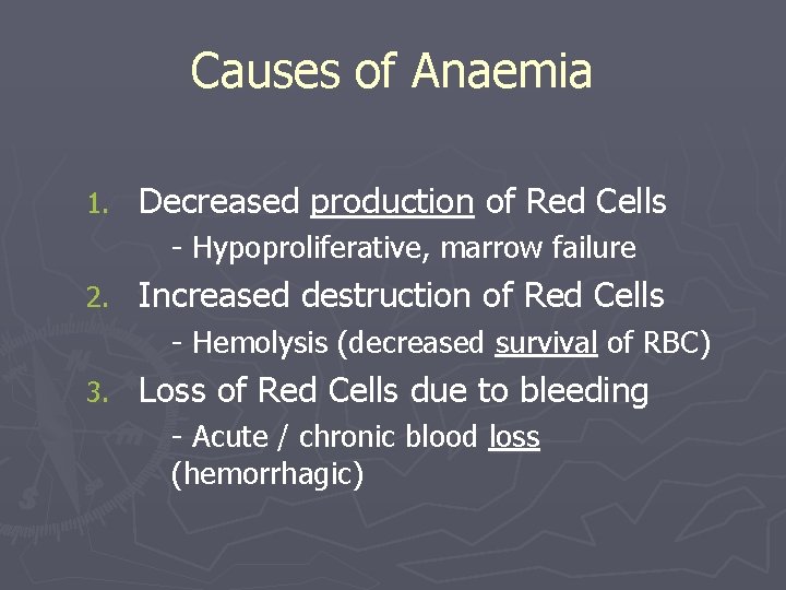
Causes of Anaemia 1. Decreased production of Red Cells - Hypoproliferative, marrow failure 2. Increased destruction of Red Cells - Hemolysis (decreased survival of RBC) 3. Loss of Red Cells due to bleeding - Acute / chronic blood loss (hemorrhagic)
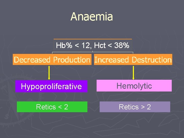
Anaemia Hb% < 12, Hct < 38% Hypoproliferative Hemolytic Retics < 2 Retics > 2
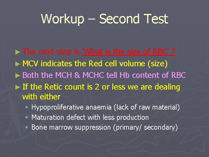
Workup – Second Test ► The next step is ‘What is the size of RBC’ ? ► MCV indicates the Red cell volume (size) ► Both the MCH & MCHC tell Hb content of RBC ► If the Retic count is 2 or less we are dealing with either § § § Hypoproliferative anaemia (lack of raw material) Maturation defect with less production Bone marrow suppression (primary/ secondary)
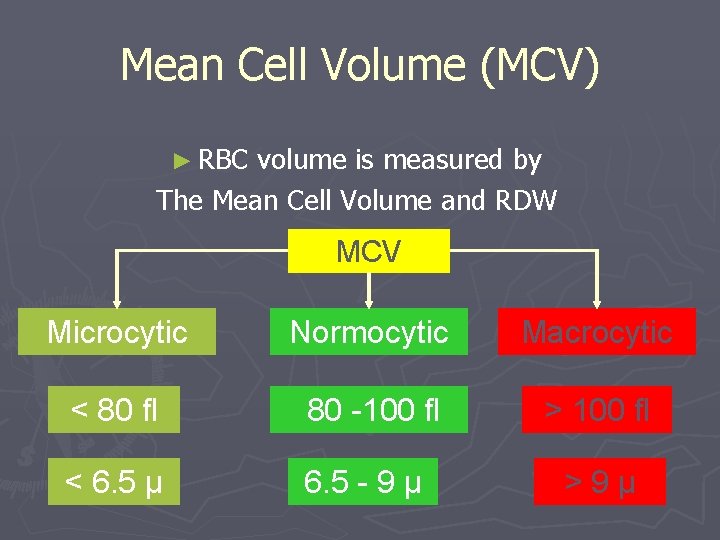
Mean Cell Volume (MCV) ► RBC volume is measured by The Mean Cell Volume and RDW MCV Microcytic Normocytic Macrocytic < 80 fl 80 -100 fl > 100 fl < 6. 5 µ 6. 5 - 9 µ > 9 µ

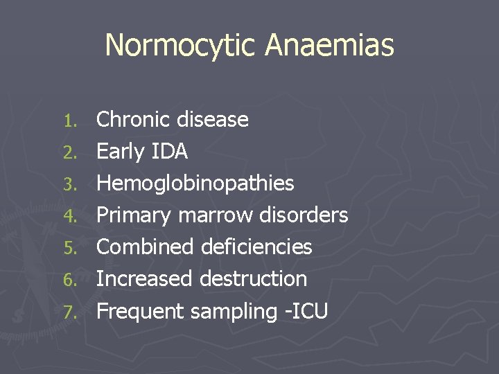
Anaemia Workup - MCV Microcytic Iron Deficiency IDA Chronic Infections Thalassemias Hemoglobinopathies Normocytic Chronic disease Early IDA Hemoglobinopathies Primary marrow disorders Sideroblastic Anemia Combined deficiencies Macrocytic Megaloblastic anemias Liver disease/alcohol Hemoglobinopathie s Metabolic disorders Increased destruction Marrow disorders
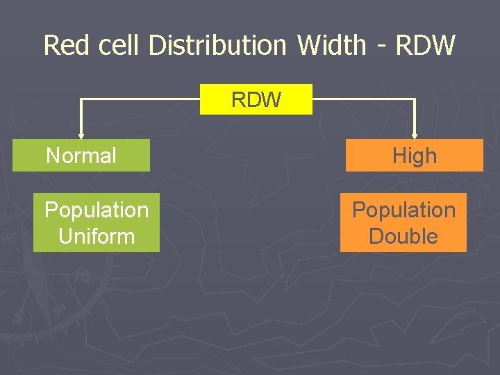
Red cell Distribution Width - RDW Normal Population Uniform High Population Double
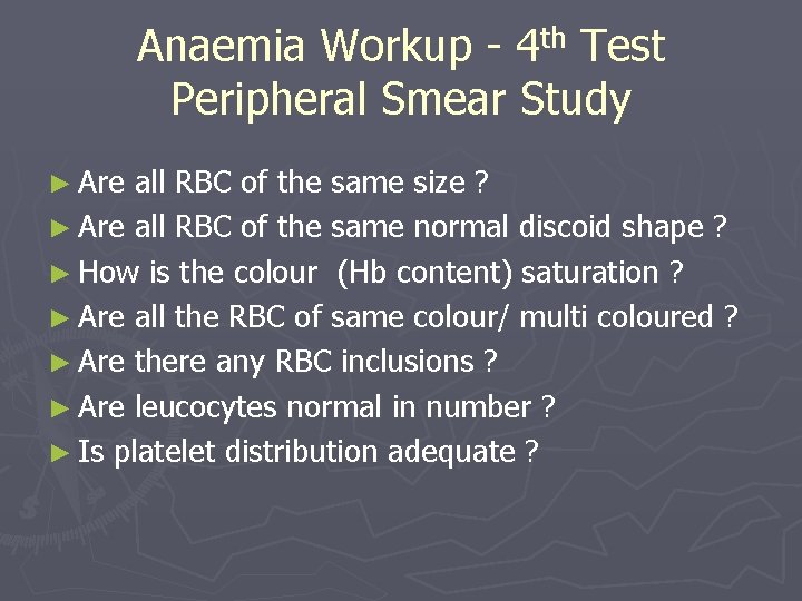
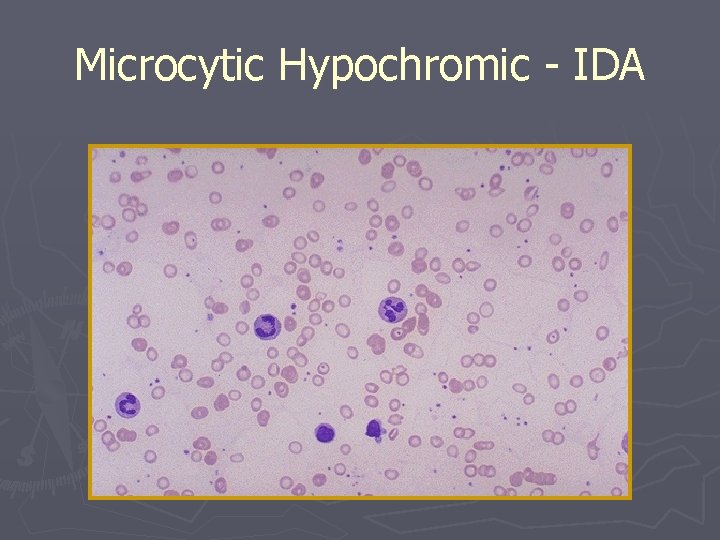
Anaemia Workup - 4 th Test Peripheral Smear Study ► Are all RBC of the same size ? ► Are all RBC of the same normal discoid shape ? ► How is the colour (Hb content) saturation ? ► Are all the RBC of same colour/ multi coloured ? ► Are there any RBC inclusions ? ► Are leucocytes normal in number ? ► Is platelet distribution adequate ?
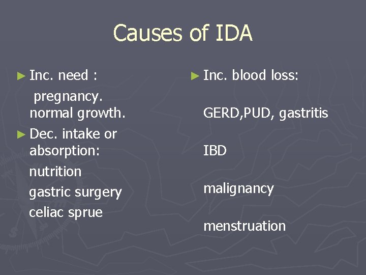
Causes of IDA ► Inc. need : ► Inc. blood loss: pregnancy. normal growth. GERD, PUD, gastritis ► Dec. intake or absorption: IBD nutrition malignancy gastric surgery celiac sprue menstruation
IDA -CBC
Microcytic Hypochromic - IDA
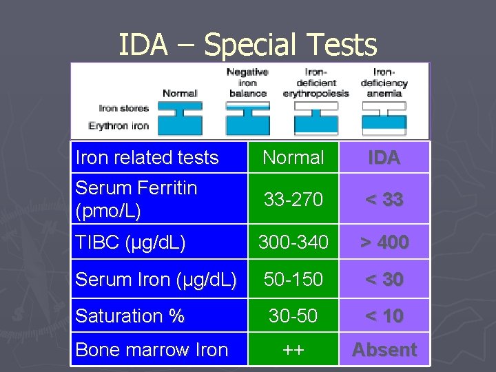
IDA – Special Tests Iron related tests Normal IDA Serum Ferritin (pmo/L) 33 -270 < 33 TIBC (µg/d. L) 300 -340 > 400 Serum Iron (µg/d. L) 50 -150 < 30 Saturation % 30 -50 < 10 ++ Absent Bone marrow Iron
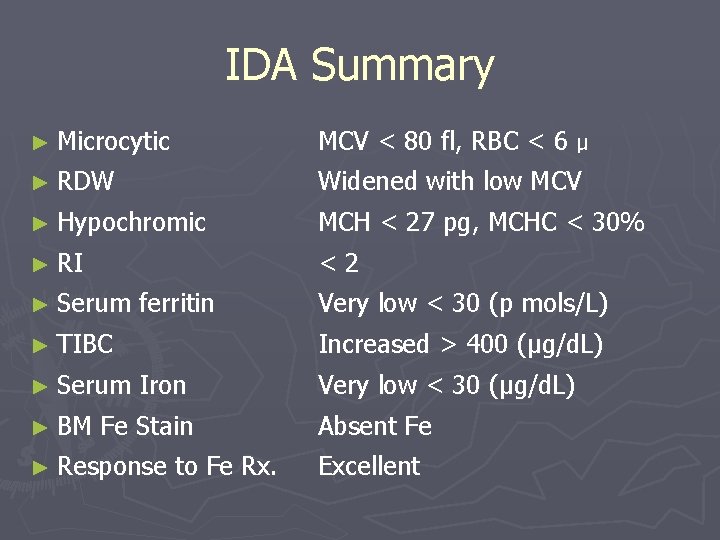
IDA Summary ► Microcytic MCV < 80 fl, RBC < 6 µ ► RDW Widened with low MCV ► Hypochromic MCH < 27 pg, MCHC < 30% ► RI < 2 ► Serum ferritin Very low < 30 (p mols/L) ► TIBC Increased > 400 (µg/d. L) ► Serum Iron Very low < 30 (µg/d. L) ► BM Fe Stain Absent Fe ► Response to Fe Rx. Excellent
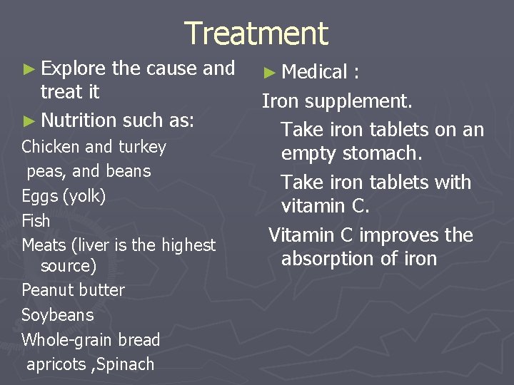
Treatment ► Explore the cause and treat it ► Nutrition such as: Chicken and turkey peas, and beans Eggs (yolk) Fish Meats (liver is the highest source) Peanut butter Soybeans Whole-grain bread apricots , Spinach ► Medical : Iron supplement. Take iron tablets on an empty stomach. Take iron tablets with vitamin C. Vitamin C improves the absorption of iron
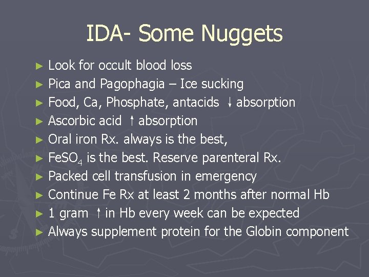
IDA- Some Nuggets ► Look for occult blood loss ► Pica and Pagophagia – Ice sucking ► Food, Ca, Phosphate, antacids ↓absorption ► Ascorbic acid ↑absorption ► Oral iron Rx. always is the best, ► Fe. SO 4 is the best. Reserve parenteral Rx. ► Packed cell transfusion in emergency ► Continue Fe Rx at least 2 months after normal Hb ► 1 gram ↑in Hb every week can be expected ► Always supplement protein for the Globin component
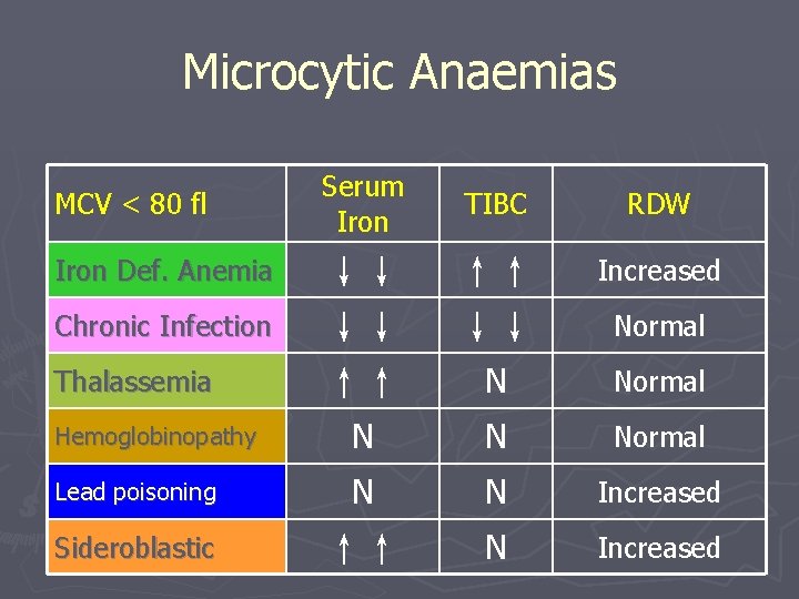
Microcytic Anaemias MCV < 80 fl Serum Iron TIBC RDW ↓↓ ↓↓ ↑↑ ↑↑ ↓↓ Increased N Normal Hemoglobinopathy N N Normal Lead poisoning N N Increased Sideroblastic ↑↑ N Increased Iron Def. Anemia Chronic Infection Thalassemia Normal
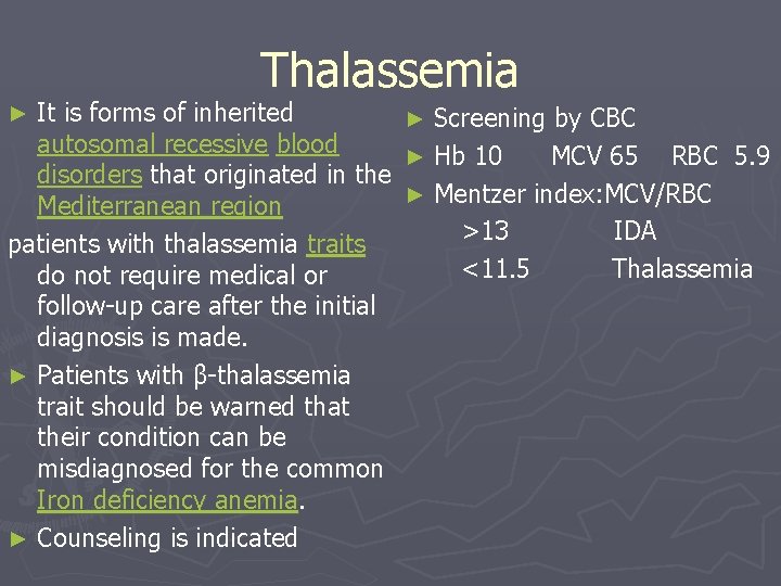
Thalassemia It is forms of inherited ► Screening by CBC autosomal recessive blood ► Hb 10 MCV 65 RBC 5. 9 disorders that originated in the ► Mentzer index: MCV/RBC Mediterranean region patients with thalassemia traits >13 IDA <11. 5 Thalassemia do not require medical or follow-up care after the initial diagnosis is made. ► Patients with β-thalassemia trait should be warned that their condition can be misdiagnosed for the common Iron deficiency anemia. ► Counseling is indicated ►
► Therapy for beta thalassemia primarily involves chelation or removal of excessive iron deposits in the blood ► Curative methods Bone Marrow Transplant ► Beta-thalassemia major requires lifelong blood transfusions. ► Alpha-thalassemia major is also referred to as hydrops fetalis ► Diagnosed by blood electrophoresis ► Hb A 2 >3. 5 ► Hb A <95 Hb F >1
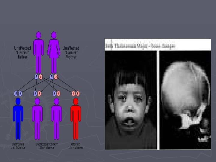
Complication Iron overload ► Infection: This is especially true if the spleen has been removed. ► Bone deformities: Thalassemia can make the bone marrow expand, which causes bones to widen especially in the face and skull. ► Enlarged spleen ► Slowed growth rates: Puberty also may be delayed in children with thalassemia. ► Heart problems: such as congestive heart failure and abnormal heart rhythms (arrhythmias), may be associated with severe thalassemia. ►
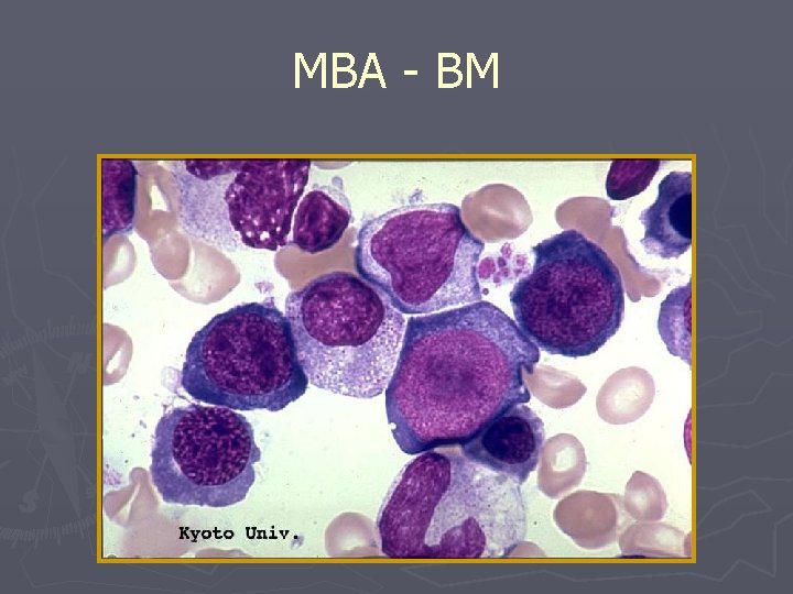
Macrocytic Anaemias Megaloblastic Macrocytic – B 12 and Folate↓ (hypersegmented neutrophil) B. Non Megaloblastic Macrocytic Anaemias 1. Liver disease/alcohol 2. Hemoglobinopathies 3. Metabolic disorders, Hypothyroidism 4. Myelodystrophy, BM infiltration 5. Accelerated Erythropoesis - ↑destruction 6. Drugs (cytotoxics, immunosuppressants, anticonvulsants) A.
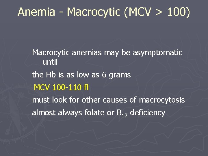
Anemia - Macrocytic (MCV > 100) Macrocytic anemias may be asymptomatic until the Hb is as low as 6 grams MCV 100 -110 fl must look for other causes of macrocytosis almost always folate or B 12 deficiency
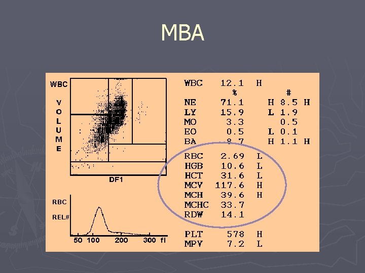
MBA
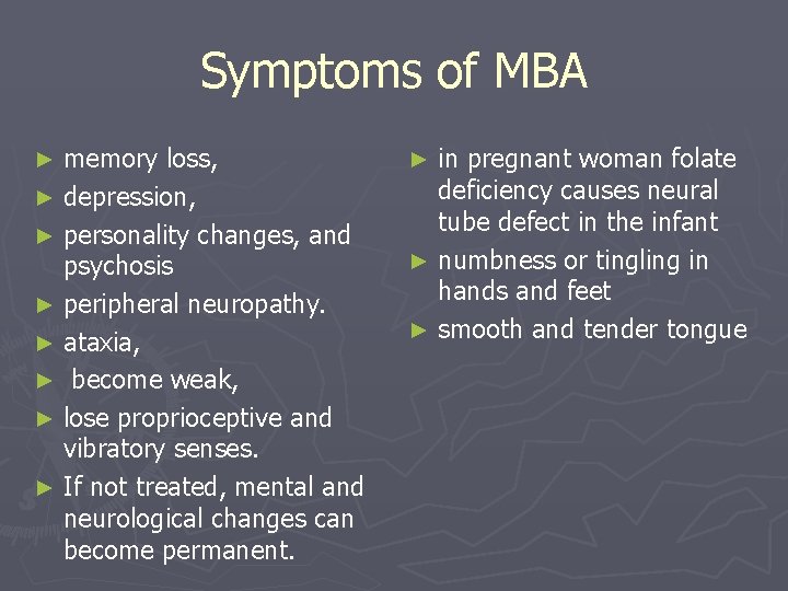
Symptoms of MBA memory loss, ► depression, ► personality changes, and psychosis ► peripheral neuropathy. ► ataxia, ► become weak, ► lose proprioceptive and vibratory senses. ► If not treated, mental and neurological changes can become permanent. ► in pregnant woman folate deficiency causes neural tube defect in the infant ► numbness or tingling in hands and feet ► smooth and tender tongue ►
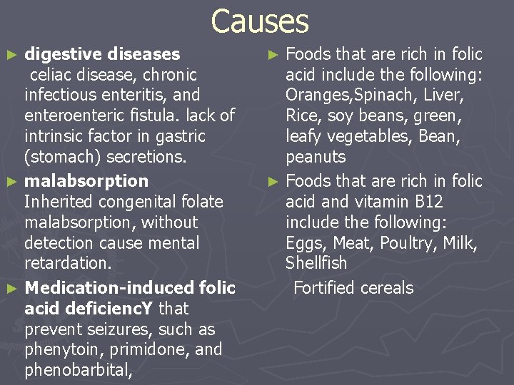
Causes digestive diseases celiac disease, chronic infectious enteritis, and enteroenteric fistula. lack of intrinsic factor in gastric (stomach) secretions. ► malabsorption Inherited congenital folate malabsorption, without detection cause mental retardation. ► Medication-induced folic acid deficienc. Y that prevent seizures, such as phenytoin, primidone, and phenobarbital, ► Foods that are rich in folic acid include the following: Oranges, Spinach, Liver, Rice, soy beans, green, leafy vegetables, Bean, peanuts ► Foods that are rich in folic acid and vitamin B 12 include the following: Eggs, Meat, Poultry, Milk, Shellfish Fortified cereals ►
TREATMENT Must be tailoret to cause ► Replacement : Folic acid : 1 mg /d Vit B 12 : 1000 micr gr/d I. M for 5 days , then q week until Hct normal, then q month for life. ►
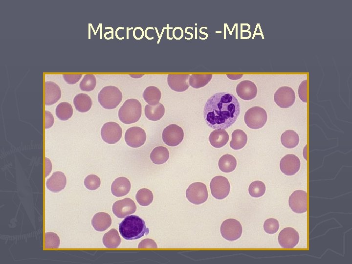
Macrocytosis -MBA
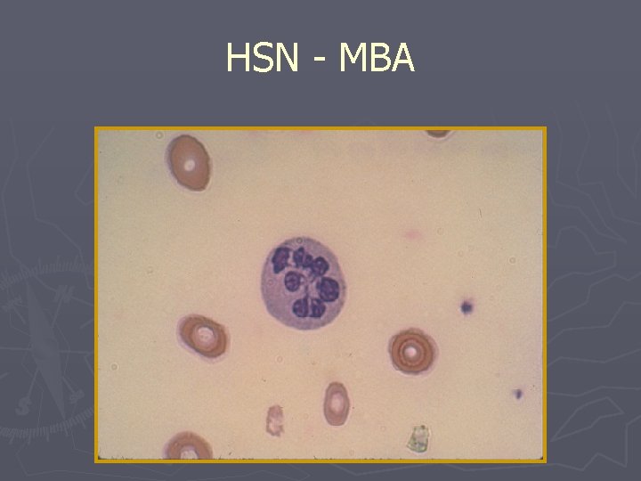
HSN - MBA
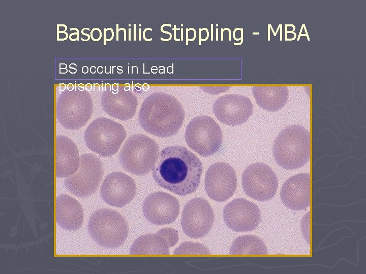
Basophilic Stippling - MBA BS occurs in Lead poisoning also
MBA - BM
Pernicious Anaemia - Tongue Bald, smooth, lemon yellowish red tongue
Normocytic Anaemias 1. 2. 3. 4. 5. 6. 7. Chronic disease Early IDA Hemoglobinopathies Primary marrow disorders Combined deficiencies Increased destruction Frequent sampling -ICU
Anaemia of Chronic Disease ► Thyroid diseases ► Malignancy ► Collagen Vascular Disease § Rheumatoid Arthritis § SLE § Polymyositis § Polyarteritis Nodosa • IBD – Ulcerative Colitis – Crohn’s Disease • Chronic Infections – HIV, Osteomyelitis – Tuberculosis • Renal Failure
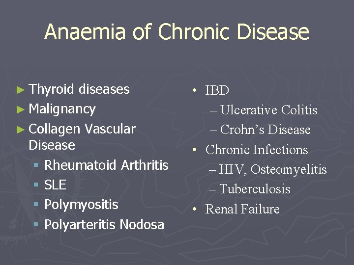
CBC in ACD Anemia of chronic disease is often a mild normocytic anemia, show a low reticulocyte production index, suggesting that reticulocyte production is impaired and not enough to compensate for the decreased red blood cell count. ► While no single test is always reliable to distinguish the two causes of disease, there are sometimes some suggestive data: ► ferritin levels should be normal or high, reflecting the fact that iron is stored within cells, and ferritin is being produced as an acute phase reactant ► TIBC should be low or normal in anemia of chronic disease ►
‘Dimorphic’ Anaemia ► Folate & Fe deficiency (pregnancy, alcoholism) ► B 12 & Fe deficiency (PA with atrophic gastritis) ► Thalassemia minor & B 12 or folate deficiency ► Fe deficiency & hemolysis (prosthetic valve) ► Folate deficiency & hemolysis (Hb SS disease) ► Peripheral smear exam is critical to assess these ► RDW is increased very much
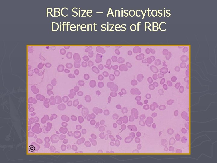
RBC Size – Anisocytosis Different sizes of RBC
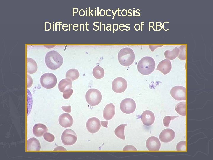
Poikilocytosis Different Shapes of RBC
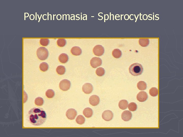
Polychromasia - Spherocytosis
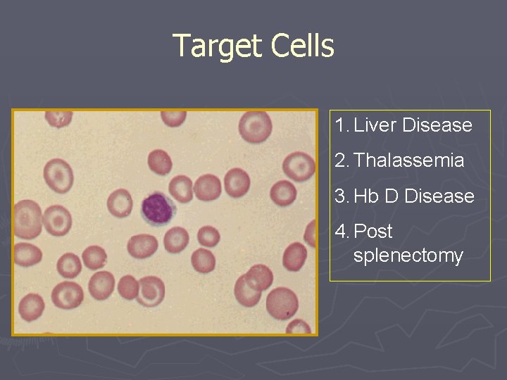
Target Cells 1. Liver Disease 2. Thalassemia 3. Hb D Disease 4. Post splenectomy
High RBCs count. Increased RBC count = Polycythemia A. Primary B. Secondary living at altitude chronic lung/heart disease tobacco use/carbon monoxide C. Relative Polycythemia dehydration
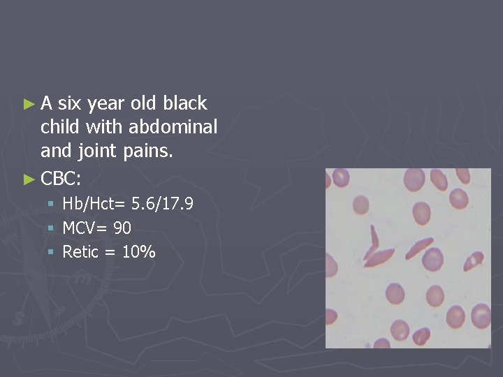
► A six year old black child with abdominal and joint pains. ► CBC: § Hb/Hct= 5. 6/17. 9 § MCV= 90 § Retic = 10%
► Anemia is present ► Sickle cells are seen on the blood smear ► This patient has Sickle Cell Anemia ► How do I confirm it? § Hemoglobin Fractionation - demonstration of Hemoglobin S (and no Hemoglobin A)
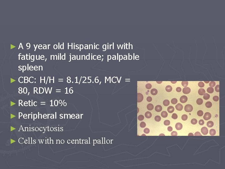
► A 9 year old Hispanic girl with fatigue, mild jaundice; palpable spleen ► CBC: H/H = 8. 1/25. 6, MCV = 80, RDW = 16 ► Retic = 10% ► Peripheral smear ► Anisocytosis ► Cells with no central pallor
► It is normochromic, normocytic Reticulocytes are increased Spherocytes are present on the smear The findings are consistent with Hereditary Spherocytosis ► osmotic fragility – confirm the spherocytes ► Anemia + reticulocyte count § Must rule out a source of bleeding ► Extravascular (in the spleen) § indirect bilirubin, urobilinogen, LDH, haptoglobin § Spherocytes on the peripheral smear ► Intravascular § indirect bilirubin, urobilinogen, LDH, haptoglobin, + free plasma hemoglobin, + hemoglobinuria, + hemosiderinuria § Schistocytes on the peripheral smear
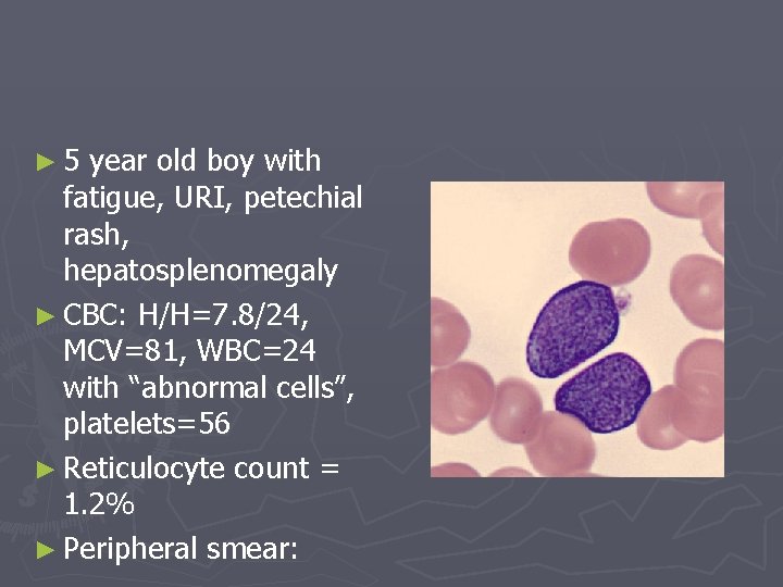
► 5 year old boy with fatigue, URI, petechial rash, hepatosplenomegaly ► CBC: H/H=7. 8/24, MCV=81, WBC=24 with “abnormal cells”, platelets=56 ► Reticulocyte count = 1. 2% ► Peripheral smear:
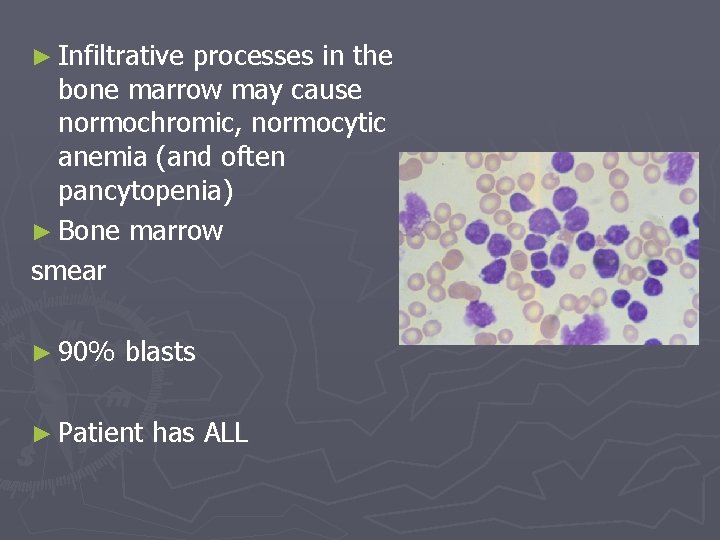
► Infiltrative processes in the bone marrow may cause normochromic, normocytic anemia (and often pancytopenia) ► Bone marrow smear ► 90% blasts ► Patient has ALL
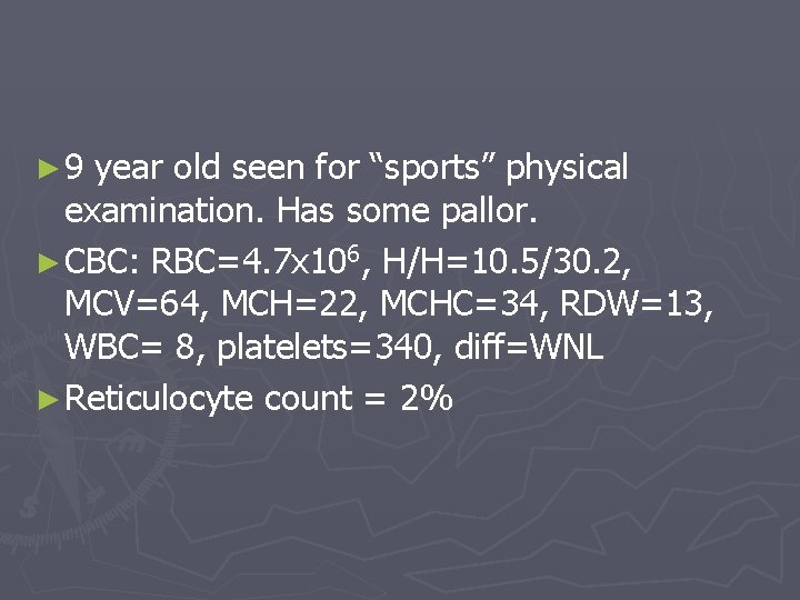
► 9 year old seen for “sports” physical examination. Has some pallor. ► CBC: RBC=4. 7 x 10⁶, H/H=10. 5/30. 2, MCV=64, MCH=22, MCHC=34, RDW=13, WBC= 8, platelets=340, diff=WNL ► Reticulocyte count = 2%
► Is anemia present? § RBC count is normal, but H/H are low ► What category of anemia? § MCV and MCH are low --> Hypo/micro ► What further tests? § Iron studies § Hemoglobin fractionation
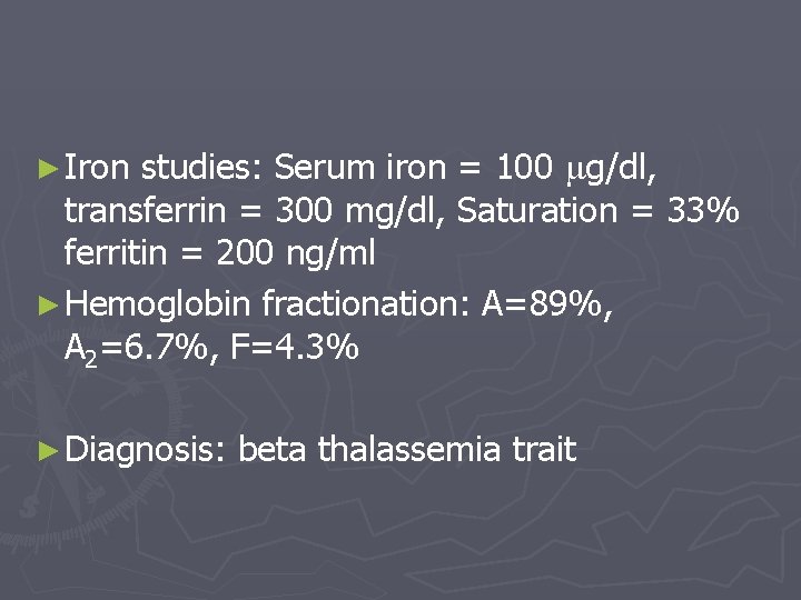
► Iron studies: Serum iron = 100 g/dl, transferrin = 300 mg/dl, Saturation = 33% ferritin = 200 ng/ml ► Hemoglobin fractionation: A=89%, A 2=6. 7%, F=4. 3% ► Diagnosis: beta thalassemia trait
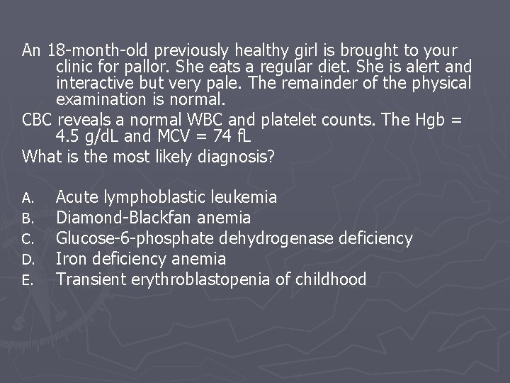
An 18 -month-old previously healthy girl is brought to your clinic for pallor. She eats a regular diet. She is alert and interactive but very pale. The remainder of the physical examination is normal. CBC reveals a normal WBC and platelet counts. The Hgb = 4. 5 g/d. L and MCV = 74 f. L What is the most likely diagnosis? A. B. C. D. E. Acute lymphoblastic leukemia Diamond-Blackfan anemia Glucose-6 -phosphate dehydrogenase deficiency Iron deficiency anemia Transient erythroblastopenia of childhood
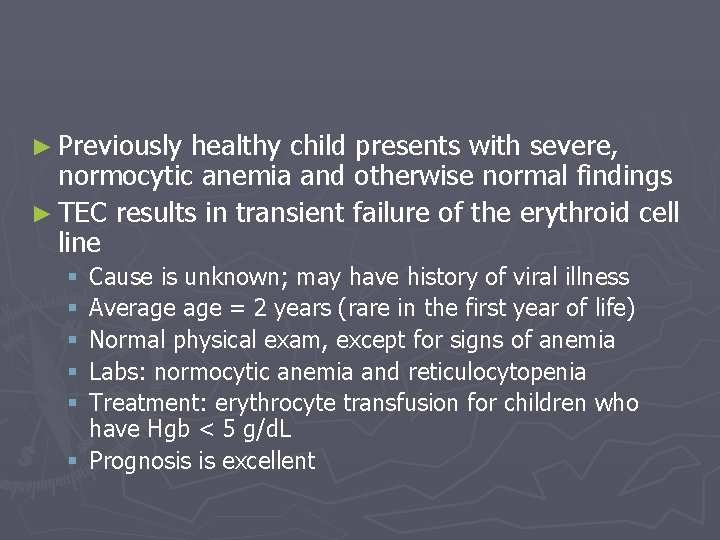
► Previously healthy child presents with severe, normocytic anemia and otherwise normal findings ► TEC results in transient failure of the erythroid cell line Cause is unknown; may have history of viral illness Average = 2 years (rare in the first year of life) Normal physical exam, except for signs of anemia Labs: normocytic anemia and reticulocytopenia Treatment: erythrocyte transfusion for children who have Hgb < 5 g/d. L § Prognosis is excellent § § §
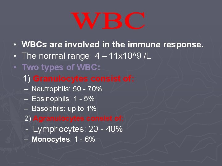
• WBCs are involved in the immune response. • The normal range: 4 – 11 x 10^9 /L • Two types of WBC: 1) Granulocytes consist of: – Neutrophils: 50 - 70% – Eosinophils: 1 - 5% – Basophils: up to 1% 2) Agranulocytes consist of: - Lymphocytes: 20 - 40% – Monocytes: 1 - 6%
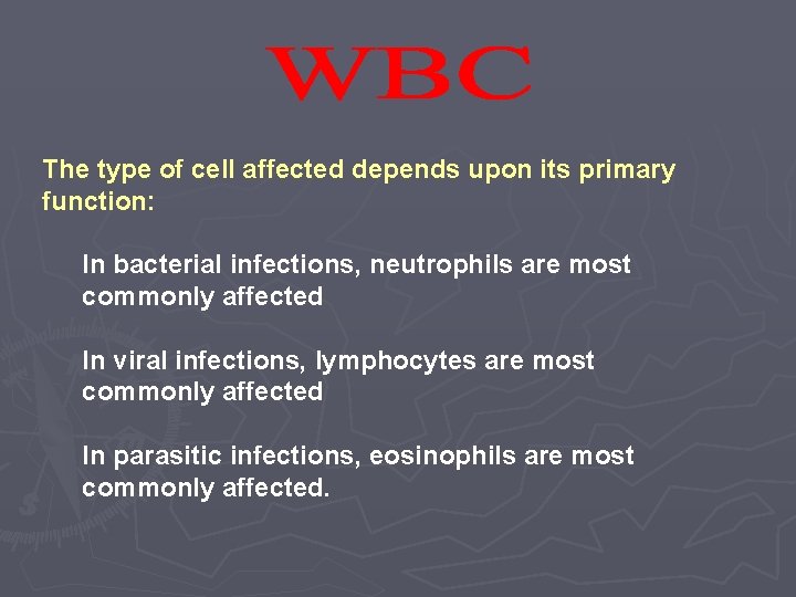
The type of cell affected depends upon its primary function: In bacterial infections, neutrophils are most commonly affected In viral infections, lymphocytes are most commonly affected In parasitic infections, eosinophils are most commonly affected.
• Neutrophil disorders – Neutrophilia – an increase in neutrophils – Conditions associated with neutrophilia are: 1 -Bacterial infections (most common cause) 2 -Tissue destruction e. g. tissue infarctions, burns. 3 - leukemoid reaction 4 -Leukemia
– Neutropenia – this may result from 1 -Decreased bone marrow production e. g. BM hypoplasia. 2 -Ineffective bone marrow production – E. g. megaloblastic anemias and myelodysplastic syndromes. 3 - post acute infection _ e. g. typhoid fever, brucellosis.
– Eosinophilia may be found in • Parasitic infections • Allergic conditions and hypersensitivity reaction
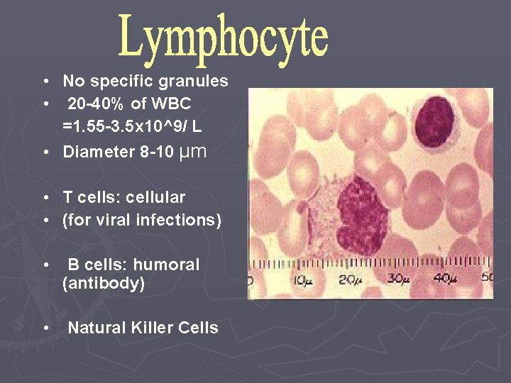
• No specific granules • 20 -40% of WBC =1. 55 -3. 5 x 10^9/ L • Diameter 8 -10 µm • T cells: cellular • (for viral infections) • B cells: humoral (antibody) • Natural Killer Cells
• Lymphocytosis – may indicate _ Viral infection e. g. Infectious mononucleosis, CMV _ Bacterial infection e. g. TB or pertussis. • Lymphopenia – caused by _Stress. _Steroid therapy _ Irradiation
• • Infection of the mouth and throat. Painful skin ulceration. Recurrent infection. Septicemia.
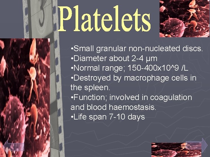
• Small granular non-nucleated discs. • Diameter about 2 -4 µm • Normal range; 150 -400 x 10^9 /L • Destroyed by macrophage cells in the spleen. • Function; involved in coagulation and blood haemostasis. • Life span 7 -10 days
• Numbers of platelets – Increased (Thrombocythemia) • • Pregnancy. Exercise. High attitudes. splenectomy – Decreased (Thrombocytopenia) • Menstruation. • Haemorrhage. • Bone marrow destruction or suppression e. g. leukemia • The values have to fit the clinical situation.
Manifestation of thrombocytopenia Petechial hemorhage. • Easy bruising. • Mucosal bleeding e. g. _ epistaxes. • _ gum bleeding
ITP condition of having an abnormally low platelet count, As most incidents of ITP appear to be related to the production of antibodies against platelets ► the spontaneous formation of purpura and petechiae , especially on the extremities, bleeding from the nostrils, gums, and menorrhagia , A very low count may result in the spontaneous formation of hematomas (blood masses) in the mouth or on other mucous membranes. Bleeding time from minor lacerations or abrasions is usually prolonged. ► Serious and possibly fatal complications due to an extremely low count may include subarachnoid or intracerebral hemorrhage , lower gastrointestinal bleeding or other internal bleeding. ►
The diagnosis of ITP is a process of exclusion. First, the clinician has to determine that there are no blood abnormalities other than low platelet count, and no physical signs except for signs of bleeding. Then, the secondary causes (usually 5– 10 percent of suspected ITP cases) should be excluded. Secondary causes could be leukemia, medications (e. g. , quinine, heparin), lupus erythematosus, cirrhosis, HIV, hepatitis C, congenital causes, antiphospholipid syndrome, von Willebrand factor deficiency ►. Treatment may not be necessary in mild cases, but very low counts or significant bleeding might prompt treatment with steroids, intravenous immunoglobulin, anti-D immunoglobulin, or stronger immunosuppressive drugs. ITP not responsive to conventional treatment may require splenectomy. Platelet transfusions may be used in severe bleeding together with a very low count. ►
TTP is a rare disorder of the blood-coagulation system, ► Classically, the following five features are indicative of TTP; ► Thrombocytopenia leading to bruising or purpura ► Microangiopathic hemolytic anemia (anemia, jaundice and a blood film featuring evidence of mechanical fragmentation of red blood cells) ► Neurologic symptoms (fluctuating), such as hallucinations, bizarre behavior, altered mental status, stroke or headaches ► Kidney failure ► Fever ►
► Secondary TTP Cancer ► Bone marrow transplantation ► Pregnancy ► Medication use: § Quinine § Platelet aggregation inhibitors (ticlopidine, clopidogrel, and prasugrel) § Immunosuppressants (cyclosporine, mitomycin, tacrolimus/FK 506, interferon-α) ► HIV-1 infection ►
Treatment ► , plasmapheresis has become the treatment of choice for TTP; the procedure has to be repeated daily to eliminate the inhibitor and abate the symptoms. ► Lactate dehydrogenase levels are generally used to monitor disease activity. ► Plasmapheresis may need to be continued for 1– 8 weeks
References ► Essentials of family medicine sixth edition ► Nelson Essentials of pediatrics fourth edition ► Kumar and clarck clinical medicine ► The american family physician (web site)november ► Family practice review course: anemia Dx and Rx Reid Blackwelder, East Tennessee State University ► http: //en. wikipedia. org/wiki/anemia ► Medicinenet. com, definition of anemia (http//www. medterms. com/script/main/art. asp? articlekey( 15491) ► Emedicine-anemia, chronic: article by Fredrick M Abrahamian, DO, FACEP (http: //www. emedicine. com/emerg/topic 734. htm#section ~clinical)
- Slides: 80