The vermiform appendix Dr Firas Majeed Acute appendicitis


")


 appendicitis")


 score. Alvarado score")


 incision for appendicitis")


 This is a rare but very serious complication of gangrenous appendicitis")


- Slides: 66
The vermiform appendix Dr. Firas Majeed
Acute appendicitis is the most common cause of an ‘acute abdomen’ in young adults. Appendicitis is sufficiently common that appendicectomy (termed ‘appendectomy’ in North America) is the most frequently performed urgent abdominal operation and is often the first major procedure performed by a surgeon in training
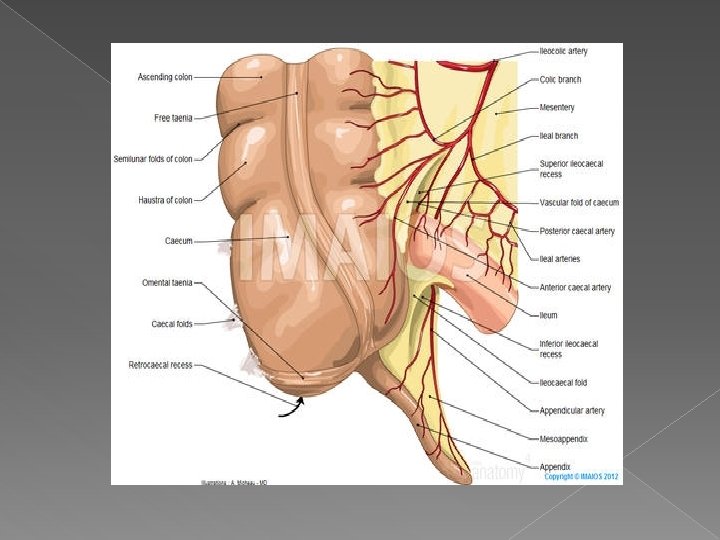
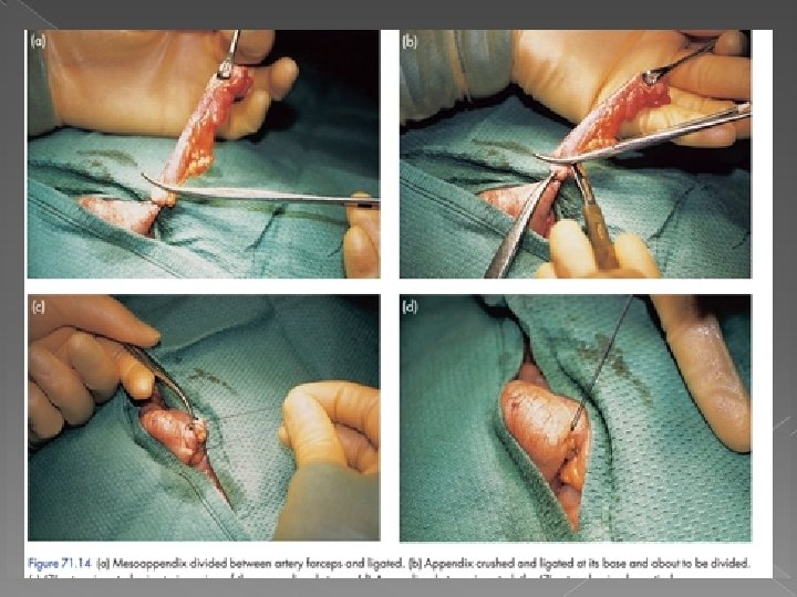
The position of the base of the appendix is constant, being found at the confluence of the three taeniae coli of the caecum, which fuse to form the outer longitudinal muscle coat of the appendix. At operation, , as gentle traction on the taeniae coli, particularly the anterior taenia, will lead the operator to the base of the appendix.
The mesentery of the appendix or mesoappendix arises from the lower surface of the mesentery or the terminal ileum and is itself subject to great variation. Sometimes, as much as the distal one-third of the appendix is bereft of mesoappendix Especially in childhood, the mesoappendix is so transparent that the contained blood vessels can be seen. In many adults, it becomes laden with fat, which obscures these vessels. The appendicular artery, a branch of the lower division of the ileocolic artery, passes behind the terminal ileum to enter the mesoappendix a short distance from the base of the appendix.
INCIDENCE Acute appendicitis is relatively rare in infants, an increasingly common in childhood and early adult life, reaching a peak incidence in the teens and early 20 s. After middle age, the risk of developing appendicitis is quite small. The incidence of appendicitis is equal among males and females before puberty. In teenagers and young adults, the male–female ratio increases to 3: 2 at age 25; thereafter, the greater incidence in males declines. .
AETIOLOGY • 1. Decreased dietary fibre and increased consumption of refined carbohydrates may be important. . . Therefore in developing countries that are adopting a more refined western-type diet, the incidence continues to rise. .
2. While appendicitis is clearly associated with bacterial proliferation within the appendix, no single organism is responsible. A mixed growth of aerobic and anaerobic organisms is usual. The initiating event causing bacterial proliferation is controversial. Obstruction of the appendix lumen has been widely held to be important, and some form of luminal obstruction, either by a faecolith or a stricture, is found in the majority of cases. .
Causes of obstruction of appendix a. A faecolith (sometimes referred to as an ‘appendicolith’) is composed of inspissated faecal material, calcium, phosphates bacteria and epithelial debris Rarely a foreigen body is incorporated into the mass. . b. A fibrotic stricture of the appendix usually. indicates previous appendicitis that resolved out surgical intervention.
c. Obstruction of the appendiceal orifice by tumour, particularly carcinoma of the caecum, is an occasional cause of acute appendicitis in middleaged and elderly patients. d. Intestinal parasites, particularly Oxyuris vermicularis (pinworm), can proliferate in the appendix and occlude the lumen.
y Risk factors for perforation of the appendix ■ Faecolith obstruction
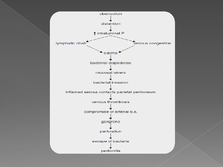
Pathology Lymphoid hyperplasia narrows the lumen of of appendix lead to luminal obstruction. Once obstruction occurs, continued mucus secretion and inflammatory exudation increase intraluminal pressure, obstructing lymphatic drainage. Oedema and mucosal ulceration develop with he submucosa. Ø Ø
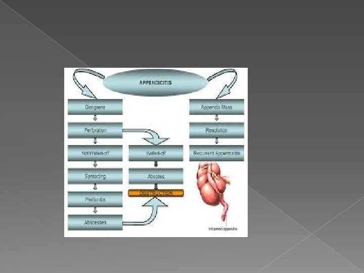
Resolution may occur at this point either spontaneously or in response to antibiotic therapy. If the condition progresses, further distension of the appendix may cause venous obstruction and ischaemia of the appendix wall. With ischaemia, bacterial invasion occurs through the muscularis propria and submucosa, producing acute appendicitis Finally, ischaemic necrosis of the appendix wall produces gangrenous appendicitis, with free bacterial contamination of the peritoneal cavity.
Alternatively, the greater omentum and loops of small bowel become adherent to the inflamed appendix, walling off the spread of peritoneal contamination, and resulting in a phlegmonous mass. Rarely, appendiceal inflammation resolves, leaving a distended mucus-filled organ termed a ‘mucocoele’ of the appendix.
Clinical diagnosis History/Clinical examination
During the first 6 hours, there is rarely any alteration in temperature or pulse rate. After that time, slight pyrexia (37. 2– 37. 7°C) with a corresponding increase in the pulse rate to 80 or 90 is usual. However, in 20 per cent of patients, there is no pyrexia or tachycardia in the early stages. In children, a temperature greater than 38. 5°C suggests other causes, e. g. mesenteric adenitis.
The classic visceral–somatic sequence of pain is present in only about half of those patients subsequently proven to have acute appendicitis. A typical presentations include pain that is predominantly somatic or visceral and poorly localised. Atypical pain is more common in the elderly, in whom localization to the right iliac fossa is unusual.
Typically, two clinical syndromes of acute appendicitis can be discerned, acute catarrhal(non obstructive) appendicitis and acute obstructive appendicitis, the latter characterized by a more acute course. The onset of symptoms is abrupt, and there may be generalized abdominal pain from the start. The temperature may be normal and vomiting is common, so that the clinical picture may mimic acute intestinal obstruction.
An inflamed appendix in the pelvis may never produce somatic pain involving the anterior abdominal wall, but may instead cause suprapubic discomfort and tenesmus. In this circumstance, tenderness may be elicited only on rectal examination and is the basis for the recommendation thata rectal examination should be performed on every patient who presents with acute lower abdominal pain. .
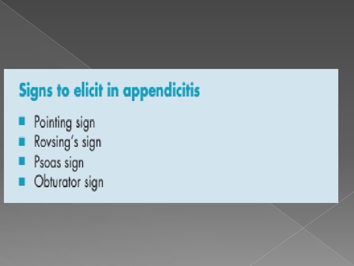
The patient is asked to point to where the pain start and where it moved (the pointing sign ). Gentle superficial palpation of the abdomen, beginning in the left iliac fossa moving anticlockwise to the right iliac fossa will detect muscle guarding over the point of maximum tenderness, classically Mc. Burney’s point. Asking the patient to cough or gentle percussion over the site of maximum tenderness will elicit rebound tenderness
Deep palpation of the left iliac fossa may cause pain in the right iliac fossa, Rovsing’s sign, which is helpful in supporting a clinical diagnosis of appendicitis. Occasionally, an inflamed appendix lies on the psoas muscle, and the patient, often a young adult, will lie with the right hip flexed for pain relief. Spasm of the obturator internus is sometimes demonstrable when the hip is flexed and internally rotated. If an inflamed appendix is in contact with the obturator internus, this manoeuvre will cause n in the hypogastrium(the obturator test
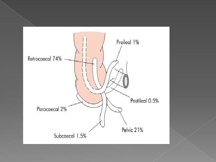
Special features, according to position of theappendix Retrocaecal Rigidity is often absent, and even application of deep pressure may fail to elicit tenderness (silent appendix), the reason being that the caecum, distended with gas, prevents the pressure exerted by the hand from reaching the inflamed structure. However, deep tenderness is often present in the always positive.
Pelvic Occasionally, early diarrhoea results from an inflamed appendix being in contact with the rectum. there is usually complete absence of abdominal rigidity, and often tenderness over Mc. Burney’s pointis also lacking. , deep tenderness can be made out just above and to the right of the symphysis pubis. , a rectal examination reveals tenderness in the rectovesical pouch or the pouch of Douglas, especially on the right side. Spasm of the psoas and obturator internus muscles may be present. An inflamedappendix in contact with the bladder may cause frequency of micturition. This is more common in children.
Postileal It presents the greatest difficulty in diagnosis because the pain may not shift, diarrhoea is a feature and marked retching may occur. Tenderness, if any, is ill defined, .
Special features, according to age Infants Appendicitis is relatively rare, the patient is unable to give a history, diagnosis is often delayed, and thus the incidence of perforation higher than in older children. Diffuse peritonitis can develop rapidly because of the underdeveloped greater omentum, whichis unable to give much assistance in localising the infection. .
. Children It is rare to find a child with appendicitis who has not vomited. usually have complete aversion to food. Elderly Gangrene and perforation occur much more frequently in elderly patients. Elderly patients with lax abdominal walls or obesity may harbour a gangrenous appendix with little evidence of it, and the clinical picture may simulate subacute intestinal obstruction.
The obese. Obesity can obscure and diminish all the local signs cute appendicitis result in delay daignosis. Pregnancy the caecum and appendix are progressively pushed to the right upper quadrant of the abdomen as pregnancy develops during the second and third trimesters. However, pain in the right lower quadrant of the abdomen remains the cardinal feature of appendicitis in pregnancy. Fetal loss occurs in 3– 5 per cent of cases, increasing to 20 per cent if perforation is found at operation.
Differential diagnosis . Children acute gastroenteritis and mesenteric lymphadenitis. In mesenteric lymphadenitis, the pain is colicky in nature and cervical lymph nodes may be enlarged. Meckel’s diverticulitis. The pain is similar; however, signs may be central or left sided. Occasionally, there is a history of antecedent abdominal pain or intermittent lower. .
intussusception. Appendicitis is uncommon before the age of two years, whereas the median age for intussusception is 18 months. A mass may be palpable in the right lower quadrant. Henoch–Schönlein purpura is often preceded by a sore throat or respiratory infection. There is nearly always an ecchymotic rash, typically affecting the extensor surfaces of the limbs and on the buttocks. The face is usually spared. The platelet count and bleeding time are within normal limits. Microscopic haematuria is common.
Lobar pneumonia and pleurisy, especially at the right base, . Abdominal tenderness is minimal, pyrexia is marked, and chest examination may reveal a pleural friction rub or altered breath sounds auscultation. A chest radiograph is diagnostic. Adults Ureteric colic character and radiation of pain differs from that of appendicitis. Urinalysis , supine abdominal radiograph. Renal ultrasound or intravenous urogram is diagnostic.
Terminal ileitis in its acute form may be indistinguishable from acute appendicitis unless a doughy mass of inflamed ileum canbe felt. history of abdominal cramping, weight loss and diarrhoea suggests regional ileitis rather than appendicitis. The ileitis may be non-specific, due to Crohn’s disease or Yersinia infection. Yersinia enterocolitica causes inflammation of the terminal ileum, appendix and caecum with mesenteric adenopathy. If suspected, serum antibody titres are diagnostic ,
Right-sided acute pyelonephritis is accompanied and often preceded by increased frequency of micturition. . tenderness confined to the loin, fever (temperature 39°C)and possibly rigors and pyuria. perforated peptic ulcer there is a history of dyspepsia and a very sudden onset of pain that starts in the epigastrium and passes down paracolic gutter. in perforated duodenal ulcer, the rigidity is usually greater in the right hypochondrium. An erect chest radiograph will show gas under the diaphragm in 70 per cent of patients. An abdominal computed tomography (CT) examination is valuable when there is diagnostic difficulty.
Testicular torsion in a teenage or young adult male is easily missed due to shyness of patient. Acute pancreatitis should be considered in the differential diagnosis of all adults suspected of having acute appendicitis and, when appropriate, should be excluded by serum or urinary amylase Rectus sheath haematoma rare. measurement. Often occur after strenuous physical exercise. Localised pain without gastrointestinal upset is the rule. Occasionally, in an elderly patient, particularly one taking anticoagulant therapy, a rectus sheath haematoma may present as a mass and tenderness in the right iliac fossa after minor trauma
Adult female. In women of childbearing age , A careful gynaecological history should be taken in all women with suspected appendicitis, concentrating on menstrual cycle, vaginal discharge and possible pregnancy. Pelvic inflammatory disease PID comprises a spectrum of diseases that include salpingitis, endometritis and tubo-ovarian sepsis. Typically, the pain is lower than in appendicitis and is bilateral. A history of vaginal discharge, dysmenorrhoea and burning pain on micturition is a helpful differential diagnostic point. The physical findings include adnexal and cervical tenderness on vaginal examination. .
Mittelschmerz Midcycle rupture of a follicular cyst with bleeding produces lower abdominal and pelvic pain, typically midcycle. Systemic upset is rare, a pregnancy test is negative, and symptoms usually subside within. . Torsion/haemorrhage of an ovarian cyst This can prove a difficult differential diagnosis. When suspected, pelvic ultrasound and inion should be sought. .
Ectopic pregnancy the pain commences on the right side and stays there. The pain is severe. Usually, there is a history of a missed menstrual period, and a urinary pregnancy test may be positive. Severe pain is felt when the cervix is moved on vaginal examination. Signs of intraperitoneal bleeding usually become apparent, and the patient should be questioned specifically regarding referred pain in the shoulder. Pelvic ultrasonography is diagnostic.
Elderly Diverticulitis Abdominal CT scanning is particularly useful in making the distinction. treatment should be conservative with intravenous antibiotics with recourse to laparoscopy or laparotomy in the face of deterioration. Intestinal obstruction. Carcinoma of the caecum When obstructed or locally perforated, carcinoma of the caecum may mimic or cause obstructive appendicitis in adults. A history of discomfort, altered bowel habit or unexplained anaemia should raise suspicion. A mass may be palpable and an abdominal CT scan
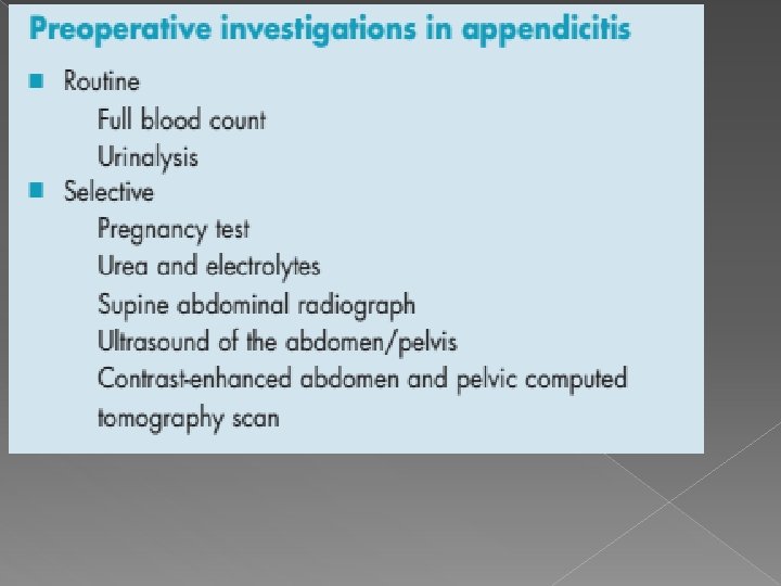
Investigation The diagnosis of acute appendicitis is essentially clinical; however, a decision to operate based on clinical suspicion alone can lead to the removal of a normal appendix in 15– 30 percent of cases. . A number of clinical and laboratory-based scoring systems have been devised to assist diagnosis. The most widely used is the Alvarado score. A score of 7 or more is strongly predictive of acute appendicitis. In patients with an equivocal score (5– 6), abdominal ultrasound or contrastenhanced CT examination further reduces the rate of negative appendicectomy. .
The Alvarado (MANTRELS) score. Alvarado score
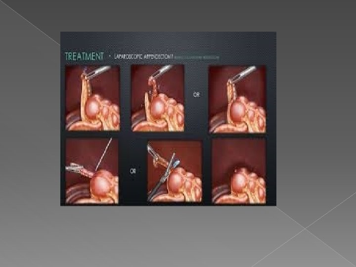
Treatment The traditional treatment for acute appendicitis is appendicectomy. a trial of conservative management in those thought not to have obstructive appendicitis. Treatment is bowel rest and intravenous antibiotics, usually metranidazole and third-generation cephalosporin. The available data indicate successful outcomes in 80– 90 per cent of patients, however there is an approximately 15 per cent recurrence rate within one year. This approach should be considered in patients with high operative.
While there should be no unnecessary delay, all patients, particularly those most at risk of serious morbidity, benefit by a short period of intensive preoperative preparation. Intravenous fluids, and appropriate antibiotics should be given. There is ample evidence that in the absence of purulant peritonitis, a single preoperative dose of antibiotics reduces the incidence of postoperative wound infection. Hyperpyrexia in children should be treated with salicylates in addition to antibiotics and. .
With appropriate use of intravenous fluids and parenteral antibiotics, a policy of deferring appendicectomy after midnight to the first case on the following morning does not increase morbidity. However, when acute obstructive appendicitis is recognised, operation should not be deferred longer n it takes to optimise the patient’s condition.
Conventional appendicectomy , the incision that is widely used for appendicectomy is the so-called gridiron incision is made at right angles to a line joining the anterior superior iliac spine to the umbilicus, its centre being along the line at Mc. Burney’s point If better access is required, it is possible to convert the gridiron to a Rutherford Morison incision by cutting the internal oblique and transversus muscles in the line of the incision.
Gridiron incision for appendicitis
Transverse or skin crease (Lanz) incision for appendicitis
Problems encountered during appendicectomy • A normal appendix is found. This demands careful exclusion of other possible diagnoses, particularly terminal ileitis, Meckel’s diverticulitis and tubal or ovarian causes in women. It is usual to remove the appendix to avoid future diagnostic difficulties, . • The appendix cannot be found. The caecum should be mobilised, and the taeniae coli should be traced to the confluence on the caecum before the diagnosis of ‘absent appendix’ is made • An appendicular tumour is found. Small tumours (under 2. 0 cm in diameter) can be removed by appendicectomy; larger tumours should be treated by a right hemicolectomy.
An appendix abscess is found and the appendix cannot be removed easily. This eventuality is rare in the era of modern diagnostic imaging. Percutaneous drainage of the abscess and intravenous antibiotic treatment is to be preferred. If found at operation, the abscess should be drained an intravenous antibiotics administered. Very rarely in the face of a frankly necrotic appendix, a caecectomy or partial right my is required.
Appendicitis complicating Crohn’s disease Providing that the caecal wall is healthy at the baseof the appendix, appendicectomy can be performed without increasing the risk of an enterocutaneous fistula. Rarely, the appendix is involved with the Crohn’s disease. In this situation, a conservative approach may be warranted, and a trial of intravenous corticosteroids and systemic antibiotics can be used to resolve the acute inflammatory process.
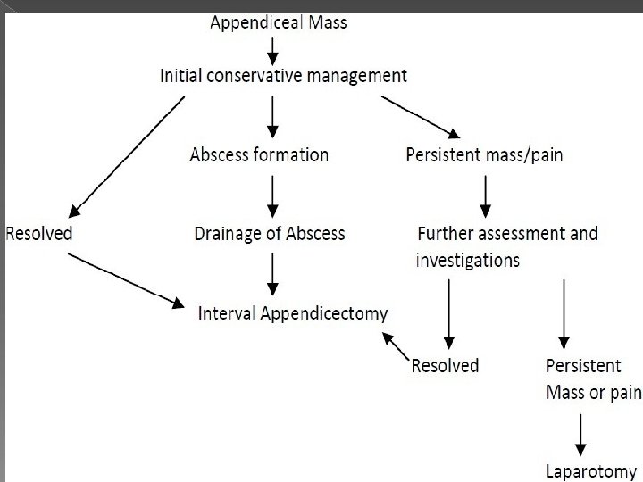
Management of an appendix mass the standard treatment is the conservative Ochsner–Sherren regimen. This strategy is based on the premise that the inflammatory process is already localised and that inadvertent surgery is difficult and may be dangerous. It maybe impossible to find the lly, a faecal fistula may form.
1. Careful recording of the patient’s condition domen regularly reexamined. 3. It is helpful to mark the limits of the mass on the al wall using a skin pencil. . 4. A contrast-enhanced CT examination of the abdomen should be performed. 5. antibiotic therapy instigated. 6. Temperature and pulse rate should be recorded 4 -hourly 7. fluid balance record maintained
Appendix abscess Failure of resolution of an appendix mass or continued spiking pyrexia usually indicates that there is pus within the phlegmonous appendix mass. Ultrasound or abdominal CT scan may identify an area suitable for the insertion of a percutaneous drain. Rarely, this is unsuccessful and laparotomy through a midline incision is
Pelvic abscess. is an occasional complication of appendicitis and can occur irrespective of the position of the appendix within the peritoneal cavity. The most common presentation is a spiking pyrexia several days after appendicitis; indeed, the patient may already have been discharged from hospital. Pelvic pressure or discomfort , loose stool or tenesmus is common. Rectal examination reveals a boggy mass in the pelvis, anterior to the rectum Dx: Pelvic ultrasound or CT scan. nder general anaesthetia , 2. guided percutaneous drainage.
Postoperative complications Wound infection usually presents with pain and erythema of the wound on the 4 th or 5 th postoperative day, often soon after hospital discharge. Treatment is by wound drainage and antibiotics when required. Intra-abdominal abscess a spiking fever, malaise and anorexia developing 5– 7 days after operationis suggestive of an intraperitonealcollection Abdominalultrasonography and CT scanning greatly facilitate diagnosis and allow percutaneous drainage. Laparotomy should be considered in in whom imaging fails to show a collection, particularly those with.
Ileus Respiratory , respiratory complications are rare following appendicectomy. Adequate postoperative analgesia and physiotherapy, when appropriate, reduce the incidence. Venous thrombosis and embolism These conditions are rare, except in the elderly and in women taking the oral contraceptive pill. Appropriate prophylactic measures should be. Adhesive intestinal obstruction
Portal pyaemia (pylephlebitis) This is a rare but very serious complication of gangrenous appendicitis associated with high fever, rigors and jaundice. It is caused by septicaemia in the portal venous system and leads to the development of intrahepatic abscesses (often multiple). Treatment is with systemic antibiotics and percutaneous drainage of hepatic abscesses as appropriate Faecal fistula This is occur if the encircling stitch has been put in too deeply or if the caecal wall was involved by oedema or inflammation. Occasionally, a fistula may result following appendicectomy in Crohn’s disease. Conservative management with low
Recurrent acute appendicitis Appendicitis is notoriously recurrent. . The attacks vary in intensity and may occur every few months, and the majority of cases ultimately culminatein severe acute appendicitis. If a careful history is taken from patients many remember having had milder but similar attacks of pain.
Neoplasms of the appendix Carcinoid tumours arise in argentaffin tissue (Kulchitsky cells of the crypts of Lieberkühn)and are most common in the vermiform appendix. The tumour can occur in any part of the appendix, but it is frequently found in the distalthird. The neoplasm feels moderately hard and, on sectioning the appendix, it can be seen as a yellow tumour betweenthe intact mucosa and the peritoneum. Unlike carcinoid tumours arising in other parts of the intestinal tract, carcinoid tumour of the ndix rarely gives rise to metastases.
Appendicectomy has been shown to be sufficient treatment, unless the caecal wall is involved, the tumour is 2 cm or more in size or involved lymph nodes are found, when right hemicolectomy is indicated. Other appendiceal tumours Goblet cell carcinoid tumour. Mucinous cystadenoma Primary adenocarcinoma of the appendix is extremely rare. It is usually of the colonic type and should be treated by right hemicolectomy (as a second-stage procedure if the condition is not recognised at the first