Appendicitis History 1800 s perityphlitis Melier 1827 Rignald




- Slides: 48
Appendicitis 외과 최 성 일
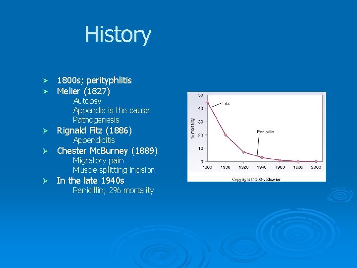
History Ø Ø 1800 s; perityphlitis Melier (1827) Ø Rignald Fitz (1886) Ø Chester Mc. Burney (1889) Ø In the late 1940 s Autopsy Appendix is the cause Pathogenesis Appendicitis Migratory pain Muscle splitting incision Penicillin; 2% mortality
Anatomy & Embryology Cecum; 5 th week Ø Appendix; 8 th Ø Outpouching of cecum Ø Appendiceal orifice Always at the confluence of cecal taenia Ø Lymphoid follicle Submucosa Increase through adolescence Ø Length; 9 cm (2 -20)
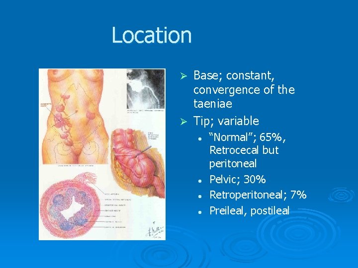
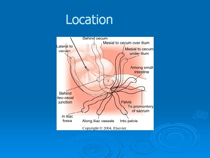
Location Base; constant, convergence of the taeniae Ø Tip; variable Ø l l “Normal”; 65%, Retrocecal but peritoneal Pelvic; 30% Retroperitoneal; 7% Preileal, postileal
Location
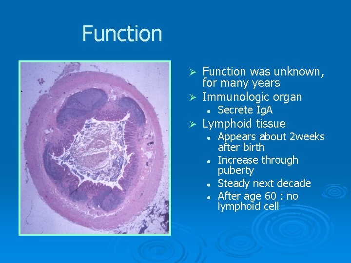
Function was unknown, for many years Ø Immunologic organ Ø l Ø Secrete Ig. A Lymphoid tissue l l Appears about 2 weeks after birth Increase through puberty Steady next decade After age 60 : no lymphoid cell
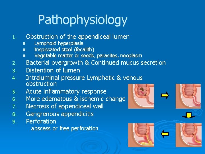
Pathophysiology Obstruction of the appendiceal lumen 1. l l l 2. 3. 4. 5. 6. 7. 8. 9. Lymphoid hyperplasia Inspissated stool (fecalith) Vegetable matter or seeds, parasites, neoplasm Bacterial overgrowth & Continued mucus secretion Distention of lumen Intraluminal pressure Lymphatic & venous obstruction Acute inflammatory response More edematous & ischemic change Necrosis of appendiceal wall Gangrenous appendicitis Perforation abscess or free perforation
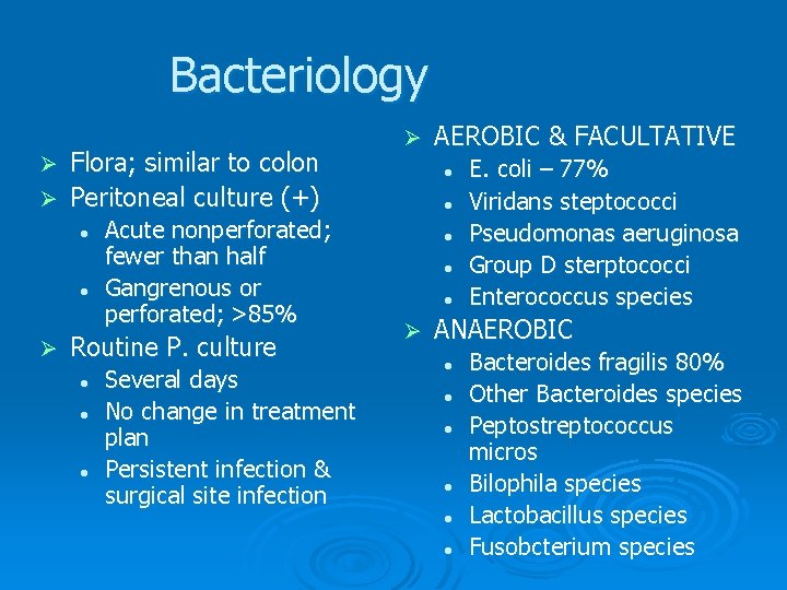
Bacteriology Flora; similar to colon Ø Peritoneal culture (+) Ø Ø l l Ø Acute nonperforated; fewer than half Gangrenous or perforated; >85% Routine P. culture l l l Several days No change in treatment plan Persistent infection & surgical site infection AEROBIC & FACULTATIVE l l l Ø E. coli – 77% Viridans steptococci Pseudomonas aeruginosa Group D sterptococci Enterococcus species ANAEROBIC l l l Bacteroides fragilis 80% Other Bacteroides species Peptostreptococcus micros Bilophila species Lactobacillus species Fusobcterium species
Diagnosis
Clinical Ø History & Physical finding l Primary Ø Laboratory & Radiology l Additional assistance Ø Typical history; 50% l Anorexia & nausea l Epigastric Periumbilical (activation of visceral afferent neurons) RLQ pain (parietal peritoneum irritation) l Migrating pain
Clinical; P/E Ø Ø Ø Diminished bowel sound Direct tenderness & muscle spasm in RLQ Rebound tenderness BT; mildly elevated(38 OC) variable Mc. Burney point Maximal tender point Ø Rectal examination l l Ø Little value Mass Rupture l l l Abdominal pain; intense & more diffuse High fever; 39 – 40 OC Quite ill appearance Obvious deterioration Pain-free interval; rare
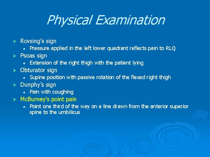
Physical Examination Ø Rovsing's sign l Ø Psoas sign l Ø Supine position with passive rotation of the flexed right thigh Dunphy’s sign l Ø Extension of the right thigh with the patient lying Obturator sign l Ø Pressure applied in the left lower quadrant reflects pain to RLQ Pain with coughing Mc. Burney's point pain l Point one third of the way on a line drawn from the anterior superior spine to the umbilicus
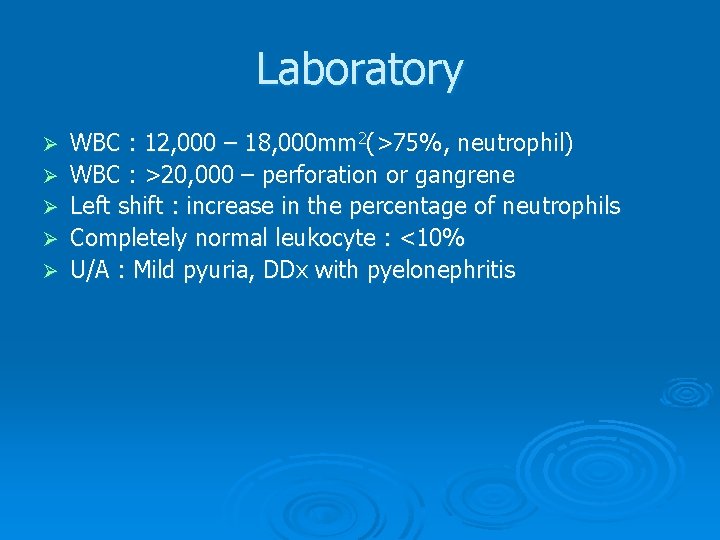
Laboratory Ø Ø Ø WBC : 12, 000 – 18, 000 mm 2(>75%, neutrophil) WBC : >20, 000 – perforation or gangrene Left shift : increase in the percentage of neutrophils Completely normal leukocyte : <10% U/A : Mild pyuria, DDx with pyelonephritis
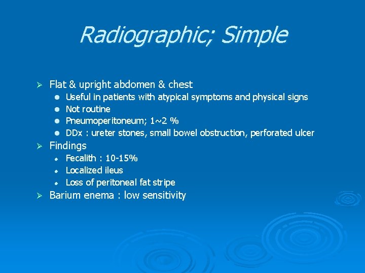
Radiographic; Simple Ø Flat & upright abdomen & chest Useful in patients with atypical symptoms and physical signs l Not routine l Pneumoperitoneum; 1~2 % l DDx : ureter stones, small bowel obstruction, perforated ulcer l Ø Findings l l l Ø Fecalith : 10 -15% Localized ileus Loss of peritoneal fat stripe Barium enema : low sensitivity
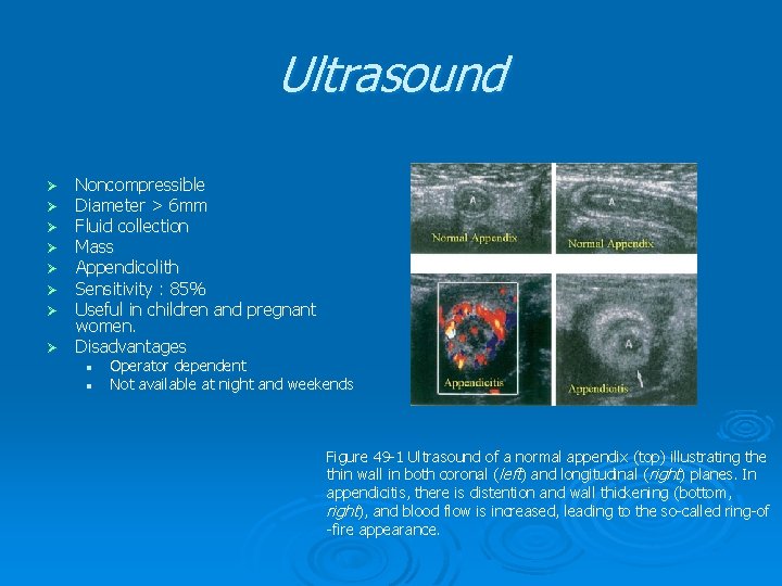
Ultrasound Noncompressible Diameter > 6 mm Fluid collection Mass Appendicolith Sensitivity : 85% Useful in children and pregnant women. Ø Disadvantages Ø Ø Ø Ø l l Operator dependent Not available at night and weekends Figure 49 -1 Ultrasound of a normal appendix (top) illustrating the thin wall in both coronal (left) and longitudinal (right) planes. In appendicitis, there is distention and wall thickening (bottom, right), and blood flow is increased, leading to the so-called ring-of -fire appearance.
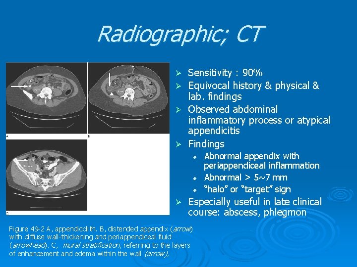
Radiographic; CT Ø Ø Sensitivity : 90% Equivocal history & physical & lab. findings Observed abdominal inflammatory process or atypical appendicitis Findings l l l Ø Abnormal appendix with periappendiceal inflammation Abnormal > 5~7 mm “halo” or “target” sign Especially useful in late clinical course: abscess, phlegmon Figure 49 -2 A, appendicolith. B, distended appendix ( arrow) with diffuse wall-thickening and periappendiceal fluid (arrowhead). C, mural stratification, referring to the layers of enhancement and edema within the wall (arrow),
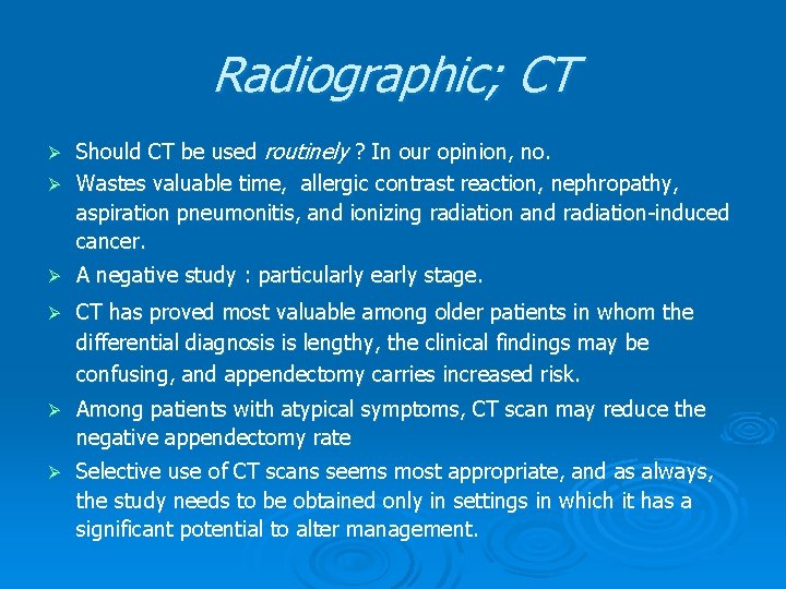
Radiographic; CT Should CT be used routinely ? In our opinion, no. Ø Wastes valuable time, allergic contrast reaction, nephropathy, aspiration pneumonitis, and ionizing radiation and radiation-induced cancer. Ø Ø A negative study : particularly early stage. Ø CT has proved most valuable among older patients in whom the differential diagnosis is lengthy, the clinical findings may be confusing, and appendectomy carries increased risk. Ø Among patients with atypical symptoms, CT scan may reduce the negative appendectomy rate Ø Selective use of CT scans seems most appropriate, and as always, the study needs to be obtained only in settings in which it has a significant potential to alter management.
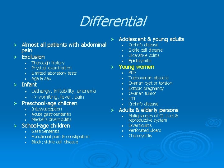
Differential Almost all patients with abdominal pain Ø Exclusion Ø Ø l l l l Ø l l Lethargy, irritability, anorexia -> vomiting, fever, pain l l Intussusception Acute gastroenteritis Meckel’s diverticulitis l l l School-age children l l l Gastroenteritis Functional pain & constipation Black; sickle cell disease l Ø Crohn’s disease Sickle cell disease Ulcerative colitis Epididymitis Young women l Preschool-age children l Ø l Infant l Ø Thorough history Physical examination Limited laboratory tests Age & sex Adolescent & young adults PID Tuboovarian abscess Ovarian cyst or torsion Ectopic pregnancy Ovarian tumor UTI Crohn’s disease Adults & elderly persons l l Malignancies of GI tract & reproductive system Diverticulitis Perforated ulcers Cholecystitis
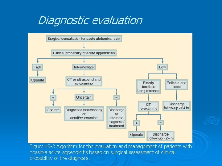
Diagnostic evaluation Figure 49 -3 Algorithm for the evaluation and management of patients with possible acute appendicitis based on surgical assessment of clinical probability of the diagnosis.
Types of Treatment Depending on the stage of disease Fluid resuscitation before surgery; 1 or 2 hours
Stage of Appendicitis Acute; Medical, Surgical Ø Perforated Ø Presentation with a mass or late complicated appendicitis Ø Chronic or Recurrent Ø
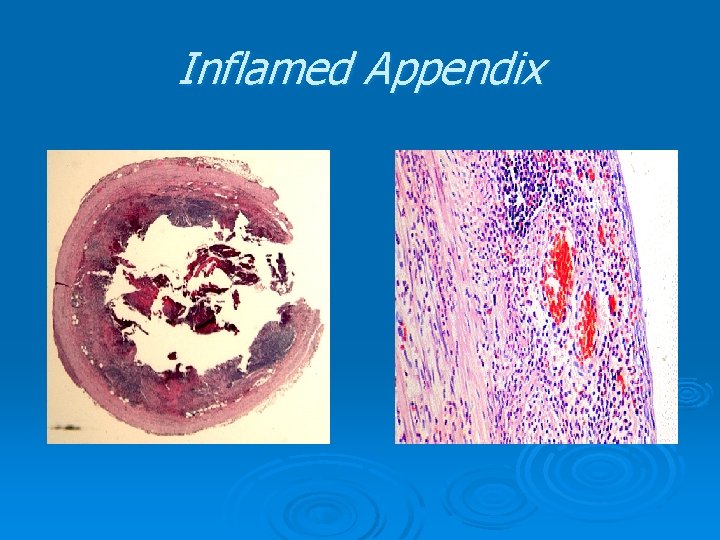
Inflamed Appendix
Medical Treatment Should undergo urgent appendectomy Ø Antibiotic therapy Ø l l l Ø Initial success; 95% Recurrence rate; 35% Used in rare situation; sailors on submarine tours Current standard; Operation Ø Prophylactic antibiotics l l Ø Acute case Single dose of cefoxitin or cefotetan Negative exploration rate l l l Past; 20% Present; ultrasound- and CTassisted diagnosis Female; still high
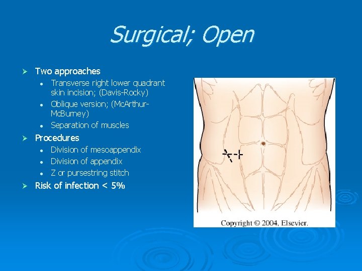
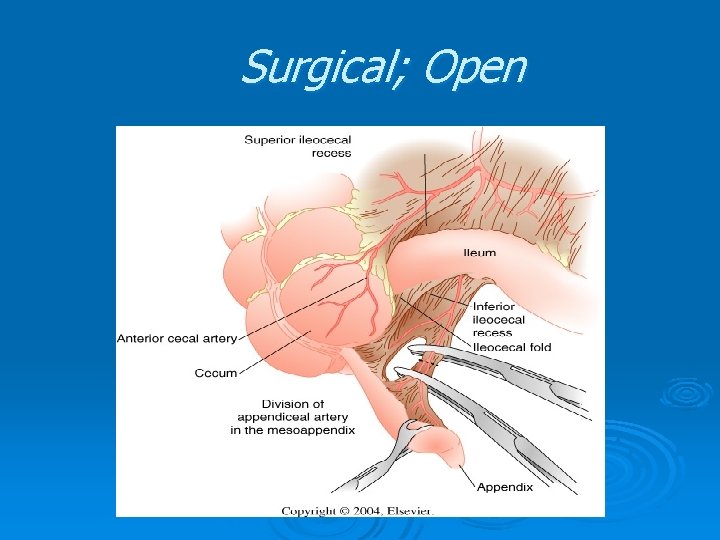
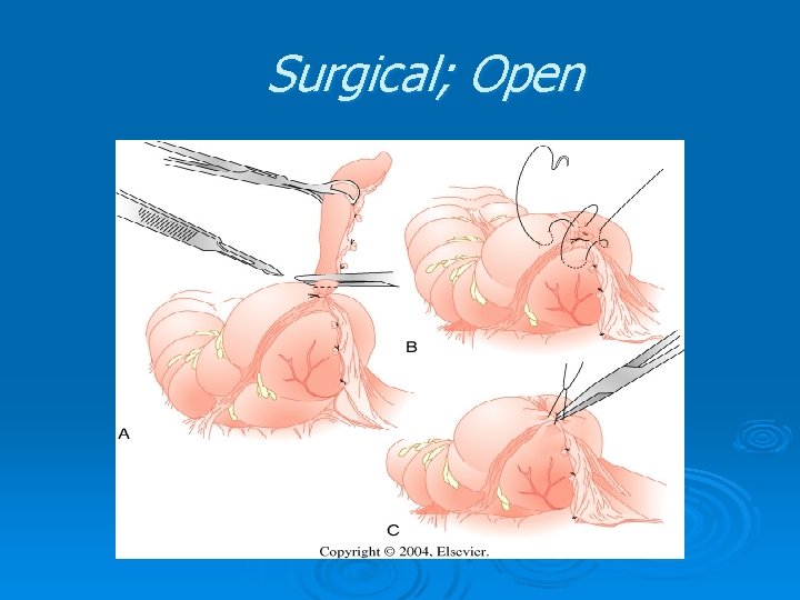
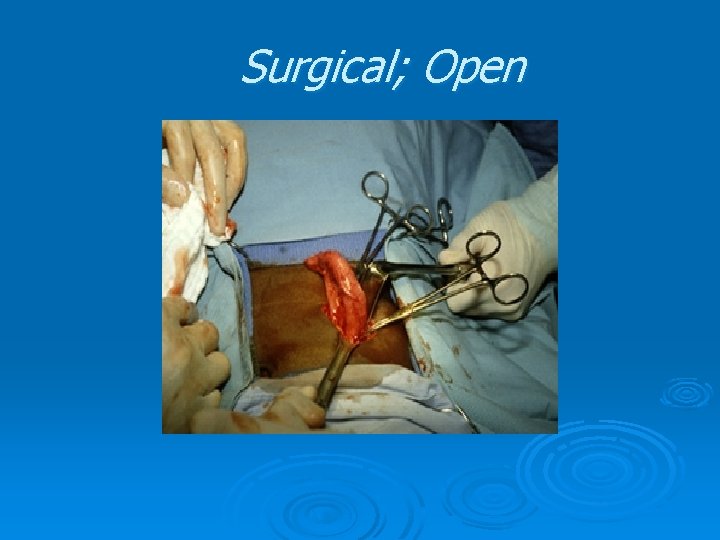
Surgical; Open Ø Two approaches l l l Ø Procedures l l l Ø Transverse right lower quadrant skin incision; (Davis-Rocky) Oblique version; (Mc. Arthur. Mc. Burney) Separation of muscles Division of mesoappendix Division of appendix Z or pursestring stitch Risk of infection < 5%
Surgical; Open
Surgical; Open
Surgical; Open
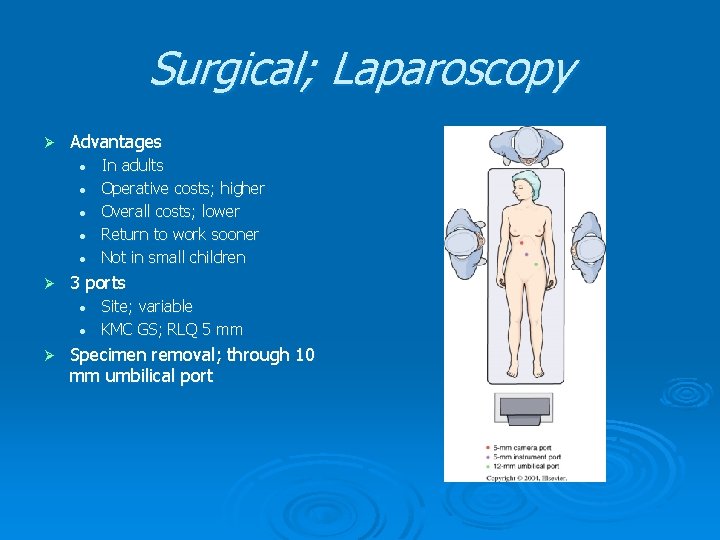
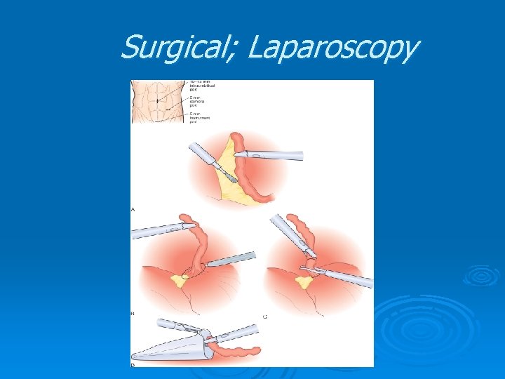
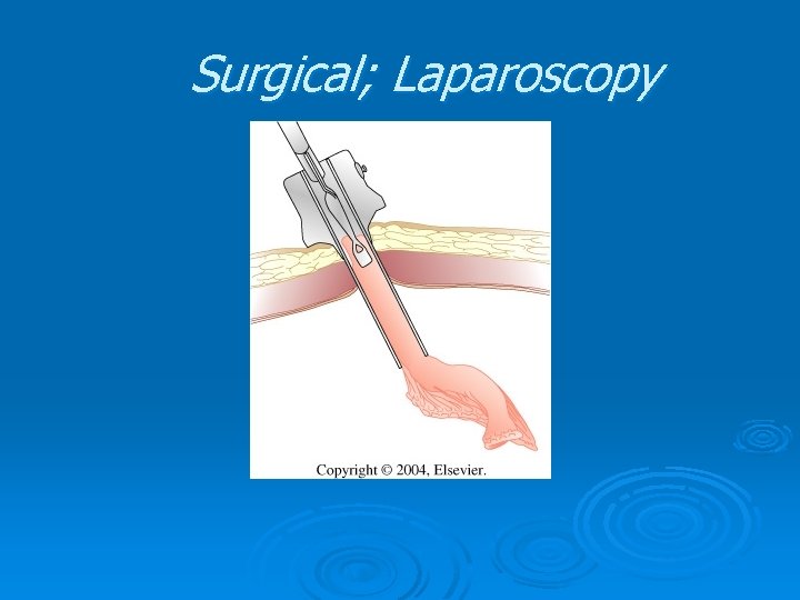
Surgical; Laparoscopy Ø Advantages l l l Ø 3 ports l l Ø In adults Operative costs; higher Overall costs; lower Return to work sooner Not in small children Site; variable KMC GS; RLQ 5 mm Specimen removal; through 10 mm umbilical port
Surgical; Laparoscopy
Surgical; Laparoscopy
Surgical; Laparoscopy
Perforated Appendicitis Perforated or gangrenous vs. acute nonperforated disease Ø Appendix; already perforated Ø Longer duration of symptoms, high fever Ø Higher WBC count Ø Ø Volume depleted; l Ø Laparoscopic appendectomy : l Ø Fluid resuscitation before operative intervention decrease postoperative wound complication and shorter hospital stay Peritonitis; Antibiotics l l l Appropriate broad-spectrum intravenous antibiotics As soon as diagnosis established Duration; controversial
Presentation With a Mass or Late Complicated Appendicitis Palpable RLQ mass; 5% Ø l l Management Ø • • Ø Discrete abscess Phlegmonous inflammation Percutaneous drainage under image guidance IV antibiotics (Aerobic gram-negative & anaerobic organisms) In late, complicated appendicitis Appendectomy; hazardous
Interval Appendectomy Ø Localized appendiceal abscess l l Ø Initially CT-guided percutaneous drainage IV antibiotics & fluid administration Laparoscopic appendectomy; 6 weeks Children; 8~12 weeks Controversy l l l 15% recur -> early appendectomy 15/17; patent to tip 1/10; appendectomy needed (long term flu-up)
Algorithm for the management of appendiceal abscess
Chronic or Recurrent Ø Controversial l Ø Mechanism l l Ø Intermittent bouts of obstruction of appendiceal lumen Spontaneous remission Mild local inflammation Chronic RLQ discomfort CT finding l Ø But, possible Similar to acute Appendectomy l l Histological abnormality; 92% Completely cured; 95%
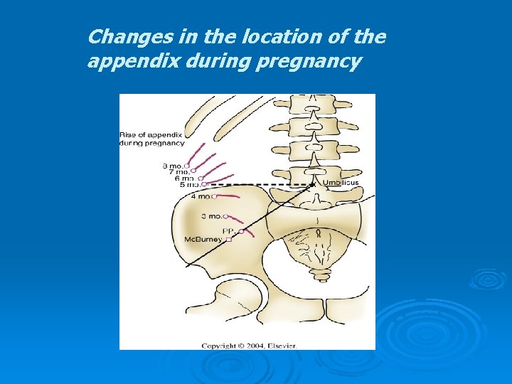
Appendicitis During Pregnancy Ø Ø Ø Ø Appendicitis & cholecystitis; most common causes of abdominal pain during pregnancy Tenderness location; varies th l 5 month of gestation; above the iliac crest, rotated medially WBC count; not helpful l Frequently elevated in pregnancy GI symptoms; not helpful Ultrasound; helpful Suspicion Early surgical intervention l Negative laparotomy; minimal fetal loss l Perforation; high fetal death, maternal death Laparoscopy; possible
Changes in the location of the appendix during pregnancy
Normal appearing appendix Ø Ø Ø Ø Inspection of T. ileum and ascending colon IBD Mesenteric adenitis Meckel’s diverticulitis Gallbladder, ovary, sigmoid colon Pancreatitis Omental infartion or torsion Appendix should be removed ? ? ? l l Little morbidity Pathological abnormality (grossly normal app. )
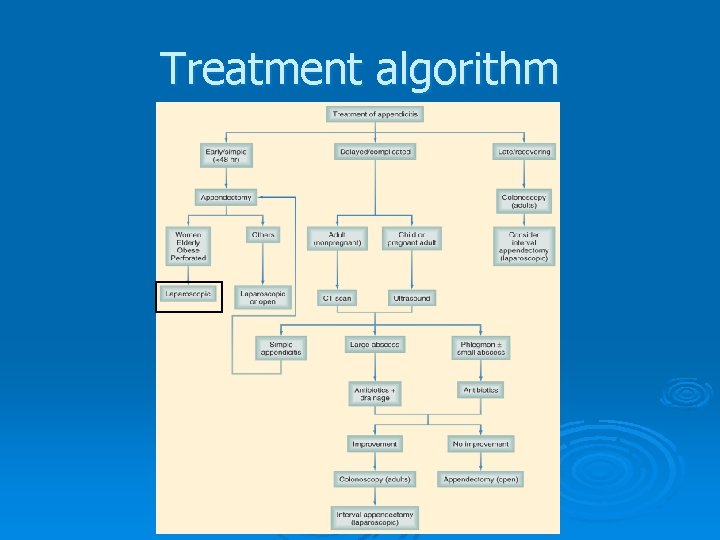
Treatment algorithm
Postoperative Complications
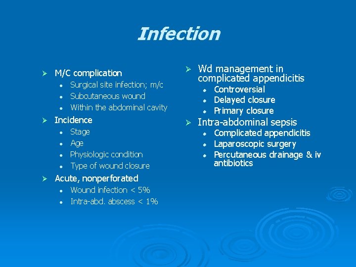
Infection Ø M/C complication l l l Ø Surgical site infection; m/c Subcutaneous wound Within the abdominal cavity Incidence l Stage Age Physiologic condition Type of wound closure Acute, nonperforated l l Ø Wound infection < 5% Intra-abd. abscess < 1% Wd management in complicated appendicitis l l l Ø Controversial Delayed closure Primary closure Intra-abdominal sepsis l l l Complicated appendicitis Laparoscopic surgery Percutaneous drainage & iv antibiotics
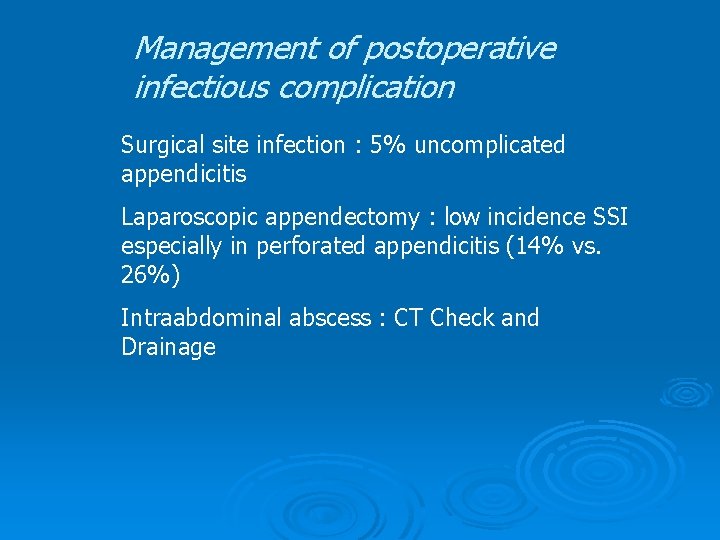
Management of postoperative infectious complication Surgical site infection : 5% uncomplicated appendicitis Laparoscopic appendectomy : low incidence SSI especially in perforated appendicitis (14% vs. 26%) Intraabdominal abscess : CT Check and Drainage
Others Ø Bowel obstruction l l Ø Infertility l l Ø 1% Within 6 months Sufficiently low Perforated > Nonperforated Miscellaneous l l l UTI Pneumonia Fecal fistula; perforated
See you, next year!