Diseases of the gastrointestinal tract Dr Nesreen Bataineh

Diseases of the gastrointestinal tract Dr. Nesreen Bataineh MD, FRCPath

Adenomas � Neoplastic � Common polyps in colon � The incidence increases with age (∼ 50% after age of 50 y*). M=F. � Sporadic or syndromatic. q. Familial predisposition to sporadic adenomas.

Pathology of adenomas � Epithelial proliferation and dysplasia*. � Dysplasia may be of low or high grade. � Increased risk of transformation to carcinoma.

Pathology of adenomas � Subtypes § based on the epithelial architecture: Tubular adenomas > 75% tubules q. Most common. q. Common in rectosigmoid. q. Usually multiple, small & pedunculated q Villous adenomas >50% villous projections q. Common in rectosigmoid. q. Most are large & sessile with cauliflower appearance. q. Invasive carcinoma is found in 40%. q Tubulovillous projections. adenomas 25 -50% villous

Malignant risk in adenomas � Depends on three features: 1. Polyp size q ↑ risk if diameter is > 4 cm*. 2. Severity of epithelial dysplasia: q High grade dysplasia often found in villous areas. 3. Histologic architecture q ↑risk in villous adenoma.

Adenomas

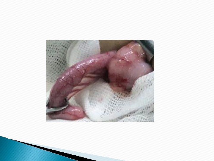
Tubular adenoma - The head is composed of neoplastic epithelium, forming rounded or tubular glands lined by dysplastic cells - Notice the contrast between dysplastic & normal tubules

Villous adenoma

Clinical picture of polyps � Most � If adenomas are asymptomatic. large can cause obstruction. � Most distal adenomas are more symptomatic: § Bleeding per rectum § May prolapse through the anus � Villous adenomas may secrete mucoid material rich in protein and potassium (producing hypoproteinemia or hypokalemia).

Familial Polyposis Syndromes � Uncommon � Most AD disorders. are due to germline mutations in tumor suppressor genes. � Increased risk of intestinal and (in some syndromes) extra-intestinal cancers.

Familial adenomatous polyposis � AD syndrome Mutation in APC gene. § Classic FAP § Attenuated FAP § Gardner syndrome § Turcot syndrome
 carpet the mucosal surface. �A minimum")
Classic FAP � Innumerable colonic adenomas (usually thousands) carpet the mucosal surface. �A minimum number of 100 polyps is required for the diagnosis. � Most polyps are tubular adenomas. � Multiple adenomas anywhere in the GI including ampulla of Vater and stomach. � A 100% lifetime risk of progression to cancer at younger age (< 30 yrs) unless prophylactic colectomy is performed.

FAP associated with adenocarcinoma
 in the proximal colon. �")
Attenuated FAP � Fewer � Mainly polyps (average 30) in the proximal colon. � Increased risk of colonic adenocarcinoma in 50% of cases

Gardner syndrome � Intestinal polyps as classic FAP � Multiple osteomas � Epidermal inclusion cysts � Fibromatosis* � Abnormal dentition* Multiple osteomas

Turcot syndrome � Adenomatous � CNS tumors. colonic polyposis as FAP
 syndrome Lynch syndrome � AD familial syndrome of DNA")
Hereditary Nonpolyposis Colorectal Cancer (HNCC) syndrome Lynch syndrome � AD familial syndrome of DNA mismatch repair genes (esp. MSH 2 & MLH 1 Most common). � This leads to genomic instability (microsatellite instability)*. � Subsequent mutations in growth regulatory genes as TGF-B receptor II & BAX � Inactivation
 syndrome � Increased risk of colorectal cancer q. Usually")
Hereditary Nonpolyposis Colorectal Cancer (HNCC) syndrome � Increased risk of colorectal cancer q. Usually multiple. q. At younger age*. q. Mainly in the cecum & proximal colon. q. Usually mucinous or poorly differentiated. q. Infiltration by intraepithelial lymphocytes. q. Of better Prognosis Compared to sporadic Colon cancer.
 syndrome �Risk of gastric and small intestinal adenocarcinoma �Risk")
Hereditary Nonpolyposis Colorectal Cancer (HNCC) syndrome �Risk of gastric and small intestinal adenocarcinoma �Risk as: of extra-intestinal malignancies § Endometrial cancer § Cancer of ureter & renal pelvis § Cancer of pancreas § Cholangiocarcinoma

Juvenile polyposis syndrome � AD syndrome � Mutation � 3 -100 of SMAD 4 gene*. juvenile polyps**. � Increased risk of adenomas and carcinomas of the GI (stomach, SI, colorectal ).
 gene*. �")
Peutz-Jeghers syndrome � AD syndrome � Mutation in STK 11 (LKB 1) gene*. � Characterized by: q. Multiple hamartomatous (Peutz-Jeghers polyps) in the small intestine (100%), colon (30%) & stomach (25%). q. Melanotic mucosal and cutaneous pigmentation around the lips, oral mucosa and genitalia � Increased risk of intestinal & extraintestinal malignancies of (uterine CX, ovary & breast)**

Cowden syndrome � AD syndrome. � Mutation of PTEN tumor suppressor gene. � Intestinal hamartomatous polyps in the GI. � Oral keratosis and oral papilloma. � Benign skin tumors (trichilemmomas). � Increased risk of thyroid and breast cancer. � No increased risk of GI cancer.

Colorectal Carcinoma � Colorectal CA has a worldwide distribution. � Age: The peak incidence is 60 to 70 ys (< 20% occur < age of 50). � Gender: M>F. � Most � Risk arises sporadically in adenomas. factors: Genetic & environmental influences

Risk factors of colorectal cancer Dietary factors: Low fiber diet High content of refined carbohydrates High fat content (as from meat) Decreased intake of micronutrients vitamins A, C, and E. such as The use of aspirin and other NSAIDs are protective Inhibition of cyclooxygenase-2 (COX-2) The presence of adenomas (the precancerous lesions) Preexisting UC or one of the familial syndromes
")
Colorectal Carcinogenesis � Two pathogenetically distinct pathways: q The APC/β-catenin pathway (the adenoma-carcinoma sequence) 80% of sporadic colorectal CA. q The mismatch repair (microsatellite instability) pathway 10% to 15% of sporadic colorectal CA.

Adenoma-carcinoma sequence

The microsatellite instability pathway

Morphology � Site § § in order of frequency: The rectosigmoid The cecum or ascending colon The transverse colon The descending colon � May be single or multiple

Gross appearance � Tumors § § in the proximal colon: Grow as polypoid, exophytic masses Obstruction is uncommon. � Tumors § in the distal colon: Annular encircling lesions (napkin-ring constrictions) and narrow the lumen. � Both forms of neoplasm directly penetrate the bowel wall and may appear as firm masses on the serosal surface.

Colon CA – polypoid mass

Microscopic apearance � Almost all colorectal CA are adenocarcinomas � Mucinous adenocarcinoma q> 50% extracellular mucin q> 50% signet ring � Signet ring adenocarcinoma Poor Px � Cancers of the anal zone are squamous cell carcinoma. � Grading: G 1 (well diff. ), G 2, G 3 & G 4 (undiff. ).

Adenocarcinoma of colon

Tumor spread � Direct spread. � Lymph � Distant bones. node metastasis to liver (mainly), lungs and � Carcinomas of the anal region: q Locally invasive and metastasize to regional lymph nodes and distant sites.

Clinical Features � Asymptomatic for years. � Symptoms develop insidiously. � Cecal and right colonic cancers: q Fatigue, weakness, & iron deficiency anemia � Left-sided cancers: q Occult or frank bleeding, changes in bowel habit (partial obstruction), or crampy left lower quadrant pain.

Staging of colorectal CA

Prognosis � The single most important prognostic indicator is the STAGE at time of diagnosis. (depth of invasion T, LNs N, distant mets M) � Microscopic type. q Mucinous & signet ring are poor � Histologic grade.

Appendix �The appendix is a normal diverticulum of the cecum. �Diseases of the appendix: q. Acute appendicitis. q. Appendiceal tumors. true

Acute Appendicitis common in adolescents and young adults, but may occur in any age group. � Most � The lifetime risk for appendicitis is 7 %. � M: F 1. 5: 1. � Need to be differentiated from other causes of acute abdomen (False positive ~ 20%)*.

Pathogenesis � In 50% - 80% luminal obstruction: � In minority, no significant obstruction. § § Small stone-like mass of stool (Fecalith). Mass of worms (oxyuriasis vermicularis). Gallstone. Tumor.

Pathogenesis Progressive increases in intraluminal pressure ⇓ Compromise of venous outflow ⇓ Ischemic injury & stasis of luminal contents ⇓ Bacterial proliferation ⇓ Inflammation with tissue edema & neutrophilic infiltration of the lumen, muscular wall & periappendiceal soft tissues

Morphology � Early acute appendicitis: q Gross § appearance: A dull, granular, erythematous surface. q Microscopic § § § appearance: Diagnosis requires neutrophilic infiltration of the muscularis propria*. Subserosal vessels are congested. Mild perivascular neutrophilic infiltrate within all layers of the wall.

Morphology � Acute suppurative appendicitis*: q The serosa is covered by fibrinopurulent exudate. q Focal abscesses may be formed within the wall. � Acute gangrenous appendicitis: q Gangrenous necrosis. q Often followed by rupture and suppurative peritonitis.

Acute suppurative appendicitis

Acute suppurative appendicitis

Perforated gangrenous appendix

Clinical Features � Periumbilical pain that ultimately localizes to the right lower quadrant. � Nausea, vomiting & low-grade fever. � Mc. Burney’s sign. � Mildly elevated peripheral white cell count. Complications: � Appendiceal perforation. � Peritonitis. � Portal venous thrombosis. � Liver abscess. � Bacteremia.

- Slides: 48