The Microscopes The Microscope 1 The microscope is


 and")


 • 1. The image is formed by aiming a")


- Slides: 54
The Microscopes
• The Microscope • 1. The microscope is an optical instrument that uses a lens or combination of lenses to magnify and resolve the fine details of an object.
• Magnifying glasses were the first microscopes. They made things look bigger because of the way light rays are bent by the glass.
• A compound microscope is used to get better magnification. They are made by using two lenses mounted at the end of a tube.
• The object is placed under the lower lens (the objective lens) and the magnified image is viewed through the upper lens (the eyepiece lens).
• The image formed by the objective lens is inverted and magnified. • The eyepiece lens magnifies it more.
• There are five types of microscopes used in forensics.
The Compound Microscope • 1. Consists of a mechanical system and an optical system.
• 2. The mechanical system is composed of a base, arm, stage, body tube, coarse adjustment, and fine adjustment.
• 3. The optical system consists of –a. An illuminator, which is a light source. For transparent objects the light source is placed underneath. This is called transmitted illumination.
When objects are not transparent the light must be placed above the object. This is vertical or reflected illumination.
• b. A condenser. The condenser collects light rays from the base illuminator and concentrates them on the object.
• c. An objective lens. Closest to the object. Most have two or three objectives (marked with the magnification). If the object stays in focus when switching objectives it is said to be parafocal.
• d. Eyepiece or ocular lens. Closest to the eye. One eyepiece = monocular, Two eyepieces = binocular.
• e. The ability of an objective lens to resolve details into 2 clear images instead of one blurry one is directly proportional to the numerical aperture value of the objective lens. The larger the N. A. , the better the detail.
• 4. To decide the power magnification you need to use always start out with the lowest power. Low magnification will give you a larger field of view. This means you will be able to see more of the object.
• The higher the magnification the smaller the field of view. • 5. Depth of focus is another function of magnifying power.
• This is the depth of the object that is in focus. The higher the magnification the shallower the depth of view.
The Comparison Microscope • 1. Gives forensic scientists the ability to do a side-by-side comparison of specimens.
• 2. It is basically two compound microscopes joined together by an optical bridge. • 3. Often used in ballistics to compare bullet marks.
The Stereoscopic Microscope • 1. The most commonly used and most versatile microscope used in forensic lab.
• 2. Not as powerful as other microscopes but it has a much larger working distance. This is the space between the specimen and the objective lens.
The Polarizing Microscope • 1. Most light vibrates in all directions perpendicular to the direction of the beam. Light can be passed through a polarizer which only allows light in one plane to pass through.
• The light that passes through is called planepolarized light. • Ex. Glare reducing sunglasses.
• If two polarizers are used the first is called a polarizer and the second is called an analyzer. • If they are held parallel to each other light will pass through them.
• If they are held perpendicular to each other no light passes through them. • 2. These microscopes are used to identify minerals and synthetic fibers.
The Microspectrophotometer • An instrument that links a microscope and a spectrophotometer. • It helps with identification and comparison of materials.
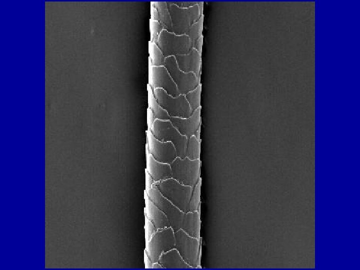
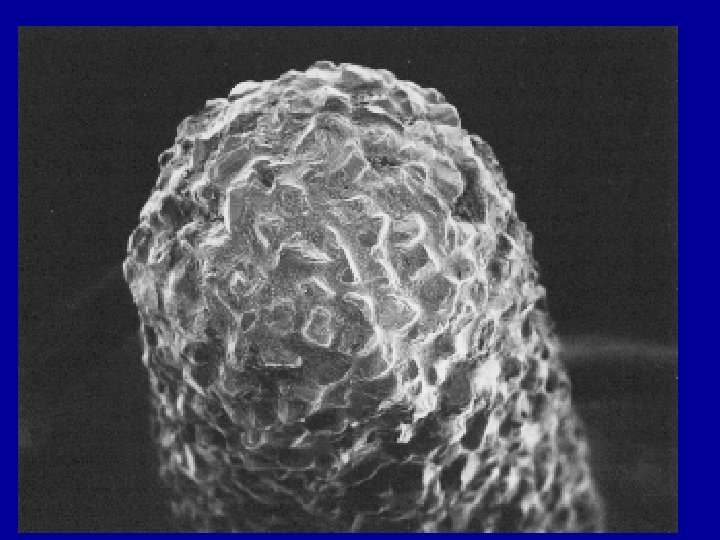
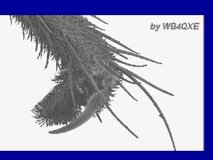
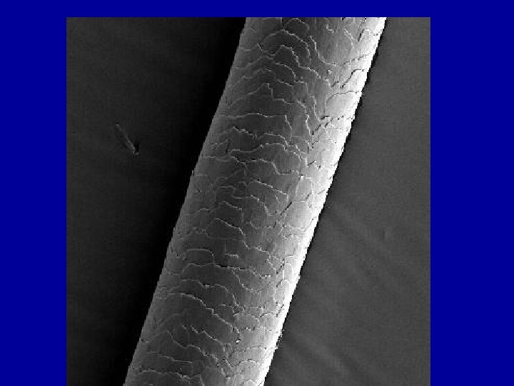
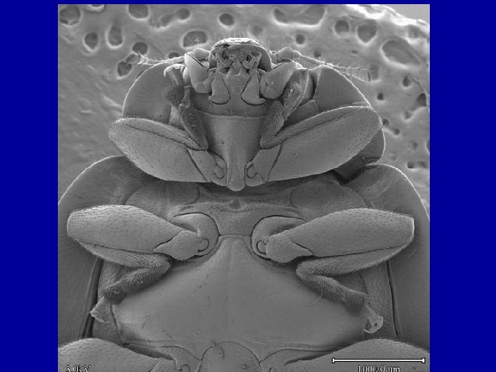
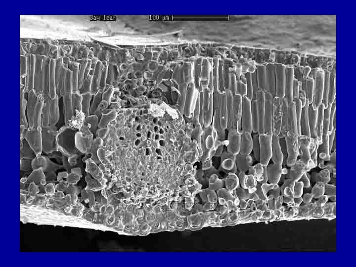
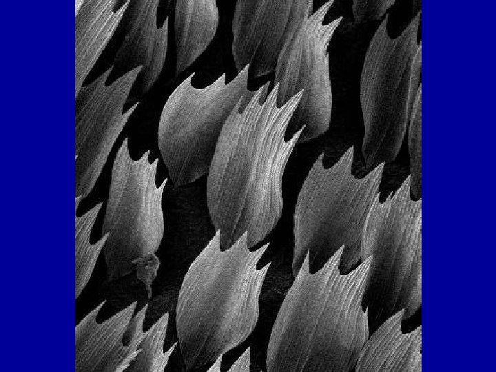
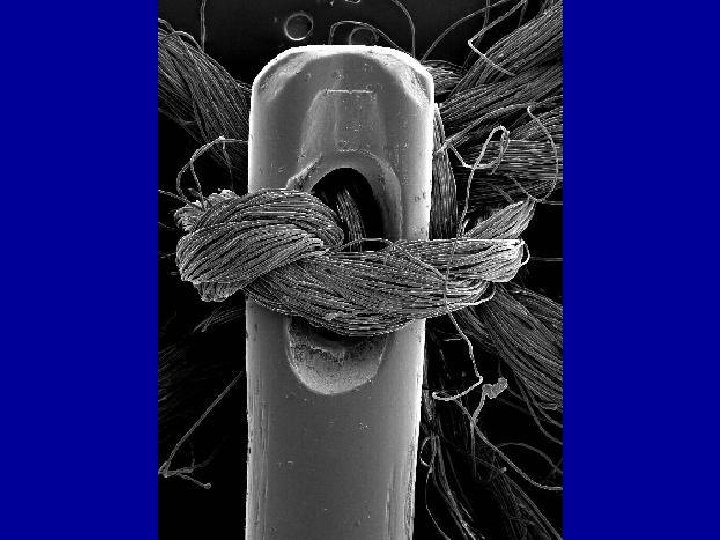
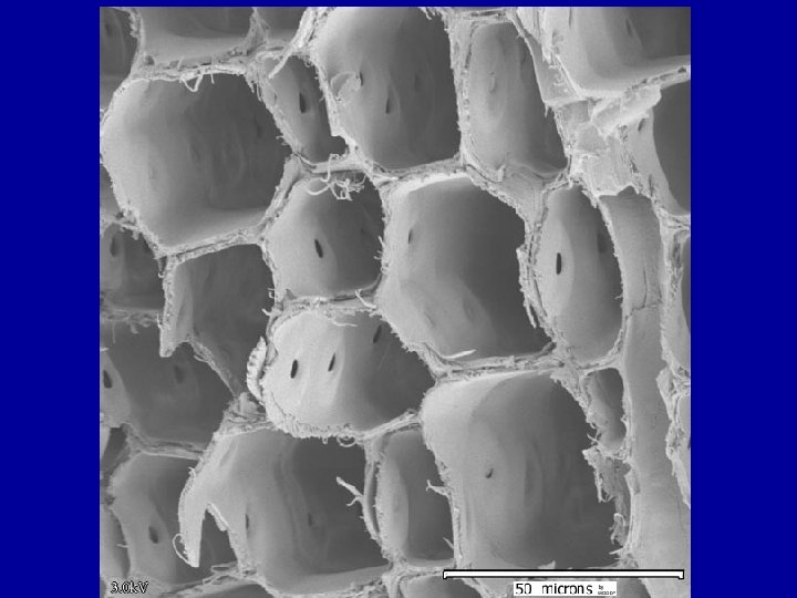
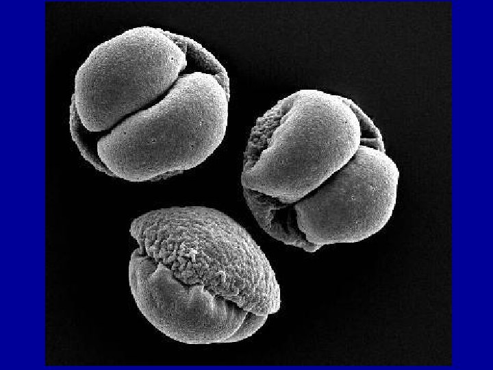
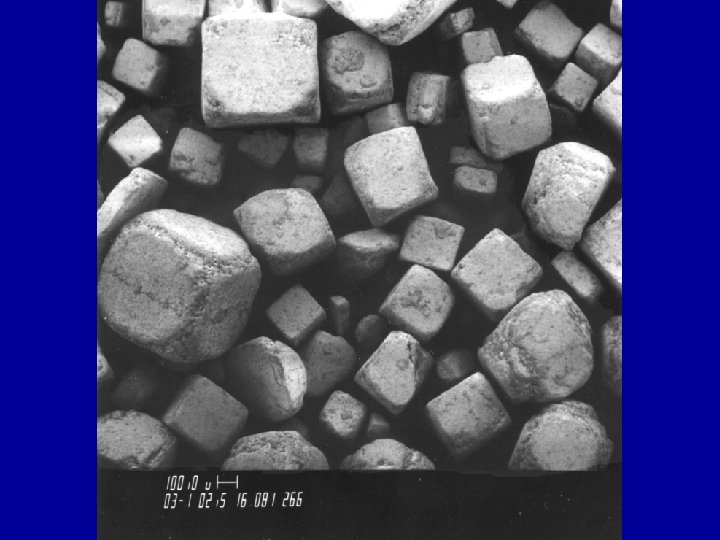
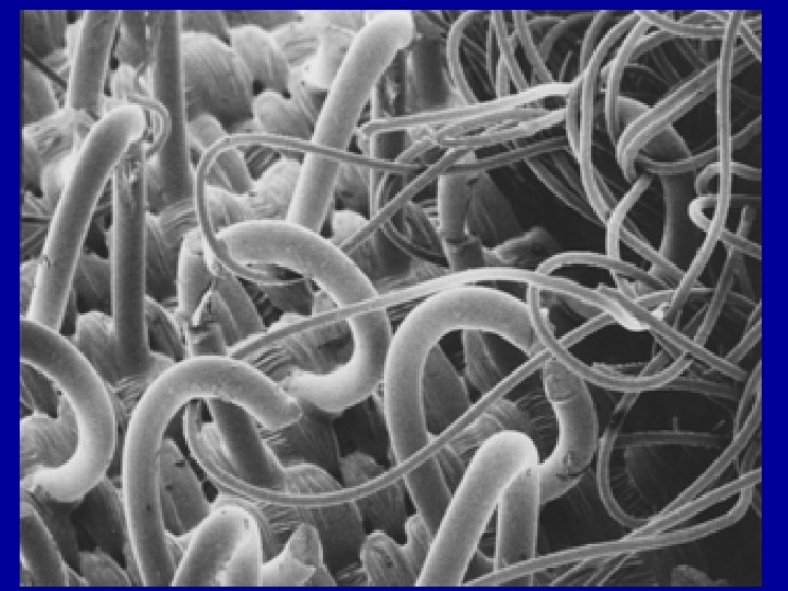
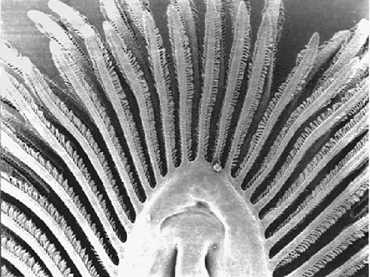
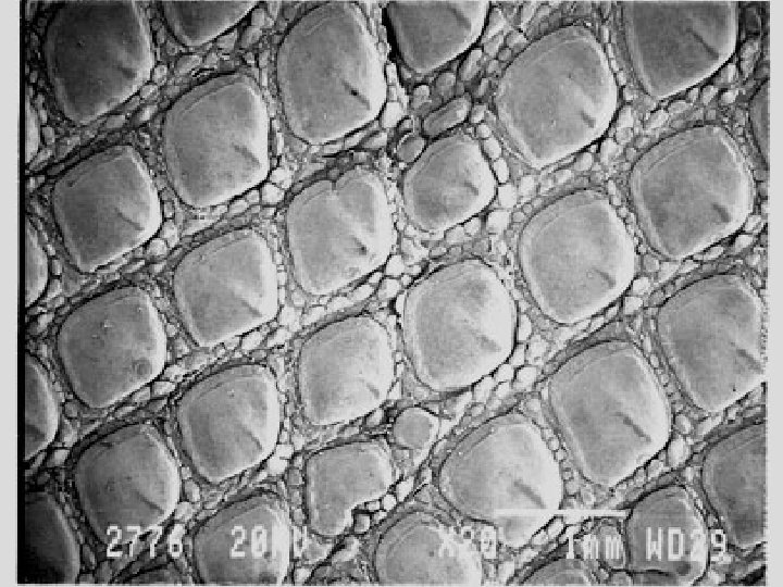
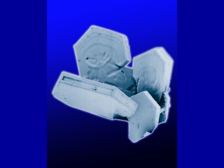
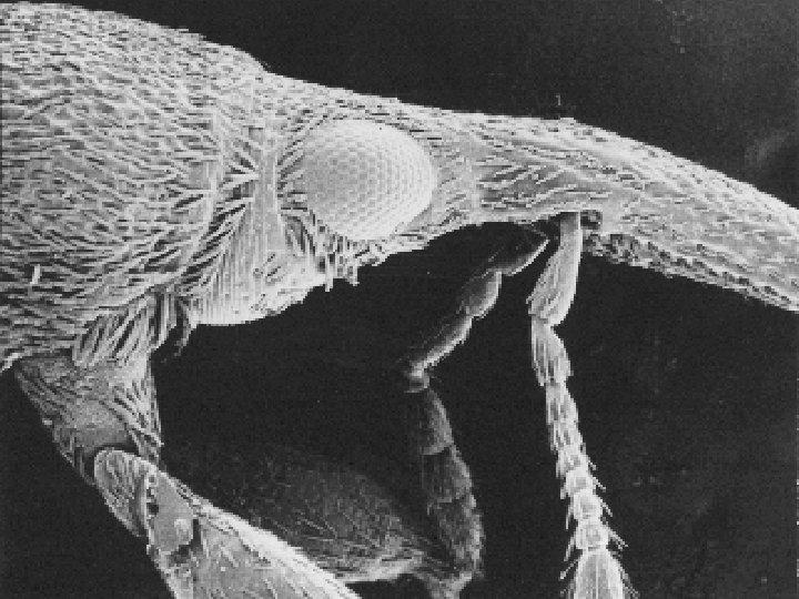
The Scanning Electron Microscope (SEM) • 1. The image is formed by aiming a beam of electrons onto a specimen and studying electron emissions on a closed circuit tv.
• 2. A beam of electrons is emitted from a tungsten filament and focused onto the specimen using electromagnets. • This is called the primary electron beam.
• When this beam hits the specimen it causes the elements on the surface to give off secondary electrons. • 20 - 30% of the electrons that hit the specimen bounce off.
• These are called backscatter electrons. • The secondary and backscatter electrons are collected and the amplified signal is displayed on a screen.
• The advantages of an SEM is the high level of magnification (up to 100, 000 times), the depth of focus is 300 times better than optical microscopes, and a superior resolution.
88888888888