REPRODUCTIVE SYSTEM MALE RO LEARNING OUTCOMES Learning Outcomes


: These are a pair of small glands")


 * Irritable and depressed. * Low")


. hormone")


 follicles matures and")

 and additional estrogen.")


![#1 FEMALE HORMONE CONTROL #7 5. When the [estrogen] secretion begins to rise, rise](https://slidetodoc.com/presentation_image_h2/aef3908db389d9716db7ed22aa5633ee/image-108.jpg "#1 FEMALE HORMONE CONTROL #7 5. When the [estrogen] secretion begins to rise, rise")


![FEMALE HORMONE CONTROL 15. When [LH] plummets, the corpus luteum begins to degenerate 16.](https://slidetodoc.com/presentation_image_h2/aef3908db389d9716db7ed22aa5633ee/image-112.jpg "FEMALE HORMONE CONTROL 15. When [LH] plummets, the corpus luteum begins to degenerate 16.")


- Slides: 125
REPRODUCTIVE SYSTEM
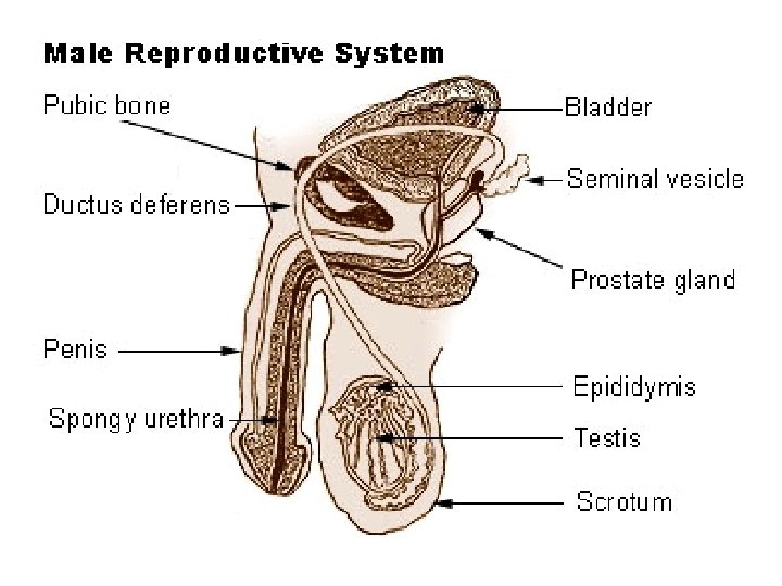
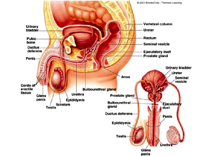
MALE RO LEARNING OUTCOMES # Learning Outcomes Text Pages P 1 P. 416 -417 Identify and give functions for each of the following: testes (seminiferous tubules and interstitial cells), epididymis, ductus (vas) deferens, prostate gland, cowper's gland, seminal vesicles, penis. P 2 Demonstrate a knowledge of the path of sperm from the seminiferous tubules to the urethral opening. P. 414 -415 P 3 List the functions of seminal fluid. P. 414 -415 P 4 Identify the tail, midpiece, head, and acrosome of a mature sperm and state their functions. P. 416 -417 P 5 Describe the functions of testosterone. P. 417 P 6 Demonstrate a knowledge of the control of testosterone levels the endocrine system. P. 417
HUMAN REPRODUCTION The goal of the reproductive system is to pass on your genetic code onto a new and unique generation. This is ultimately accomplished via fertilization
HUMAN REPRODUCTION Human reproduction employs internal fertilization, fertilization and depends on the integrated action of hormones, the nervous system, and the reproductive system. Male gonads are the testes, testes which produce sperm and testosterone estrogen Female gonads are the ovaries which produce eggs and 2 hormones: estrogen and progesterone.
http: //www. youtube. com/watch? v=WEzc. K-OKd 10
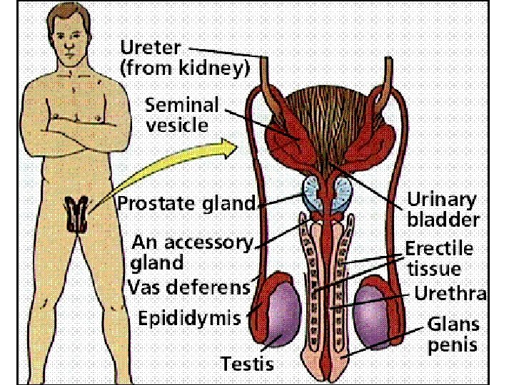
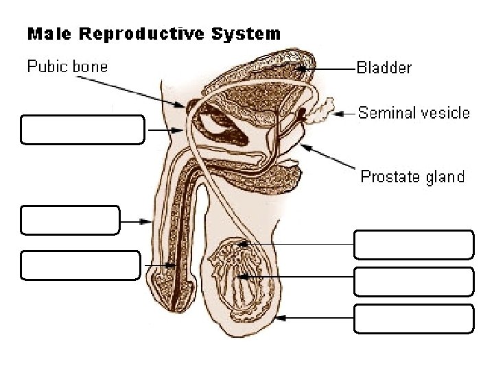
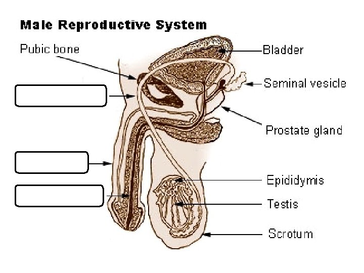
MALE REPRODUCTION Male Anatomy The male external genitalia are the scrotum and penis The scrotum is a fold of skin that encloses the male gonads (testes)
MALE REPRODUCTION scrotum testes
MALE REPRODUCTION Male fruit bat of the species Rousettus aegyptiacus have testes that are 2. 15 % of body mass, whereas their brains are only 1. 70 %. The males in some bat species can have testes that are up to 8. 5 % of their body mass.
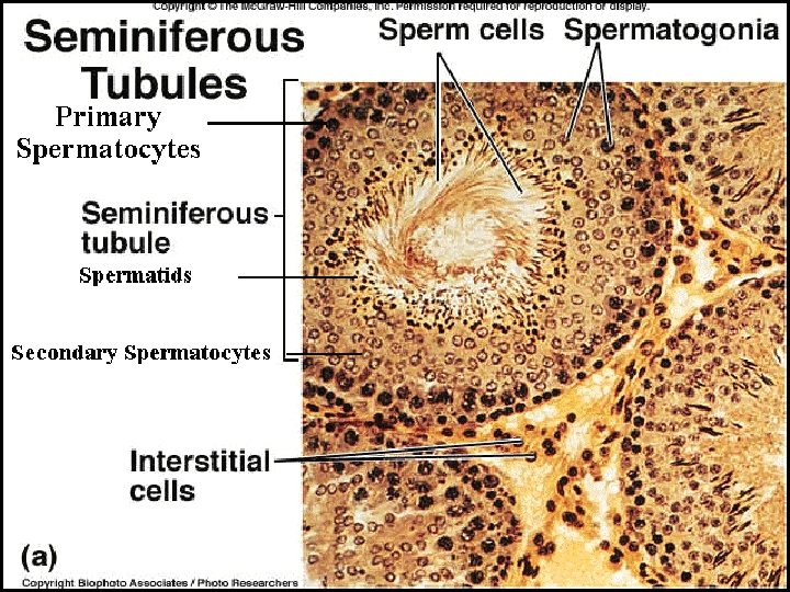
MALE REPRODUCTION The testes are a pair of tightly coiled tubes surrounded by several layers of connective tissues. These tubes are the SEMINIFEROUS TUBULES, TUBULES where sperm are produced by meiosis. About 250 meters of tubules are packed into each testis.
MALE REPRODUCTION
MALE REPRODUCTION
MALE REPRODUCTION
MALE REPRODUCTION
MALE REPRODUCTION The INTERSTITIAL CELLS are scattered between the seminiferous tubules in the testes. These cells produce testosterone
MALE REPRODUCTION Sperm production cannot occur at normal body temperature. So just before birth, birth the testes descend to hang outside the abdominal cavity in the scrotum, a fold of skin. The temperature in the scrotum is ~2 o. C below body temperature
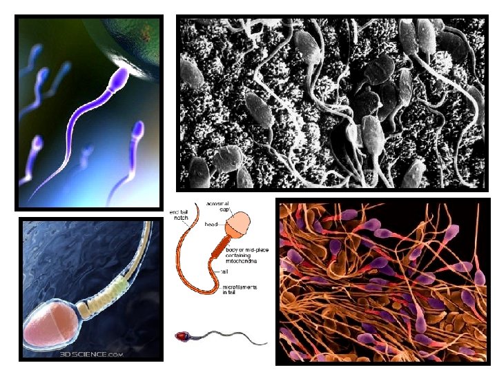
MALE REPRODUCTION SPERMATOGENESIS The process from spermatogonia to motile sperm, sperm takes 65 -75 days in the human male. Each ejaculation of a human male contains about 400 million sperm cells. Sperm production begins at puberty and continues throughout life, with several hundred million sperm being produced each day.
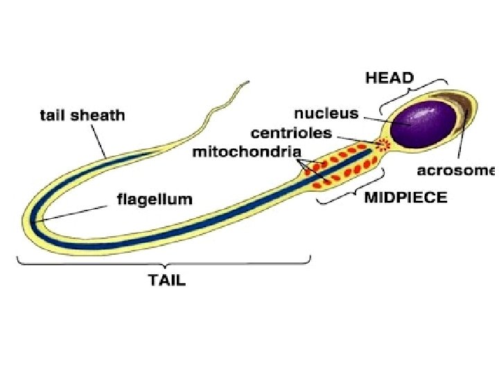
PARTS OF SPERM CELLS The structure of a sperm cell fits function. The thick head contains the haploid nucleus and is tipped with a special body, the acrosome The acrosome contains enzymes that help the sperm penetrate the egg. The neck contains large numbers of mitochondria that provide ATP for movement. The tail is a flagellum which moves the sperm.
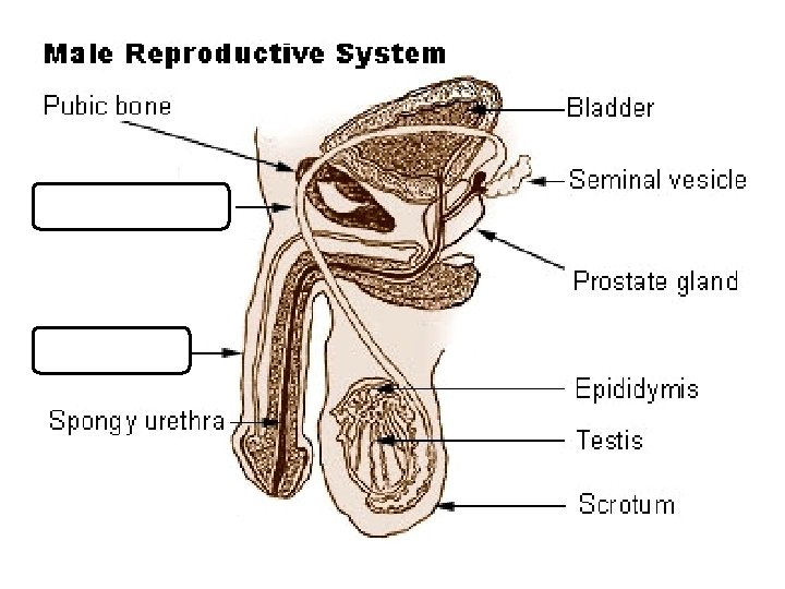
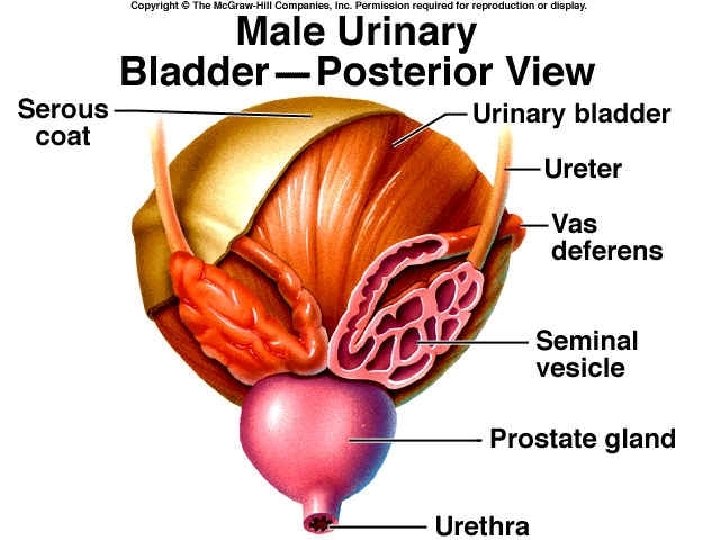
MALE REPRODUCTION During ejaculation, the sperm are propelled from the epididymis through the muscular VAS DEFERENS ducts, which run from the scrotum around and behind the bladder, where they join to form a short EJACULATORY DUCT This duct opens into the URETHRA, URETHRA the tube which drains both the excretory and reproductive systems (never both at the same time). The urethra runs through the penis and opens to the outside at the tip of the penis.
SEMINAL FLUID In addition to the testes and ducts, the male reproductive system contains three sets of glands that add their secretions to the SEMEN (the fluid ejaculated). 1. SEMINAL VESICLES: VESICLES contribute about 60% of the total volume of semen This pair of glands lies below and behind the bladder and empties into the ejaculatory duct. The fluid is thick and clear and contains mucous, amino acids, and large amount of fructose (which provides energy for the sperm). These vesicles also secrete PROSTAGLANDINS, PROSTAGLANDINS which once in the female reproductive tract, stimulate contractions of the uterine muscles that help move the semen up into the uterus. Proteins in the seminal fluid cause the semen to coagulate after it is deposited in the female, thus, making it easier for uterine contractions to move the semen.
MALE REPRODUCTION
MALE REPRODUCTION 2. PROSTATE GLAND: GLAND This is the largest of the accessory glands. Prostatic fluid is thin, milky, and quite alkaline, alkaline which balances the acidity of any residual urine in the urethra and the natural acidity of the vagina
MALE REPRODUCTION 2. PROSTATE GLAND: This gland is the sources of some of the most common medical problems of men over 40. A benign enlargement of the prostate occurs in more than ½ of all men in this age group. That is why it is important to get tested annually after the age of 40.
MALE REPRODUCTION
MALE REPRODUCTION 3. BULBOURETHRAL GLANDS (cowper’s gland): These are a pair of small glands along the urethra below the prostate. They secrete a viscous fluid before emission of the semen It has been suggested that this fluid lubricates the penis and vagina, vagina but the volume (just one or two drops) seems insufficient to be very effective for this function. This fluid does carry some sperm released before ejaculation, . This is one factor in the low success rate of the withdrawal method of birth control.
MALE REPRODUCTION
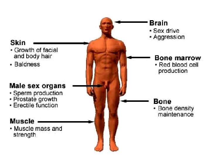
HORMONES MALE SEX HORMONES The principal male sex hormones are the androgens, androgens of which, testosterone is the most important. Androgens are steroid hormones produced by the interstitial cells of the testes, and are directly responsible for the primary and secondary sex characteristics of the male. Androgens are also potent determinants of behaviour in mammals. In addition to specific sexual behaviour and libido (sex drive), androgens increase general aggressiveness
CONSEQUENCES OF USE: *ROID RAGE (extreme uncontrollable aggression) * Irritable and depressed. * Low sex drive * Possible cancer & liver damage * Feminizing effects in males (growth of breast tissue)… * Shrunken testicles… * Limb loss… * Heart disease/heart attacks… * HIV/AIDS from the sharing of needles…
CONSEQUENCES OF USE: * Reduced sperm count… * Impotence & Infertility… * Baldness… * Pain & difficulty urinating… * Enlarged prostate… * Adolescents experience stunted growth…
MALE REPRODUCTION MALE SEX HORMONES Primary sex characteristics: development of the vas deferens and other ducts, the eternal genitalia, genitalia and sperm production All babies start as female If testosterone is present, they will become male, if not, they will be born female. Sex is determined by 13 weeks, sex is determined.
MALE REPRODUCTION MALE SEX HORMONES Secondary sex characteristics: features we associate with maleness such as: deepening of the voice, the male distributions of axillary (armpit), facial, and pubic hair, and muscle growth (androgens stimulate protein synthesis).
HOMEOSTATIC REGULATION The hypothalamus makes a hormone called Gn. RH (the gonadotropin-releasing hormone). hormone Gn. RH control the release of two hormones from the anterior pituitary: pituitary 1. Follicle-stimulating hormone (FSH) 2. Luteinizing hormone (LH)
HOMEOSTATIC REGULATION LH stimulates the interstitial cells in the seminiferous tubules to secrete testosterone Testosterone has a role in sperm production and developing male secondary sex characteristics FSH acts on the epidymis to help in sperm maturation
HOMEOSTATIC REGULATION Negative feedback by testosterone controls the actions of Gn. RH.
HOMEOSTATIC REGULATION Hypothalamus - Gn. RH
HOMEOSTATIC REGULATION Hypothalamus - Gn. RH Anterior Pituitary LH & FSH
HOMEOSTATIC REGULATION Hypothalamus - Gn. RH Anterior Pituitary LH & FSH Follicle stimulating hormone
HOMEOSTATIC REGULATION Hypothalamus - Gn. RH Anterior Pituitary LH & FSH Follicle stimulating hormone Epidydimis
HOMEOSTATIC REGULATION Hypothalamus - Gn. RH Anterior Pituitary LH & FSH Follicle stimulating hormone Epidydimis Matures the sperm (makes it motile)
HOMEOSTATIC REGULATION Hypothalamus - Gn. RH Anterior Pituitary LH & FSH Follicle stimulating hormone Epidydimis Matures the sperm (makes it motile) Fish swim
HOMEOSTATIC REGULATION Hypothalamus - Gn. RH Anterior Pituitary LH & FSH LH luteinizing hormone FSH Follicle stimulating hormone Epidydimis Matures the sperm (makes it motile)
HOMEOSTATIC REGULATION Hypothalamus - Gn. RH Anterior Pituitary LH & FSH LH luteinizing hormone FSH Follicle stimulating hormone Interstitial Cells Epidydimis Matures the sperm (makes it motile)
HOMEOSTATIC REGULATION Hypothalamus - Gn. RH Anterior Pituitary LH & FSH LH luteinizing hormone FSH Follicle stimulating hormone Interstitial Cells Epidydimis Testosterone Matures the sperm (makes it motile)
HOMEOSTATIC REGULATION Hypothalamus Gn. RH - Anterior Pituitary LH & FSH LH luteinizing hormone FSH Follicle stimulating hormone Interstitial Cells Epidydimis Testosterone Sperm production 2 o sex characteristics deep voice, body hair, more muscle, libido… Matures the sperm (makes it motile)
HOMEOSTATIC REGULATION Hypothalamus Gn. RH - Anterior Pituitary LH & FSH Teens have a Love Hate relationship with testosterone LH luteinizing hormone FSH Follicle stimulating hormone Interstitial Cells Epidydimis Testosterone Sperm production 2 o sex characteristics deep voice, body hair, more muscle, libido… Matures the sperm (makes it motile)
MALE REPRODUCTION
MALE REPRODUCTION
FEMALE RO LEARNING OUTCOMES # Learning Outcome Text Pages P 7 P. 418 -419 Identify and give a function for each of the following: ovaries (follicles and corpus luteum), oviducts (fallopian tubes), uterus, cervix, vagina, clitoris. P 8 Describe the functions of estrogen. P. 421 -424 P 9 Describe the sequence of events in the ovarian and uterine cycles. P. 420 -423 P 10 Demonstrate knowledge of the control of the ovarian and uterine cycles by hormones. P. 420 -423 P 11 Demonstrate knowledge of a positive feedback mechanism involving oxytocin. P. 208, 394 P 12 Describe the hormonal changes that occur as a result of implantation. Page 424
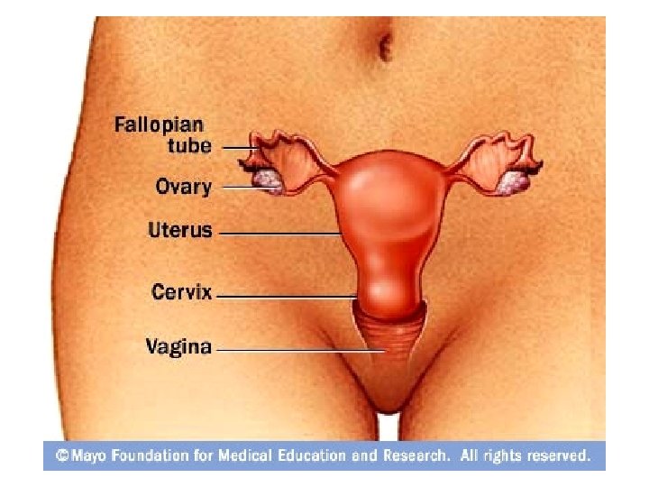
FEMALE RO LEARNING OUTCOMES The female gonads, ovaries are located within the lower abdominal cavity, below most of the digestive system. Each ovary is enclosed in a tough protective capsule and contains many follicles.
FEMALE REPRODUCTION A FOLLICLE consists of one egg cell surrounded by one or more layers of follicle cells, which nourish and protect the developing egg cell. All of the 400, 000 follicles a woman will ever have are formed at birth Of these, only several hundred will be released during the woman’s reproductive years.
FEMALE REPRODUCTION After puberty, puberty one (or rarely two or more) follicles matures and releases its egg during each menstrual cycle The cells of the follicle also produce the primary female sex hormones, ESTROGEN
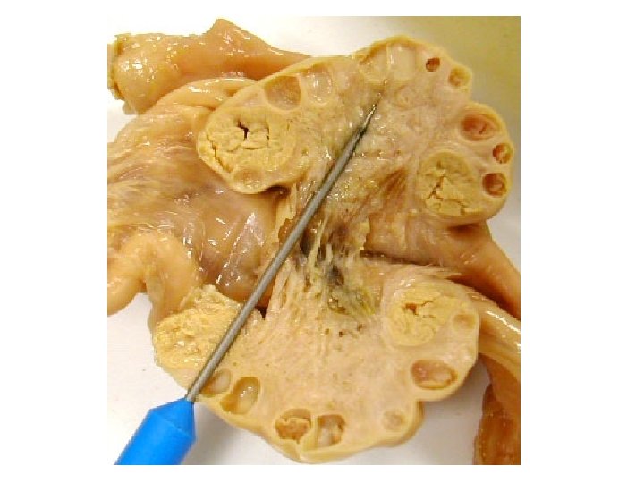
FEMALE REPRODUCTION The remaining follicular tissue grows within the ovary to form a solid mass called the CORPUS LUTEUM
FEMALE REPRODUCTION The corpus luteum secretes PROGESTERONE (the hormone of pregnancy) and additional estrogen. If the egg is not fertilized, fertilized the corpus luteum degenerates and a new follicle matures during the next cycle.
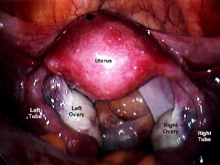
FEMALE REPRODUCTION The female reproductive system is not completely closed. The egg cell is expelled into the abdominal cavity near the opening of the OVIDUCT (or fallopian tube).
FEMALE REPRODUCTION The fimbriae on the oviduct sweep the ovary for the egg, egg and the cilia on the inner epithelium lining the duct help collect the egg cell by creating a current to draw fluid into the duct.
FEMALE REPRODUCTION
Ovarian Cysts
FEMALE REPRODUCTION
FEMALE REPRODUCTION The oviduct is the site of fertilization The cilia in this duct also convey the egg cell down the duct to the UTERUS (womb).
FEMALE REPRODUCTION The UTERUS is a thick, muscular organ (shaped like an upside down pear). It is remarkably small; the uterus of a woman who has never been pregnant is about 7 cm long and 4 -5 cm wide at its widest.
FEMALE REPRODUCTION The unique arrangement of muscles that make up the uterine wall allow it to expand to accommodate a 4 kg (? lb) fetus.
FEMALE REPRODUCTION The inner lining of the uterus, the ENDOMETRIUM, ENDOMETRIUM is richly supplied with blood vessels, vessels in which a fertilized egg implants and develops.
FEMALE REPRODUCTION The narrow neck of the uterus is the CERVIX, CERVIX which opens into the VAGINA The vagina is a thin-walled chamber that forms the birth canal through which the baby is expelled; it is also the receptacle for the man’s penis.
FEMALE REPRODUCTION
FEMALE REPRODUCTION
FEMALE REPRODUCTION
FEMALE REPRODUCTION
FEMALE REPRODUCTION
FEMALE REPRODUCTION
FEMALE REPRODUCTION
FEMALE REPRODUCTION
FEMALE REPRODUCTION
FEMALE REPRODUCTION
FEMALE REPRODUCTION
FEMALE REPRODUCTION
FEMALE REPRODUCTION
FEMALE REPRODUCTION The lack of estrogen in males prevents the development of both the secretory apparatus and the fat deposits, so the breasts remain small and the nipple is not connected to the ducts
FEMALE REPRODUCTION THE FEMALE HORMONE CYCLES Two different types of cycles occur in the female mammals: 1. The menstrual cycle 2. The ovarian cycle
FEMALE REPRODUCTION The Menstrual Cycle The human menstrual cycle refers specifically to the changes that occur in the uterus It lasts an average of 28 days, days but only ~30% of women have cycle lengths within a day or two of the statistical 28 days. Cycles vary from about 20 to 40 days. In some women the cycles are usually very regular, regular but in other individuals the timing varies from cycle to cycle.
FEMALE REPRODUCTION MENSTRUAL CYCLE : This cycle occurs in the uterus. There are 3 phases in this cycle. Day 1 is the first day of a woman’s ‘period’ period (1 st day of menstruation) menstruation THREE PHASES OF THE MENSTRUAL CYCLE 1. The Flow phase: phase Menstrual bleeding usually persists for a few days (day 2 -8). 2. The Proliferative phase: phase the endometrium begins to thicken for a week or two (until ovulation). ovulation 3. The Secretory phase: phase usually about 2 weeks long, the endometrium continues to thicken, thicken becomes more vascularized and secretes a fluid rich in glycogen to prepare for a fertilized egg
FEMALE REPRODUCTION MENSTRUAL CYCLE *If an embryo has not implanted in the uterine lining by the end of the secretory phase, a new menstrual flow commences *If an embryo is implanted before the end of the secretory phase, the endometrium will remain until birth
FEMALE REPRODUCTION THE OVARIAN CYCLE: This cycle occurs in the ovaries. There are THREE phases in this cycle THREE PHASES OF THE OVARIAN CYCLE: 1. This cycle begins with the FOLLICULAR PHASE, PHASE during which several follicles in the ovary begin to grow and release estrogen The maturing follicle develops an internal fluid-filled cavity and grows very large, forming a bulge near the surface of the ovary.
FEMALE REPRODUCTION THREE PHASES OF THE OVARIAN CYCLE: 2. The follicular phase ends with OVULATION when the follicle and adjacent wall of the ovary rupture, releasing the egg cell. The egg cell is transported into the oviduct and is transported into the uterus.
FEMALE REPRODUCTION THREE PHASES OF THE OVARIAN CYCLE: 3. The last phase is the LUTEAL PHASE During this phase, the follicular tissue that remains in the ovary after ovulation is transformed into the corpus luteum The corpus luteum is an endocrine tissue that synthesizes and secretes hormones to prepare the uterus for pregnancy
FEMALE REPRODUCTION The next cycle begins with growth of new follicles. These cyclic phases are interrupted only by pregnancy and continue until menopause, menopause when reproductive capability ends.
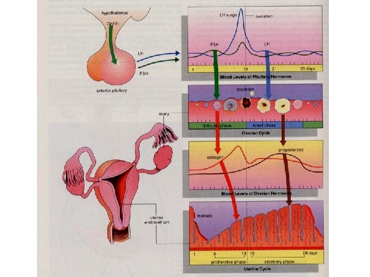
FEMALE REPRODUCTION THE FEMALE SEX HORMONES Hormones coordinate the menstrual and ovarian cycles in such a way that growth of the follicle and ovulation are synchronized with preparation of the uterine lining for possible implantation of an embryo. 5 hormones participate in both (+) and (-) feedback cycles: 1. Gonadotropin-releasing hormone (Gn. RH): secreted by the hypothalamus 2. Follicle stimulating hormone (FSH): secreted by anterior pituitary 3. Luteinizing hormone (LH): secreted by anterior pituitary 4. Estrogen: Estrogen secreted by ovary (follicle) 5. Progesterone: Progesterone secreted by ovary (corpus luteum)
FEMALE REPRODUCTION
#1 FEMALE HORMONE CONTROL 1. During the follicular phase of the ovarian cycle, the hypothalamus releases Gn. RH 2. This causes the anterior pituitary to secrete small quantities of FSH and LH. #2
FEMALE HORMONE CONTROL 3. At this time, the follicles in the ovary have receptors for FSH, FSH but not for LH. #3 The FSH causes the follicles to grow. 4. The follicles release estrogen as they grow. The amount of estrogen secreted during this time is small. #4
#1 FEMALE HORMONE CONTROL #7 5. When the [estrogen] secretion begins to rise, rise it causes the hypothalamus to release more Gn. RH (positive feedback). 6. Gn. RH causes even more LH and FSH to be released from the anterior pituitary. 7. This causes the LH SURGE. #6 #5
FEMALE HORMONE CONTROL 8. By now, the follicles have receptors for LH and can respond to this hormonal cue. The LH SURGE induces final maturation of the follicle 9. Ovulation occurs on day 14, 14 about one day after the LH surge #9 #8
FEMALE HORMONE CONTROL 10. After ovulation, LH will stimulate the follicle to become the corpus luteum. 11. LH will also stimulate the corpus luteum to start secreting estrogen and progesterone #10 #11
FEMALE HORMONE CONTROL 12. The corpus luteum reaches its maximum development about 8 -10 days after ovulation At this point there is very high [progesterone] and [estrogen] in the blood. 13. High levels of these hormones exerts negative feedback on the hypothalamus 14. This causes a severe decrease in the amounts of LH and FSH #13 #14 #12
FEMALE HORMONE CONTROL 15. When [LH] plummets, the corpus luteum begins to degenerate 16. This causes a sharp decrease in the [estrogen] and [progesterone]. 17. Without these hormones, the endometrium layer is sloughed off and day one of the cycle begins again. #14 #15 #16 #17
FEMALE HORMONE CONTROL 18. As these hormones drop off, the hypothalamus and pituitary are not inhibited anymore 19. So the anterior pituitary begins to secrete enough FSH to stimulate growth of new follicles in the ovary. 20. This initiates the follicular phase of the next ovarian cycle. #18 #19 #20
FEMALE REPRODUCTION How is the ovarian cycle synchronized with the menstrual cycle? 1. Follicular phase/proliferative phase: phase Estrogen (which is released by follicles in the follicular phase) is a hormonal signal to the uterus, which stimulates the development of the endometrium (proliferative phase). FOLLICULAR PHASE e s t r o g e n PROLIFERATIVE PHASE
FEMALE REPRODUCTION How is the ovarian cycle synchronized with the menstrual cycle? 2. Luteal phase/secretory phase: phase After ovulation, estrogen and progesterone secreted by the corpus luteum stimulate continued development and maintenance of the endometrium (secretory phase) as the body prepares for the possibility of a fertilized egg. LUTEAL PHASE p r o g e s t e r o n e SECRETORY PHASE
FEMALE REPRODUCTION How is the ovarian cycle synchronized with the menstrual cycle? 3. A rapid drop in the level of estrogen and progesterone hormones when the corpus luteum degenerates causes spasms of the arteries in the uterine lining that deprive the endometrium of blood. Degeneration of the endometrium results in menstruation and the beginning of a new menstrual cycle. In the meantime, ovarian follicles that will stimulate renewed thickening of the endometrium are just beginning to grow. Corpus luteum dies L o w E & P MENSES Day 1
FEMALE REPRODUCTION In addition to their role in coordinating reproductive cycles, estrogens are also responsible for the secondary sex characteristics of the female: 1. Deposition of fat in the breasts and hips 2. Increase in water retention 3. Affects calcium metabolism 4. Stimulates breast development 5. Mediates female sexual behavior 6. Initiates female libido 7. PMS 8. Mood swings 9. Cravings (chocolate, cheese, milk…) 10. Growth of hair (not as much as men)
WHAT HAPPENS IF THE EGG IS FERTILIZED?
WHAT HAPPENS IF THE EGG IS FERTILIZED?
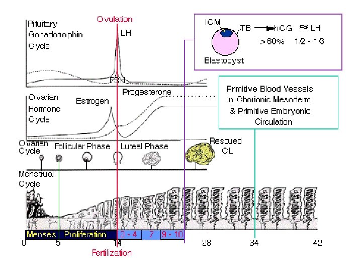
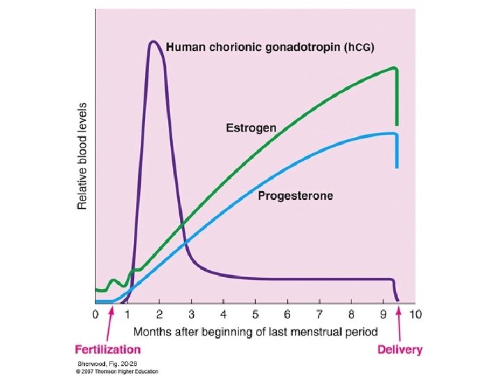
WHAT HAPPENS IF THE EGG IS FERTILIZED? If the egg is fertilized, the resulting embryo will start to release a hormone called human chorionic gonadotropin (HCG). HCG acts like LH to maintain the corpus luteum and this allows the levels of progesterone and estrogen to remain high Thus, the endometrium layer is NOT sloughed off and is maintained by the high levels of hormones. The levels of HCG are so high, that some is excreted in the urine, urine where it can be detected in pregnancy tests
HCG
WHAT HAPPENS IF THE EGG IS FERTILIZED? Other changes for the mother in the first trimester include: • Increased mucous in the cervix to form a protective plug. • Growth of the placenta • Enlargement of the uterus • Morning sickness • Mood swings • Weight gain of ~1 kg • Cessation of ovulation and menstrual cycling • Breasts become large and tender