Male Reproductive System Male reproductive system consists of


- Slides: 31

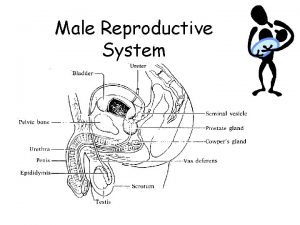
Male Reproductive System Male reproductive system consists of: 1. The testes surrounded by tunica vaginalis, 2. The epididymis, 3. The ductus deferentes/ductus deferens, 4. The accessory glands (vesicular, prostate, and bulourethral), 5. The urethra, and 6. The penis surrounded by the prepuce.

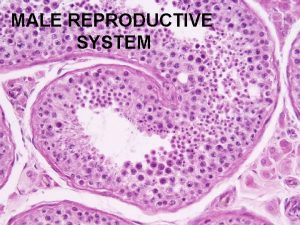
Histology of Testis • Covered by Tunica albuginea, • Convoluted Seminiferous Tubules, and • Interstitial cell/Leydig cell.

Histology of Testis-Tunica albuginea q It is a solid capsule of dense connective tissue. q Consist of collagen fiber, elastic fibers and myofibroblasts. q In cat, interstitial endocrine cells (secretes testosterone) also found in the tunica albuginea.

Histology of Testis- Seminiferous Tubules Convoluted seminiferous tubules: § Tortuous two ended loops with a diameter between 150 -200 μm. § The seminiferous tubules have 3 components: 1. Lamina propria 2. Somatic cells (Sustentacular, Supporting, or Sertoli, s cells) 3. Spermatogenic cells.

1. Lamina Propria of Seminiferous Tubules Ø The lamina propria surrounds the seminiferous tubules and consists of collagen and elastic fibers. Ø Peritubular smooth muscle (in boar), and myofibroblast (in bull) present which cause contraction of seminiferous tubules for the ejection of spermatozoa. ØLymphocytes and monocytes are also present in the lamina propria.

2. Sertoli Cells of the Seminiferous Tubules

2. Sertoli Cells of the Seminiferous Tubules Ø Also known as Sustentacular cell/ Supporting cell. Ø These cells are resting on the base of seminiferous tubules and having large nucleus with one prominanat nucleoli. Ø The cells are irregularly outlined, elongated cells. Ø Sustentacular cells are closely associated with the spermatocytes, and spermatids.

Function of Sertoli Cells Important functions of Sertoli cells: • Nutritive, Protective and Supportive functions for the spermatogenic cells. • Phagocytize regressive and spermatogenic cells. • Detached residual bodies of spermatid. • They mediate the action of FSH and testosterone on the germ cells. • They secretes inhibin which blocks the secretion of pituitary-FSH.

3. Spermatogenic Cells of Seminiferous Tubules Spermatogenic cells are: 1. Spermatogonia: They are spherical cells resting on the basement memb- -rane with round one or two nuclei. The cells are located in between Serto-li cells. There are two types of sperma -togonia. Type A and Type B undergoes mitosis and differentiated into primary spermatocyte. 2. Primary spermatocytes, secondary spermatocytes, and spermatids are the next generation of the spermatogonia.

Histology of Testis- Interstitial Cell/Leydig cell The intertubular space contain loose connec- tive tissue, blood and Lymph vessels, fibrocyte Free mononuclear cells, and interstitial endocrine (Leydig) cells.

Interstitial or Leydig cells of the testis • Embryologically interstitial cells develop from mesenchymal cells. • Interstitial cells comprises 1% of the entire testicular volume in the adult ram, 5% in the bull, and 20 -30% in the boar. • These cells found in the form of cords or clusters. The cells are spherical with large round cells, enough endoplasmic reticulum and mitochondria for the transformation of cholesterol (important component of the testosterone hormone).

Function of Interstitial or Leydig cells of the testis • Promote normal sexual behaviour of male animals. • Growth, maintenance, and initiate function of the male accessory glands and secondary sex characters. • Control spermatogenesis. • General anabolic effect.

Spermatogenesis Definition: It is the process of formation of spermatozoa from spermatogonium. Usually one mitotic and two meiotic stage occurs during the process. The sequence of event takes place by 3 successive process: 1. Spermatocytogenesis: In this process spermatogonia developed into primary spermatocytes. From 1 Bspermatogonium 1 primary spermatocytes formed (by mitosis). 2. Meiosis: By 2 meiotic division 1 primary spermatocyte divide into 2 secondary spermatocyte and then finally into 4 haploid spermatid. 3. Spermiogenesis: Transformation of spermatid into spermatozoa.

Images for Spermatogenesis

Morphology of Bull Spermatozoa Head Neck Middle Piece Principal Piece Tail Spermatozoa is a single cell having nucleus (head), cytoplasm and cell membrane around the centriole and axonema (tail). It consist of Head, Neck, and Tail. Neck is short. Tail is further divided into: Middle piece, Principal piece and End piece. Middle piece is characterized by the presence of Mitochondria for energy during motility. The principal piece is the longest. Tail is the terminal part.

Histology of Epididymis §The ductus epididymis is extremely tortuous and coiled. In bull and boar the length is 40 meter, and in stallion it is 70 meter in length. § Epididymis is lined by a pseudostratified epithelium, surrounded by a small amount of Loose connective tissue and circular smooth muscle fiber. § Two cell types are present: tall columnar ciliated cells (principal cells) and short absorptive cells. Function: Storage and maturation of the spermatozoa.

Histology of Ductus Deferens § The mucosa is lined by Pseudostratified columnar epithelium. § Lamina propria consist of loose connective tissue. § Propria submucosa in Stallion, bull and boar have circular, longitudinal, and oblique Smooth muscle layers. q. The terminal portion of the ductus deferens contains simple branched Tubulo-alveolar gland in the proria-submucosa. In the stallion, bull, and ram these glands occupy the entire propria-submucosa surrounded by smooth muscle, whereas, in dog the gland is not surrounded by muscles. The gland is lined by simple cuboidal to columnar epithelium. In man no such type of gland.

Male Accessory Gland • Male accessory glands of animals are: 1. Vesicular gland or Seminal vesicle 2. Prostate gland 3. Bulbourethral gland

Histology of Vesicular Gland • The paired vesicular gland is a compound tubular or tubulo-alveolar gland. • The glandular epithelium is pseudostratified with tall cell. • Lamina propria-submucosa is continuous. • Tunica muscularis is of smooth muscle and it is followed by tunica serosa or adventitia.

Images for Vesicular Gland of Animals

Vesicular Gland in Different Animals • Stallion Boar Ruminants • Epithelium is Same as stallion Columnar cell Pseudostratified. • It is true vesicular. Vesicular Gland is compact. • Interlobular septa is thin Smooth The interlobular septa irregularly arranged muscle is few is thick with predominant muscles. in the interlobular smooth muscle. septa.

Histology of Prostate Gland • Tubulo-alveolar gland derived from the epithelium of pelvic urethra. • Having two parts: 1. Compact or External portion (corpus prostate), and internal portion (pars disseminate). The external portion entirely surrounds part of the pelvic urethra. The disseminate portion is located in the propria -submucosa of the pelvic urethra. • The secretory tubules/alveoli is lined by a simple cuboidal or columnar epithelium.

Histology of Prostate Gland-cont • Occasionally, concentriclly laminated concretions of secretory materials are found in the tubules of the alveoli. • The prostate is surrounded by a capsule of dense irregular connective tissue that contain many smooth muscle. • From the capsule connective tissue and smooth muscle enters the gland divide the gland into lobes and lobules. Therefore aound the small secretory unit there is connective tissue and smooth muscle, contraction of which empties the secretory materials into the duct system. • •

Images for Prostate Gland of Animals

Prostate Gland in Different Animals • Carnivors Stallion Ruminants Boar • External portion is Only external The well developed portion is external and separated into present. No absent. Internal portion 2 -lobes. The Internal internal gland. portion is well is plate portion consist of few developed and like and glandular lobules. encircles the internal uretra. portion is well developed.

Histology of Bulbourethral Gland • Paired gland, and located dorsolaterally from the bulbar portion of the urethra. It is compound tubular (boar, cat, and buck) or tubulo-alveolar (stallion, bull, ram). It is absent in dog. • The secretory portion of the gland are lined with a tall simple columnar epithelium and occasional basal cells. • The gland is covered by a fibroelastic capsules with striated muscle. • Trabeculae extending from the capsule into the gland devide it into many lobes and lobules. Trabeculae consists of both smooth and striated muscle.

Images for Bulbourethral Gland of Animals

Bulbourethral Gland in Different Animals • Carnivors Stallion Ruminants In the cat, the The gland Same as sinus like duct is completely stallion and short, narrow surrounded by unbranched tubular bulbocavernous end piece. Muscle.

Histology of Penis of Different Animals • Penile muscles are two types: • Corpus cavernosum penis and • Corpus spongiosum. Corpus cavernosum penis muscle: Paired muscle originates from ischiatic tuberosities and situated dorsal to the urethra of animals. § Covered by tunica albuginea of connective tissue. § Trabeculae enters the muscle as numerous septa. § There are numerous cavernous space within this muscle, lined by endothelium, and are filled up by blood during erection.

Histology of Penis of Different Animals- cont. • Corpus spongiosum penis muscle: This muscle is situated around the urethra. Note please: • Corpus cavernosum muscle is thicker and spacious than the spongiosum muscle. • In the horse Corpus cavernosum muscle is more thicker and spacious than the ruminants. • In the dog os penis (bones of the penis) is present in the glans. • In the cat the glans is provided with the spines.

Mechanism of Erection of Penis • Erection of penis depends on the pheromonal (in insect and lower animals), psychological, neurological, and blood flow into the penis. • Relaxation of the muscles of helicine arteries of the penis causes increased blood flow into the penis and cavernous spaces resulting increases in the length of penis, which compress the veins and subsequently decreases the outflow resulting persistent of the erection of penis until semen ejaculation. • Detumescence is initiated by contraction of the musculature of the helicine arteries and thus decrease in arterial inflow causes the penis to return to flaccid state. • High sugar and cholesterol level of the blood decrease inflow of blood resulting erectile dysfunction of man and animals.
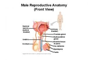
 Male reproductive system from front
 Male reproductive system and its function
 Seminal tubules
 Exercise 42 review male reproductive system
 Lymphatic drainage of breast
 Female anatomy
 Female and male reproductive system
 Male reproductive system diagram
 Reproductive physiology
 Male reproductive system of a plant
 Art-labeling activity: the male reproductive system, part 1
 Male reproductive system information
 Where is sperm located
 Male cow reproductive system diagram
 Prostate glands
 Asexual reproduction
 Disease traductor
 Diagram of male and female reproductive system of fish
 Pila reproductive system
 Bladder fetal pig
 Male plant reproductive system
 Figure 28-1 the male reproductive system
 Primary sex organ of the male reproductive system? *
 Chapter 20 reproduction and pregnancy
 Male reproductive system of cattle
 Figure 28-2 the female reproductive system
 Colon function in male reproductive system
 Figure 16-1 male reproductive system
 Differences between male and female reproductive organ
 Male reproductive system labeled
 Pathway of semen
 Drawing of the male and female reproductive system


