Clean up Clean up your materials and work


 Urinary system is made-up of the kidneys, ureters, bladder,")


- Slides: 99
Clean up: Clean up your materials and work area after each day. Leave dissection equipment with your pig, if you leave it dirty, you will find it dirty the next period. On the last day of your final IF you leave it for me to clean, or dumped in a sink, you will lose points and be assigned a sink cleaning detention thoroughly wash your hands with soap when finished- thanks for all your hard work- 99% of you have been AWESOME! 5% off the total final grade if any of your supplies/tools are not cleaned up
http: //www. napavalley. edu/people/briddell/Documents/BIO%20105/Fetal %20 Pig%20 Dissection%20 Directions%20100501. 1. pdf Fetal Pig Dissection Anatomy Final 2016
Day 1 -External Anatomy • Name your pig – label your dissection tray with the pig’s name, your initials and your class period • Pour pig juice in bucket, then rinse your pig thoroughly- you don’t want to breathe extra fumes all week • Open up the Fetal Pig Dissection on Quia PPT file OR follow along on the screen • Record your Team’s Information on Final Packet- in YELLOW- INSTRUCTIONS in PINK packet and on-line (PPT)
Let the water run over your pig for at least two to three minutes. Lay the pig on its side in the dissecting pan and locate & pin dorsal, ventral, & lateral surfaces. Also locate the anterior and posterior ends, pin- record in packet (be prepared to identify). Anterior/Cephalic
neck trunk tail head Mammillary Papillae Umbilicus Snout with nares Digits
A fetal pig has not been born yet, but its approximate age since conception can be estimated by measuring its length. Measure your pig's length from the tip of its snout (please use a piece of string) to the base of its tail and record this on your data sheet (number 1 on data sheet). Use the length/age chart on the data sheet to determine the age of your fetal pig & record this (number 2 on the data sheet). Fill in A-F **Hint- use labels on your string your fetal and metric pig diagram ruler or -using this measuring picture as tape to a guide -(the measure first page of your pig’s group length dissection packet)
Examine the pig's head. Locate the eyelids and the external ears or pinnae. Find the external nostrils (nares). Label these parts on figure 1, external fetal pig diagram. Examine the exterior of the fetal pig for hair. Describe what was found (number 3 on data sheet).
Pigs have four toes on each foot. Each toe has a hoof. The middle hoofs are divided.
Study the pig's appendages and examine the pig's toes. Count and record the number of toes and the number of hooves the pig has one on appendage (number 4 on data sheet). Label the toes and hooves on figure 1, external fetal pig diagram.
Locate the umbilical cord. With scissors, cut across the cord about 1 cm from the body. Examine the 3 openings in the umbilical cord. The largest is the umbilical vein, which carries blood from the placenta to the fetus. The two smaller openings are the umbilical arteries which carry blood from the fetus to the placenta. Label the umbilical cord on figure 1, external fetal pig diagram. Use labeling pins to mark each of the structures on your fetal pig that are in the picture to the right (H, I, and J). Prepare all group members to be able to i. d. the umbilical vein and two openings for the umbilical arteries
Locate the umbilical cord. With scissors, cut across the cord about 1 cm from the body. Examine the 3 openings in the umbilical cord. The largest is the umbilical vein, which carries blood from the placenta to the fetus. The two smaller openings are the umbilical arteries which carry blood from the fetus to the placenta. Label the umbilical cord on figure 1, external fetal pig diagram. • H = umbilical artery • I = umbilical vein • J = allantoic stalk *Prepare your group- using your textbook- to explain 3 differences between the fetal blood circulation and that of a newborn
Lift the pig's tail to find the anus. Anterior from the anus along the ventral surface of the pig study and note the tiny bumps called mammary papillary. These are present in both sexes. Count the number of mammary papillary and record it on the data sheet (number 5 on the data sheet). In the female these structures connect to the mammary glands.
Congratulations it’s a baby girl! Congratulations it’s a baby boy! Does your group have a baby Boy or Girl? Determine the sex of your pig by locating the urogenital opening through which liquid wastes and reproductive cells pass. In the male, the opening is on the ventral surface of the pig just posterior to the umbilical cord. In the female, the opening is ventral to the anus.
Sex determination Male 6= umbilical cord 8= urogenital orifice 9= scrotum 10= Mammillary papilla 7= genital papilla
Sex determination Female Male 6= umbilical cord 7= genital papilla 8= urogenital orifice 9= scrotum 10= mammillary papilla 11= anus
External features 1. Pinna 2. External auditory meatus 3. Nictitating membrane 4. Rooter 5. Vibrissae 6. Umbilical cord 7. Genital papilla 8. Urogential orifice 9. Scrotum 10. Mammary papilla 11. Anus Prepare ALL group members throughout the lab- information on all slides up to slide #20 will be verbally quizzed upon!
Group Check Time #1 for External Anatomy & Physiology When your team feels prepared to answer both structure and function questions- please call me over- and I will sign off on your external anatomy final grade after verbally quizzing you on the major features of the pig’s external anatomy, distinguishing features between male and female pigs, and some of the structures within the pig’s mouth- if your team is not ready I will not return until I have given all other teams their FIRST chance at passing the verbal quiz
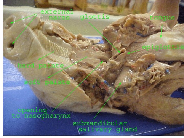
With scissors, make a 3 -cm incision in each corner of the pig's mouth. Your incision should extend posteriorly through the jaw. Spread the jaw open and examine the tongue. This will take some cutting and force. Observe the palate on the roof of the mouth. The anterior part of the palate is the hard palate, while the posterior part is the soft palate.
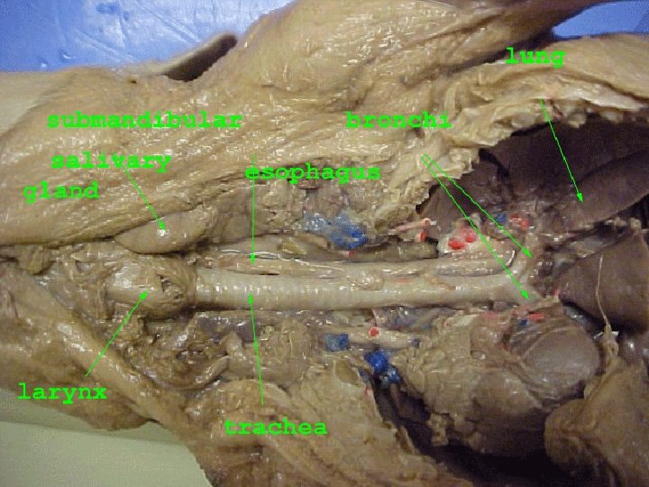
Locate the epiglottis, a cone-shaped structure at the back of the mouth. Above the epiglottis, find the round opening of the nasopharynx. This cavity carries air from the nostrils to the trachea, a large tube in the thoracic cavity which supplies air to the lungs.
Dorsal to the glottis, find the opening to the esophagus. Examine the tongue and note tiny projections called sensory papillae. Examine the teeth of the pig. Canine teeth are longer for tearing food, while incisor are shorter and used for biting. Pigs are omnivores, eating plants and animals. External nares tongue glottis epiglottis Hard palate Soft palate Opening to the nasopharynx Submandibular salivary gland
Pig Teeth • Canines vs. incisors
Digestive System Place the fetal pig ventral side up in the dissecting tray. Tie a string or use a rubber band to secure the front limbs. Run the string (or rubber band) under the tray, pull it tight, and tie it to the other front limb. Repeat this procedure with the hind limbs to hold the legs apart so you can examine internal structures.
Digestive System Note: You will keep your pig in this tray for the remainder of the dissection…. placing the pig (tray and all) into a plastic sack and adding moist paper towels at the end of each period. Label your plastic sack with your period and names/initials!!!!!!
The Incisions The lines numbered 1 -4 show the first set of incisions that you will make. To find the exact location for the incision marked 3, press along the thorax with your fingers to find the lower edge of the ribs. This is where you will make incision 3. With scissors, make the incisions in order, beginning with 1. Be sure to keep the tips of your scissors pointed upward because a deep cut will destroy the organs below. Also, remember to cut away from yourself.
The Incisions The first incision should be shallow- just enough to skin the pig. The muscles should now be exposed in the abdominal cavity. **you will need to use this pig for a week---if you damage structures it will make it harder to identify and finish- cut carefully!
Muscular System The scalpel was used to initially separate the skin from the subcutaneous tissue. Once a separation was begun it was easiest to clamp the skin with a pin and continue the separation with a blunt probe (handle end of tweezer). Another helpful technique for separating out layers or structures is using the sharp scissors closed in a scraping motion. Segmental veins and arteries were visible piercing the skin from below; it is ok to cut through these. The most difficult separation was on the ventral side near the umbilicus. The skin around the arm was cut off around the wrist.
Muscular System The fetal pig will not have fully developed musculature. For identification of muscles use a finger to smooth out the tissue and find the direction of the muscle striations. Where these change direction is a good indication of new musculature or a different section of the same musculature. Once the borders of the muscle were found use a blunt probe (handle portion of tweezers) and scissors to separate it from surrounding tissue. It was necessary to reflect some muscles in order to view musculature deep to them. Sever the muscles near their midlines so that it can be pieced back together.
Latissimus dorsi Deltoids External obliques Triceps brachii Rectus abdominus
trapezius Latissimus dorsi External obliques Biceps femoris deltoids masseter triceps gastrocnemius
Dissection of the Mouth, Oropharynx and Salivary Glands • To expose the salivary glands, remove the skin on one side of the head inferior to the ear and trim away the connective tissue in that are between this and the masseter muscle. Look for tiny lymph nodes (beanshaped) in this are. The parotid gland is on the cheek area inferior to the ear. The smaller submandibular gland is inferior and a little posterior to the parotid gland.
Dissection of the Mouth, Oropharynx and Salivary Glands The sublinguinal gland, just anterior to the submandibular gland, is the smallest salivary gland is more difficult to find. The latter is more granular and not as firm.
Today’s targets Pin & Study the basic anatomy and physiology of the internal organs of the thoracic and abdominal cavities; Liver, Lungs, Large Intestine, Small Intestine, Stomach, Spleen, Diaphragm, and Heart Verbal Quiz 1 Dissect out the digestive system-get quizzed on mouth to anus - all structures and functions Verbal Quiz 2
The Incisions Now make a deeper incision along the same lines- be careful not to damage the organs. Use pins to secure the flaps of tissue down to the tray- see next slide.
If you’ve forgotten what a “peritoneum” is- use your textbook or smart phone to look this up!! After you have made your incisions through the body wall, you will see the peritoneum, a thin layer of tissue that lines the body cavity. Cut through the peritoneum along the incision lines. Spread the flaps of the body wall apart. Cut the umbilical vein, which extends through the liver.
Once the vein is cut, carefully pull the flap of skin, including the end of the umbilical cord between the hind legs. You are now able to see the organs of the abdominal cavity. Locate AND PIN each of the structures labeled in the picture
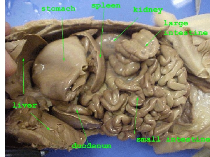
Heart Lungs Liver stomach Gall bladder…usually greenish Pyloric sphincter duodenum
Locate the diaphragm, a sheet of muscle that separates the abdominal cavity from the thoracic cavity. Find the most obvious structure in the abdominal cavity, the brownish-colored liver. Count the number of lobes. Record the number of lobes on the data sheet (number 1). What is the function of the liver in a living pig? If your group has forgotten the livers function- look it up in textbook- give detailed answers- it’s your final grade
lung diaphragm spleen liver
Digestive System Find the tube-like esophagus, which joins the mouth and the stomach. Food moves down the esophagus by muscular contractions after being softened by saliva in the mouth. Follow the esophagus and locate the soft, sac-like stomach beneath the liver. Insert pins to identify each of the structures – for group check time
Digestive System With scissors cut along the outer curve of the stomach. Open the stomach and note the texture of its inner walls. Make a drawing of the observations (number 3). These ridges inside the stomach are called rugae and increase the area for the release of digestive enzymes. The stomach may not be empty because fetal Check that you have pins in each pigs swallow amniotic of these structures- make a legendfluid. before removing any of the organs! It is much more of a challenge to identify OUTSIDE of the body
Pigs are ruminants, animals with multiple stomachs. Locate the entrance to the stomach or esophageal area, the cardiac region, which is largest, and the pyloric region where the stomach narrows to join to the small intestine. What is the function of the small Insert pins into each of these intestine (number structures- be prepared to identify and 4)? be able to explain their FUNCTIONS during the group quiz time
At the end of the stomach, there is a sphincter, or ring-shaped muscle to control food leaving the stomach and entering the duodenum. Locate the cardiac sphincter at the junction of the stomach and esophagus, and the pyloric sphincter at the junction of the stomach and small intestine. Fetal pigs receive their nourishment from their mother through the umbilical cord. Insert pins into the cardiac and pyloric sphincters Record pin # onto lab record sheet
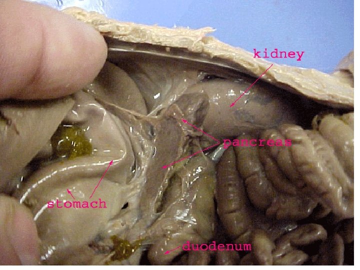
Identify the first part of the small intestine, the U-shaped duodenum, which connects to the lower end of the stomach. Pancreatic juice, made by the pancreas, and bile, made by the liver and stored in the gall bladder, are added to food here to continue digestion. What specific macromolecule(s) is bile used to breakdown during digestion (number 5)?
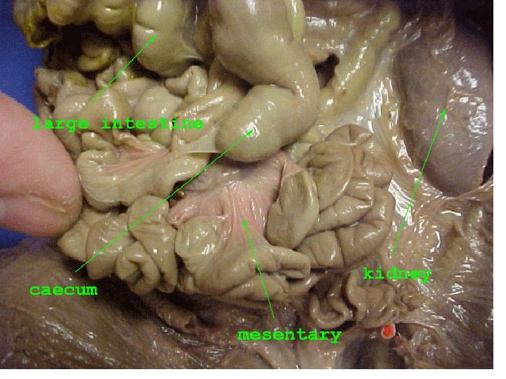
Study the rest of the small intestine. Notice that it is a coiled, narrow tube, held together by tissue called mesentery. The soupy, partly digested food that enters the small intestine from the stomach is called chyme. Carefully cut through the mesentery and uncoil the small intestine. The mid-section is called the jejunum, while the last section is called the ileum. What is the importance of the mesentery (number 6)? Note and record the small intestine’s length in centimeters on the data sheet (number 7)- this takes some patience and time…your recorder should get some of the diagrams completed during this time
Observe the inner surface of the small intestine. Run your finger along it and note its texture. Using a dissection scope, examine the villi, the tiny projections that line the small intestine and increase the surface area for absorption. Make a drawing of the observations made (number 8). Follow the small intestine until it reaches the wider, looped large intestine. Cut the mesentery and unwind the large intestine or colon. Measure and record its length on the data sheet (number 9).
Small Intestine…. Histology Cut out a small section 1 -cm section of small intestine Cut the section open to the interior of the intestine and place on microscope slide Using the dissection scope, focus on the interior surface-get checked off
At the junction of the large and small intestine, locate a blind pouch called the caecum. The caecum has no known function in the pig. Notice that the large intestine leads into the rectum, a tube that runs posteriorly along the dorsal body wall. The rectum carries wastes to the opening called the anus where they are eliminated.
Locate thin, white pancreas beneath the stomach and duodenum. Pancreatic juice flows through pancreatic ducts to the duodenum. Between the lobes of the liver, find the small, greenish-brown gall bladder. Locate the hepatic duct, which carries bile from the liver to the gall bladder. Find the spleen, a long, reddish-brown organ wrapped around the stomach. The spleen filters out old red blood cells and produces new ones for the fetus. Label the diagram of the digestive system on the data sheet figure 2. Don’t forget that each of these internal structures need to be labeled with pins and recorded on your group final sheet
Group Check Time Call me over when your group is ready to be quizzed on the ENTIRE Digestive System Be prepared to explain the travels of food from mouth to anus: including amylase, pepsin, trypsin, chymotrypsin Be prepared to explain the secretions and processes happening in the stomach, small intestine, pancreas and gall bladder Explain the roles of amylase, pepsin, HCl, microvilli
Group Check of the Internal Organs By the end of the next 2 slides- your team should be ready to identify the following structures and their basic functions: Liver, Lungs, Large Intestine (ascending, transverse, descending, sigmoid colon) , Small Intestine (duodenum, jejunum, ileum), Stomach, Pancreas, Spleen, Diaphragm, and Heart, Pancreas, Gall bladder Place pins in each of the structures- record the numbers onto your group final lab sheet- for example #8 = heart **I recommend having your reader/recorder look up functions- and write them down for your team to study before verbal quiz
Clean up: Clean up your materials and work area after each day. Keep your pig attached to your lab tray. Wrap your lab tray and pig in moist wet towels – pour the preservative that you saved- on top of the paper towels. Place this entire set-up inside two plastic grocery sack. Label with your period and name(s) to allow for you to find your pig Monday!! Return your lab equipment to the front tub and then thoroughly wash your hands with soap. Look Ahead: tomorrow we will dissect out the respiratory & cardiovascular system
Verbal Quiz Hints Digestion of food from mouth to anus ü Including-enzymes like amylase, pepsin, trypsin ü Names/locations of sphincters- controlling food into and out of the stomach ü Parts of small (3) and large intestine (4) ü Roles & Secretions from the pancreas and gall bladder ü A&P of: bolus formation, rugae, villi ü Maintenance of homeostasis-3 specific functions
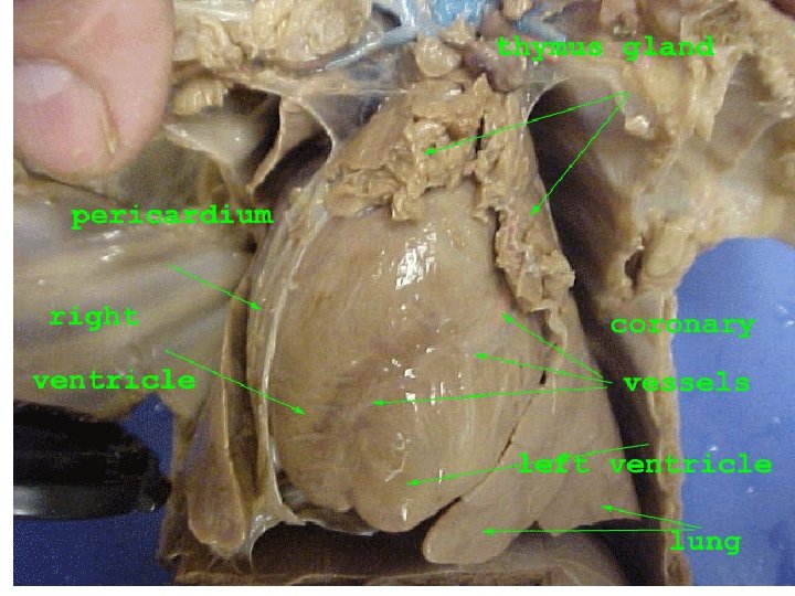
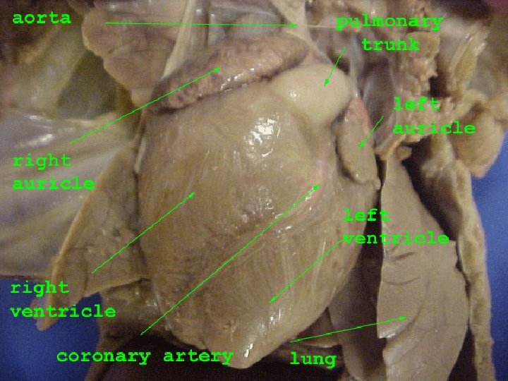
Cardiovascular Systems Circulation- each individual in team must tell me about the travels of RBCs from right atriumtricuspid valve-right ventricle…etc. First look at external anatomy of this sysstem by locating, pinning and describing: Coronary arteries, auricles, ventricles, percardium (pericardial sac), aorta, pulmonary trunk – find arteries and veins going to the lungs
Circulatory System Locate the heart. It is covered by a thin tissue called the pericardium. Gently remove this membrane to study the heart. Pigs, like all mammals, have four-chambered hearts. The right side of the heart pumps blood to the lungs, while the left side of the heart pumps blood to all other parts of the body. Locate the right and left sides of the heart. Would the left or right side of the pig heart have more cardiac muscle? Explain your answer (number 1).
Circulatory System Surgeons and Assistant surgeons- don’t forget to insert pins in each of the structures that are part of the circulatory system Reader/Recorders don’t forget to record the pin # and the functions of each of the structures for your group to reference before the verbal quiz Each side of the heart has an upper and a lower chamber. Upper chambers are called atria and receive blood, while lower chambers are called ventricles and pump blood out of the heart. Locate the right and left atria and ventricle.
Notice that the surface of the heart is covered with blood vessels. These are part of the coronary circulation, a set of arteries and veins whose only job is to nourish the heart tissue. Blockage in these vessels causes heart attacks. Anterior to the heart, locate another large vein that enters the right atrium. This vein, the anterior vena cava, brings blood to the right atrium from the anterior part of the body. Now lift the heart to view its dorsal surface. Observe the posterior vena cava that carries blood from the posterior part of the body and empties it into the right atrium. Our pigs are only singleinjected- so you will only see red latex for the arteries, not the blue latex seen in the picture to the right
Find the pulmonary artery which leaves the right ventricle. After birth, this vessel carries blood to the lungs. However, in a fetus, a shunt called the ductus arteriosus allows fetal blood to bypass the lungs and go directly to the aorta, the largest artery of the body. Locate the pulmonary veins that enter the left atrium. After birth, these vessels carry oxygenated blood from the lungs to the heart. Identify the aorta, a large artery that transports blood from the left ventricle. Many arteries that carry blood throughout the body branch off of the aorta. Place pins in each of these structures record the numbers Be prepared as a group to explain the function of the ductus arteriosus
Remove the heart by severing the blood vessels attached to it. Hold the dorsal and ventral surfaces of the heart with your thumb and forefinger and rest the ventricles on your dissecting tray. With a scalpel, cut the heart into dorsal and ventral halves. Caution: The scalpel is very sharp. Use it carefully and always cut away from yourself. Remove any material inside the heart and expose the walls of the atria and the ventricles.
Study the internal features of these chambers and note where vessels leave or enter each chamber. Locate the valves between each atrium and ventricle. These structures prevent blood from flowing backward in the heart. Label the parts of the heart on the data sheet part E, number 2 (figure 4). Label your dissected heart with pins- find all four valves, all four chambers
Respiratory System Examine the diaphragm, a sheet of muscle that stretches across the abdominal cavity and separates it from the thoracic cavity where the lungs are located. The diaphragm isn't used by the fetal pig because gas exchange occurs through the umbilical cord. What is the function on the diaphragm in an adult pig (number 1)?
Respiratory System In order to see the upper part of the respiratory system, you will need to extend cut #1 up under the pig's throat and make two more lateral incisions in order to fold back the flaps of skin covering the throat. In the thoracic cavity, carefully separate the pericardium or sac surrounding the heart and the diaphragm from the body wall.
Respiratory System Locate the two, spongy lungs that surround the heart. The tissue that covers and protects the lungs is called pleura. The lungs haven't been used by the fetus so they have never contained air. Find the trachea, a large air tube that lies anterior to the lungs. The trachea is easy to identify because of the cartilaginous rings that help keep it from collapsing as the animal inhales and exhales. Label each of the structures involved in the respiratory system with numbered pins.
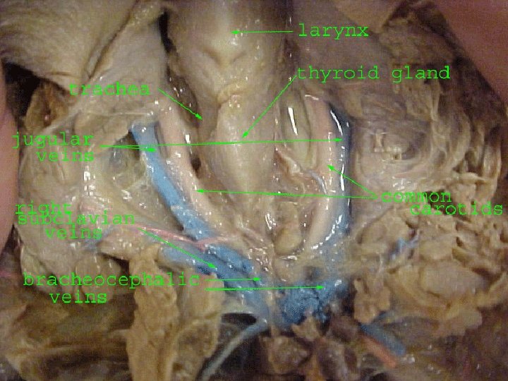
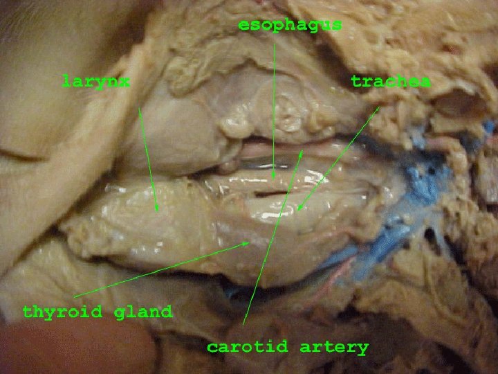
Notice that the trachea branches into each lung. These two tubes are called bronchial tubes. Inside the lungs these branch into smaller bronchioles that end with a grape -like cluster of air sacs or alveoli where oxygen and carbon dioxide are exchanged with capillaries. Draw a picture of the trachea leading to Label the thymus and thyroid- be the two bronchial tubes and then the prepared to explain the functions of each lungs (number 2). of these structures.
Lying ventral to the trachea or windpipe locate the pinkishbrown, V-shaped structure called the thyroid gland. This gland secretes hormones that control metabolism.
At the top, anterior end of the trachea, find the hard, light-colored larynx or voice box. This organ contains the vocal cords that enable the animal to produce sound. Locate the epiglottis at the top of the trachea. This flap of skin closes over the trachea whenever you swallow. Find the area called the pharynx at the back of the nasal cavity. Air enters an adult pig through the mouth or nose before passing through the pharynx and down the trachea to the lungs. Label the diagram of the respiratory system on the data sheet figure 3.
Respiratory System From mouth/nares to alveoli Gas Exchange Lobes of the Lung- right vs. left Locate, pin and check with recorder for description of pharynx, larynx, trachea, bronchi, lungs, alveoli, diaphragm
Group Quiz Time When your group is ready, call me over to verbally quiz you on the anatomy and physiology of the structures of the respiratory system. Once you are signed off begin the clean up. Clean up: Clean up your materials and work area after each day. Wrap the pig in damp paper towels and put it in a ziplock plastic bag. Obtain a piece of masking tape and label your bag with your names. Store your fetal pig in the lab station cabinet. Return your lab equipment to the lab station cabinet and then thoroughly wash your hands with soap. Please make sure you are spraying the pins, scalpels, scissors, and tweezer with the disinfectant sprays each day- dry off with paper towels.
Group Check Time Call me over when your group is ready to be quizzed on the Circulatory System If you have plenty of time left in the period- move on to the next slide on the reproductive system. Clean up: Clean up your materials and work area after each day. Keep your pig attached to your lab tray. Wrap your lab tray and pig in moist wet towels. Place this entire set-up inside a plastic grocery sack. Label with your period and name(s) to allow for you to find your pig tomorrow!! Return your lab equipment to the black bin and then thoroughly wash your hands with soap.
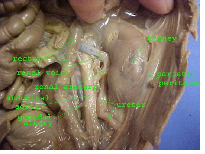
Excretory System: focus on Urinary System Kidney extraction be careful to keep a section of the ureter attached Make a longitudinal slice (see purple and white packet diagram- pin each of those parts) Keep one kidney within the fetal pig to trace the steps of urine formation and flow Identify: kidney, ureter, bladder, urethra Identify: w/in kidney (cortex, capsule, medulla, pelvis)
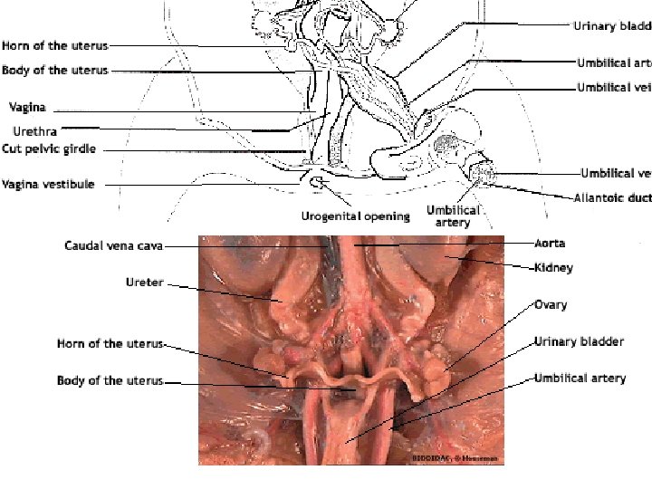
Excretory/Urinary System: Learning Targets: 1) Urinary system is made-up of the kidneys, ureters, bladder, and urethra 2) Waste is filtered from the blood and collected as urine in each kidney. Urine leaves the kidneys by ureters, and collects in the bladder. The bladder can distend to store urine that eventually leaves through the urethra.
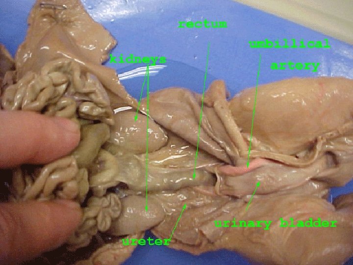
Urogenital System Find the ureters, tubes which extend from the kidneys to the bag-like urinary bladder. The urinary bladder lies between the umbilical arteries and temporarily stores liquid wastes filtered from the blood. Don’t forget to label each of these structures with numbered pins Be prepared to explain the basic functions of each.
Urogenital System Lift the urinary bladder to find the urethra, the tube which carries urine out of the body. Follow the urethra to the urogenital opening on the outside of the pig's body.
Urogenital System Remove the digestive organs to study the excretory and reproductive organs that make up the urogenital system.
Urogenital System Locate the large, bean-shaped kidneys lying against the dorsal body wall. Notice that they are covered by the peritoneum. Kidneys filter wastes from blood. Draw a picture of the kidneys on the data sheet (part f, number 1 data sheet).
Pin the parts labeled below- check with recorder for info about these parts of the kidney Take out one kidney and make a coronal cut
Group Check Time Call me over when your group is ready to be quizzed on the Urinary/Excretory System If you have plenty of time left in the period- move on to the next slide on the reproductive system. Clean up: Clean up your materials and work area after each day. Keep your pig attached to your lab tray. Wrap your lab tray and pig in moist wet towels. Place this entire set-up inside a plastic grocery sack. Label with your period and name(s) to allow for you to find your pig tomorrow!! Return your lab equipment to the black bin and then thoroughly wash your hands with soap.
Reproductive System • Fill out diagrams on last page- pin these structures in your pig- for verbal quiz- be prepared to discuss both parts of both sexes- and to ID them on another team’s pig (that has the opposite sex)
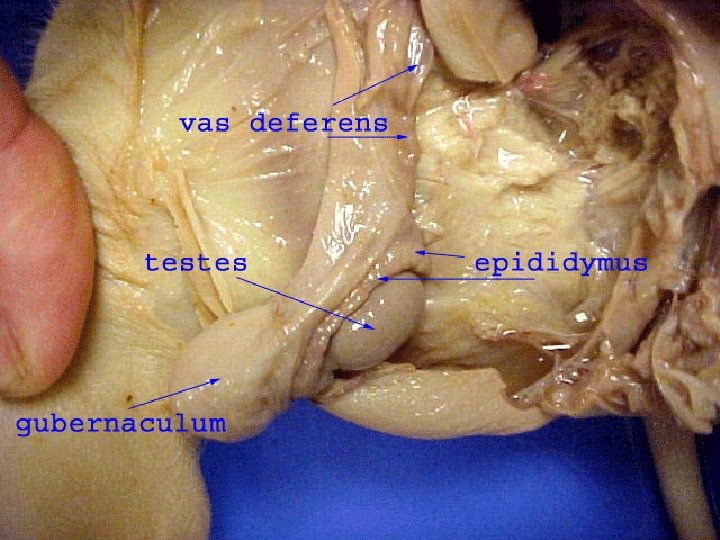
Male Reproductive System In the male pig, locate the two scrotal sacs at the posterior end of the pig. If the pig is in the later stages of development, you will find a testis in each sac. If the pig is in an early stage of development, the oval-shaped testes will be in the abdominal cavity. These testes have not yet descended into the scrotal sacs. If you have a female pig, move ahead to the appropriate slides Your group will need to check in with another group to see their dissection of the male reproductive system.
gubernaculum tes´tis = the fetal ligament attached at one end to the lower end of the epididymis and testis and at its other end to the bottom of the scrotum; it is present during the descent of the testis into the scrotum
Male Reproductive System On each testis, find the coiled epididymis. Sperm cells produced in the testis pass through the epididymis and into a tube called the vas deferens. This tube crosses over a ureter and enters the urethra. Who remembers the significance of the word “vascetomy” in humans- and its relationship to an anatomical structure of the male reproductive system?
Male Reproductive System Follow the urethra to the penis, a muscular tube lying just below the skin posterior to the umbilical cord. In mammals, the penis is the organ that transfers sperm. Label the diagram of the male urogenital system on the data sheet figure 5, male. If necessary- use a textbook for reference.
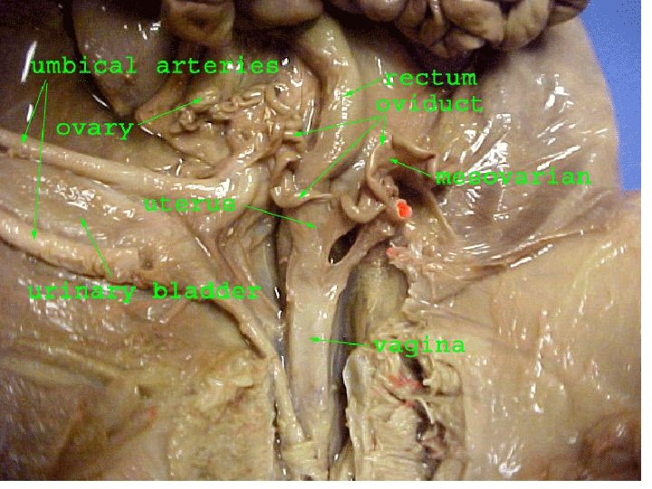
Female Reproductive System In the female pig, find the two bean-shaped ovaries at the posterior end of the abdominal cavity. Observe the coiled Fallopian tube attached to each ovary, which carries eggs from the ovary. Follow the Fallopian tube to the uterus. The uterus is dorsal to the urinary bladder and the urethra. Label each of these structures with the numbered pins. Be prepared to explain the function/physiology of the uterus, ovaries and fallopian tubes.
Female Reproductive System Trace the uterus to a muscular tube called the vagina. The vagina will appear as a continuation of the uterus. Sperm from the male are deposited into this organ during mating. The vagina and the urethra open into a common area called the urogenital sinus. This cavity opens to the outside at the urogenital opening. Label the diagram of the female urogenital system on the data sheet figure 5, female.
Female Reproductive System When you have finished with the fetal pig dissection make sure to clean all dissection tools return them to the appropriate storage place and wipe up the lab station. Dispose of the fetal pig by putting it in the plastic bag and placing it in the trash. Label the vagina, urogenital sinus, and urethra on your pig.
CONGRATULATIONS As soon as your station and all dissection supplies are cleaned up- your group earned a 100% on your final!! After completing the lab- you are ready to start the individual portion of your final.