THE FEMALE REPRODUCTIVE SYSTEM OVARIES AND ACCESSORY STRUCTURES


- Slides: 13

THE FEMALE REPRODUCTIVE SYSTEM: OVARIES AND ACCESSORY STRUCTURES Pages 547 -557

Reproductive Structures Ovaries (endocrine function) Duct system (exocrine function) Fallopian tubes Uterus Vagina External genitalia © 2015 Pearson Education, Inc.

FIGURE 16. 8 B THE HUMAN FEMALE REPRODUCTIVE ORGANS. Uterine (fallopian) tube Ovary Infundibulum Fimbriae Body of uterus Ureter (b) Cervix Vagina Uterine tube

Ovaries: Exocrine Function- egg production Ovaries contain many ovarian follicles (saclike structures) These contain oocytes (immature eggs) The lifetime supply of oocytes (about 1 million) is established by birth At puberty oocytes undergo meiosis and become functional eggs cells layers of supportive follicle cells surround the oocyte © 2015 Pearson Education, Inc.

Stages of oocyte production and the Ovarian Cycle Follicle Stimulating Hormone initiates the ovarian cycle: The oocyte matures within the follicle Luteinizing Hormone initiates release from the ovary This is called Ovulation Occurs about every 28 days Penetration of the egg by a sperm will trigger completion of meiosis II thereby creating the © 2015 Pearson Education, Inc. haploid gamete

Path of the Oocyte Released by the ovary Fimbriae of the infundibulum sweep the oocyte into the uterine (fallopian) tubes this is accomplished by way of cilia and peristalsis Fertilization takes place in the fallopian tube The tubes empty into the uterus A fertilized egg will implant here © 2015 Pearson Education, Inc.

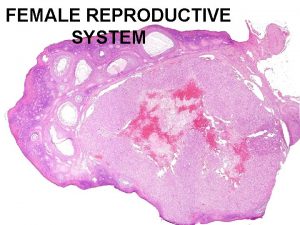
FIGURE 16. 7 SAGITTAL VIEW OF A HUMAN OVARY SHOWING THE DEVELOPMENTAL STAGES OF AN OVARIAN FOLLICLE. 1. Primary follicle 2. Growing follicles 8. Degenerating corpus luteum Blood vessels 3. Mature follicle Germinal epithelium 7. Corpus luteum 6. Developing corpus luteum 5. Ruptured. Ovulation follicle 4. Secondary oocyte

The Uterine (Menstrual) Cycle: Endocrine Function Changes in the mucosa of the uterus (endometrium) coincide with ovulation Days 1 -5: Endometrium is sloughed off Days 6 -14: Follicles grow, endometrium rebuilds Days 15 -28: Blood supply (carrying nutrients) to the uterus increases in preparation for implantation Stimulated by increasing estrogen released by follicles of ovaries Lack of implantation starts the cycle all over The ruptured follicle transforms into a corpus luteum Produces progesterone & estrogen until ovulation ceases Disintegrates if no fertilization transpires

Uterine Layers (deep to superficial) Endometrium: Mucosal layer Allows for implantation of a fertilized egg Sloughs off if no pregnancy occurs (menses) Myometrium: smooth muscle; contracts during labor Perimetrium: (visceral peritoneum) outermost serous layer of the uterus © 2015 Pearson Education, Inc.

FIGURE 16. 8 B THE HUMAN FEMALE REPRODUCTIVE ORGANS. Suspensory Uterine (fallopian) tube ligament of ovary Lumen (cavity) Fundus Ovarian of uterus blood vessels Ovary Infundibulum Broad ligament Fimbriae Ovarian ligament Round ligament of uterus Body of uterus (b) Endometrium Myometrium Perimetrium Ureter Uterine blood vessels Cervical canal Uterosacral ligament Cervix Vagina Wall of uterus Uterine tube

External Genitalia and Female Perineum Vagina from cervix to exterior of body between urinary bladder and rectum Clitoris erectile tissue that corresponds to the male penis Greater Vestibular Glands Swollen with blood during sexual excitement Produce mucus to lubricate the vagina during intercourse Perineum Diamond-shaped region between the anterior ends of the labial folds, anus posteriorly, and ischial tuberosities laterally © 2015 Pearson Education, Inc.

FIGURE 16. 9 EXTERNAL GENITALIA OF THE HUMAN FEMALE. Labia majora Clitoris Urethral orifice Vaginal orifice Opening of duct of greater vestibular gland Labia minora Perineum Anus

The Ovarian and Uterine Cycles http: //www. sumanasinc. com/webcontent/anim ations/content/ovarianuterine. html
 Accessory structures of female reproductive system
 Ovary duct
 Drawing of the male and female reproductive system
 Female and male reproductive system
 Similarities between male and female reproductive system
 Oviduct funnel
 Pearson education
 What are primary sexual characteristics
 90/2
90/2 Similarity between male and female reproductive system
 Is croaker a cartilaginous fish
 Fetus reproductive system
 Epilization
 Lesson 3 the female reproductive system
