Chapter 28 MALE and Female Reproduction System The


")


SYMPATHETIC nervous system stimulates PERISTALSIS in smooth muscle of vas deferens")


 stimulates anterior pituitary")


")

 : Inability to maintain erection CAUSES:")


- Slides: 34
Chapter 28 “MALE” and Female Reproduction System
• • • The Male Reproductive System Only system not essential to the life of the individual Secrete hormones that play major roles in normal sexual function Male PRODUCES 1/2 BILLION SPERM (500, 000) PER DAY WOW!!! primary sex organs (testes- gonads) - produce gametes (sperm) secondary sex organs – other than the gonads that are necessary for reproduction: system of ducts, glands, penis deliver sperm cells • 22 pairs of autosomes - 1 pair of sex chromosomes (XY or XX) – Male sperm carries EITHER an X or Y: determines sex of child • Sexual determination does not end at fertilization but requires interaction between hormones and genetics – SRY gene (sex-determining region of Y chromosome) codes for protein, testesdetermining factor (TDF)= initiates development of testes; interacts with other chromosomes to produce male characteristics / affects testosterone production Male development occurs due to THE PRESENCE OF ANDROGENS NOT THE ABSENCE OF ESTROGEN • Urology: study of the urinary system and diagnosis and treatment of diseases and disorders of the male reproductive system.
Androgen-Insensitivity Syndrome • Testes produce normal male levels of testosterone BUT TARGET CELLS LACK RECEPTORS; Androgen receptor mediates the effects of androgens in the human body producing male characteristics; without the stimulation by androgens individual will produce female characteristics. • Lack of response to androgens does not significantly impair female genital or sexual development. • External genitalia develop female anatomy as if no testosterone were present; generally normal, though labia and clitoris can be underdeveloped • Develop little to no public or axillary hair • Vaginal DEPTH varies, typically shorter; extreme cases vagina resembles a "dimple • Hormone from testes Sertoli cells that produce sperm, STOP development of fallopian tubes, cervix, and uterus • Total infertility – no viable sperm, unable to produce ovum • Complete AIS affects 2 -5 per 100, 000 genetically male. Partial insensitivity as common as complete androgen insensitivity. Mild androgen insensitivity much less common.
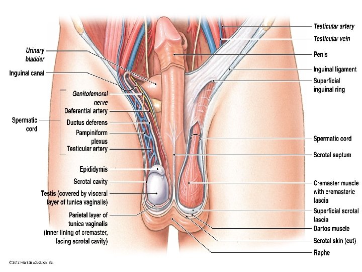
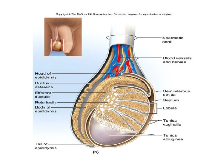
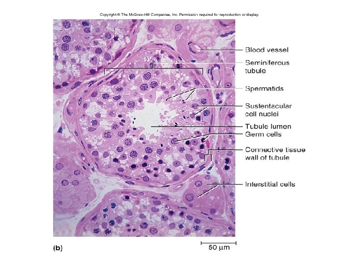
Testes Development • Gubernaculum testis connective tissue fibers extends from testis to peritoneum locking testes in position • Vaginal process – fold of the peritoneum forms a sac = tunica vaginalis that LINES SCROTAL CAVITY; reduces friction between scrotal-sac and testes. § Inguinal canal – gubernaculum and vaginal process create pathway allowing testes to moved into scrotal sac. • Tunica albuginea - dense connective tissue capsule surrounding testes; forms septa that divides testes into a series of 200 -300 compartments =lobules (within by tunica vaginalis) • Seminiferous tubules – 1/2 mile of long, tiny coiled tubes packed inside lobules/ testes where spermatogenesis occurs through entire length of tubule. Forms a loop connected to rete testis, a network of passageways; in the rete testis sperm become CONCENTRATED. Unconcentrated sperm entering the epididymis could result in possible infertility. • Developing germ cells begin their development near the periphery of the tubules get released as spermatozoa in the lumen of the tubule
Scrotum and Spermatic Cord SCROTUM connective tissue pouch of skin divided into two sacs. • scrotal septum -Divides the scrotum into two sacs each containing a testis; a tissue barrier that prevents cross bacterial contamination from one testes to the other. Externally marked with by the raphe that separates the scrotum into two sections. • Testes descend into the scrotum through the inguinal canal then closes testes are suspended in the scrotal sac by a spermatic cord and cremaster muscles • SPERMATIC CORD - Bundle of fibrous connective tissue containing the ductus (vas) deferens (sperm duct), blood and lymphatic vessels, and testicular nerve; covered by cremaster muscle.
Cryptorchidism • Testes move into the scrotum within 1 st year of infancy. Descent begins as early as the 6 th week of fetal development • Cryptorchidism 3% of boys born with undescended testes. Testes remain in the abdomen results in no sperms formation and greater risk of testicular cancer • Treatment: injection of h. GH, surgery
Scrotum Temperature 2 -3°LOWER than body; optimal 35° C or 95° F; will NOT produce sperm at (98. 6° F or 37° C) body temperature • cremaster muscle – bands of INTERNAL skeletal that surrounds spermatic cord Attaches testes to abdominal wall – In cold temperature contracts; draws testes upward toward body / warm temperatures relaxes suspending the body using (genitofemoral nerve) • pampiniform plexus – network of VEINS that surround testicular artery and spermatic cord – Countercurrent heat exchanger- removes heat from the descending arterial blood preventing warm arterial blood from inhibiting sperm production • dartos muscle – subcutaneous layer of smooth muscle – contracts when it is cold, wrinkling the scrotum, holding testes against warm body reducing surface area of the scrotum 27 -8 decreasing heat loss
The Testes • LEYDIG / INTERSTITIAL CELLS OUTSIDE the seminiferous tubules; secrete hormones (TESTOSTERONE) to regulate spermatogenesis, development of male reproductive tissues such as the testis and prostate as well as promote secondary sexual characteristics such as increased muscle and bone mass, and body hair. • SERTOLI, SUSTENTACULAR OR NURSE CELLS INSIDE seminiferous tubules cells support, nourish, protect developing gamete germ cells 1. phagocytize germ cell debris 2. secrete hormone (inhibin) decreases FSH release regulates sperm production 3. secrete androgen binding protein (ABP) when bound to testosterone: • increases spermatogenesis in the seminiferous tubules • produce sperm maturation in the epididymis. • MAINTAINS BLOOD–TESTIS BARRIER; tight junctions in cells isolates seminiferous tubule from blood; Male body capable of inducing an immune action against his own sperm!!
The blood–testis barrier
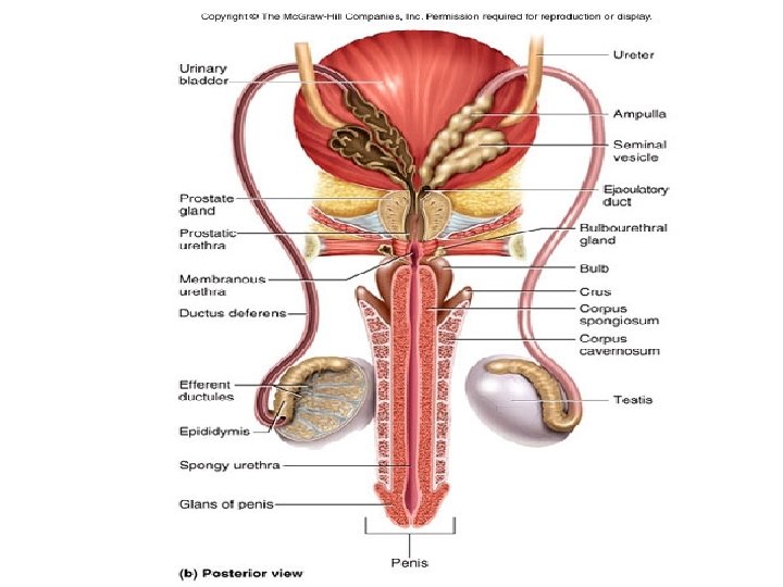
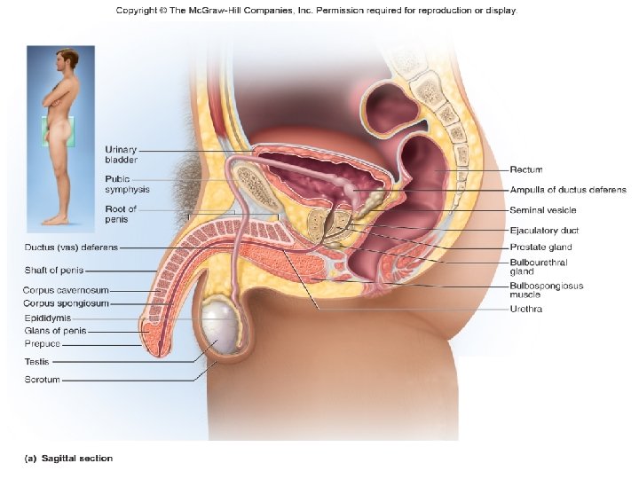
Testes to the urethra • Efferent ductules - collect sperm and transport to epididymis • Epididymis - Start of male reproductive tract – Monitors/adjusts fluid produced by seminiferous tubules – site of sperm maturation and storage for 2– 3 months. – Sperm are immature; inactive lacking motility till reaching prostate gland, move via peristalsis through ducts – if not ejaculated once matured, disintegrate; reabsorbed by epididymis • Ductus (vas) deferens transport sperm from scrotum through inguinal canal loops over bladder to the ampulla into ejaculatory duct • Ejaculatory duct FORMED FROM ductus deferens and seminal vesicle pass through prostate to empty into urethra 27 -12
Accessory Glands • seminal vesicles – empties into ejaculatory duct – forms 60% -70% of semen – Prostaglandins, fructose, fibrinogen • prostate gland – surrounds urethra and ejaculatory duct just inferior to the bladder – empty through about 20 pores in the prostatic urethra – thin milky slightly acidic secretion forms 30% of semen – release substances that activate sperm enabling them to swim. • bulbourethral (Cowper) glands – near bulb of penis – during sexual arousal, produce a clear slippery fluid that lubricates the head of the penis in preparation for intercourse – protects the sperm by neutralizing the acidity of residual urine in the urethra 27 -14
Terminal portion of the male duct system = 18 cm long Three regions: § Prostatic urethra- passes through prostate gland § Membranous urethra – urogenital diaphragm § urethra passes through muscular floor of pelvic cavity § Spongy (penile) urethra –runs through penis and opens to the outside at the external urethral orifice. Accounts for 75% of urethral length. § Passes through corpus sponginosum- mass of spongy tissue Urethra
Penis http: //getanchorednow. com/yahoo_site_admin/assets/images/penisdiagram. 17851350_std. gif • Common passage for urine and semen • Three cylindrical bodies of tissue which protect urethra and fill with blood during sexual arousal accounting for its enlargement and erection • 1) corpus spongiosum – a mid-ventral, sinus rich NON-erectile tissue that wraps around the spongy urethra. Forms bulb to anchor the penis to the abdomen; prevents compression of urethra during erection. • Expands at the tip of the penis to form glans penis (more sensitive area of the penis) which is covered by prepuce- the foreskin. • Preputial glands secrete waxy material (smegma) that can support bacterial growth. Circumcision, surgical removal of prepuce, can help prevent infection – 2) corpora cavernosa - two dorso-lateral, capillary sinus-rich masses of erectile tissues. Injury can cause fracture and rupture of tissue.
Forms bulb to anchor penis to abdominal wall; prevents compression of the urethra during erection
Male Reproductive System • Erection is the enlargement and stiffening of the penis resulting from engorgement of the erectile tissue (corpora cavernosa) with blood. When not sexually aroused, the arterioles supplying the erectile tissue are constricted and the penis is flaccid. • During sexual excitement, a parasympathetic reflex triggers release of nitric oxide from endothelial cells lining the blood vessels leading to arteriole blood vessel dilation. • Vascular spaces of erectile tissues fill with blood enlarging and erecting the penis. • Erection one of the rare parasympathetic controls of arterioles along with stimulation of the bulbourethral glands.
Male Reproductive Ejaculation 1)SYMPATHETIC nervous system stimulates PERISTALSIS in smooth muscle of vas deferens propelling sperm from epididymis to ampulla. 2)Seminal vesicles secretions added to semen. Ampulla contracts propelling sperm through ejaculatory duct into prostatic urethra 3)Prostatic gland contractions force fluid into the urethra. 4)Closing of internal urethral sphincter at the base of urinary bladder prevents release of urine System 5)The semen is ejected through the urethra with rhythmic contractions generated by the bulbospongiosus muscle.
Male Reproductive System “Little more than Sperm DNA with a propeller!” Head § Haploid nucleus 22 X or 22 Y chromosomes § Membrane sac contains hydrolytic enzymes Mid-piece – contains mitochondria that provide ATP for motility of the sperm Tail – is the flagellum that provides movement • 300 - 500 million sperms are produced daily; 20. 8 million hour “ 3, 000 sperm with each heartbeat” • Formed in seminiferous tubules released and mature in epididymis mix with seminal secretions from 3 glands § Semen: sperm + seminal fluid Slightly alkaline – p. H 7. 2 – 7. 7 § 2. 5 – 5. 0 ml per ejaculate (1/2 -1 tsp) § About 50 – 150 million -300 million sperms/ml per ejaculation § Less than 20 million sperms/ml may be a cause for infertility
Spermatogenesis • The process of sperm production begins at outermost cell layer in seminiferous tubules proceeds toward lumen • 2 N = 22 autosomes pairs + 1 pair of sex chromosomes (XY males: XX females) • 1 N = 22 autosome chromosomes + 1 X or 1 Y Spermatogonia (stem cells) divide by mitosis to produce two identical daughter cells • One remains as a primary spermatogonia Type A =LIFETIME supply of stem cells; Spermatogonia Type B = migrate to produce sperm – Primary spermatocytes (2 N) begin meiosis and form two (2) secondary spermatocytes (1 N) • Secondary spermatocytes differentiate into four (4) spermatids (immature gametes) (1 N) – Each spermatids’ transformation called spermiogenesis [the final stage] produces single spermatozoon/sperm (4) (1 N) » Spermatozoa - Lose contact with wall of seminiferous tubule and enter fluid in lumen to epididymis for final maturation process
Type A remain as stem cells Type B produce sperm
Male Reproductive System-Hormonal regulation Hypothalamus secretes Gonadotropin Releasing Hormone (Gn. RH) stimulates anterior pituitary to secrete: § FSH stimulates sertoli/sustentacular cells to produce 1) Androgen binding protein (ABP) concentrate testosterone 2) FSH supports seminiferous tubules to carry out spermatogenesis (sperm production 3) Inhibin inhibits the secretion of FSH § Luteinizing hormone (LH) stimulates Leydig cells secrete testosterone (T) stimulates spermatogenesis, secondary male characteristics, metabolism, bone and muscle growth – Without FSH and ABP, testosterone IS UNABLE TO SUFFICIENTLY SUSTAIN SPERMATOGENESIS. TESTOSTERONE Establishes male secondary sex characteristics – Distribution of facial hair Sexual behaviors Sexual drive – Increased muscle mass and body size – On average, LEVELS of testosterone in adult males is about 7– 8 x higher than in adult females. – Albumins carry 1/3 of testosterone
Sperm Pathway Once the sperms are formed in the testes Moves into rete testes to the efferent ductules into the epididymis remain in epididymis for 10 -14 days, mature (gain motility, ability to fertilize) if not ejaculated: sperms break down and get reabsorbed by the body; if ejaculated: sperms move with the help of peristalsis to vas deferens that goes through the spermatic cord and enters the abdomen to the ampula of the vas deferens Then mix with the secretion of seminal vesicle enter ejaculatory duct that passes through the prostate gland released into the prostatic urethra mix with the secretion of prostate gland pass through the membranous urethra enter spongy urethra mix with the secretion of Cowper’s gland pass through the spongy urethra of the penis ejaculated at the urethral orifice
Seminal Fluid Mixture of fluid secreted by seminal vesicle, prostate gland Cowper’s gland Fluid Contains: • Alkaline buffers to neutralize acidic environment of urethra and vagina (lactic acid) (putrescine, cadaverine). Responsible for odor and taste of semen. • Fructose acts as spermatozoa energy source (secreted by seminal vesicle) • Seminal plasmin that acts as an antibiotic; vagina has actively patrolling immune cells • Enzymes that activate sperms after ejaculation • Fibrinogen that rapidly “coagulates” semen after its deposition in vagina; holds together; once ejaculated coagulates within 5 minutes (seminal vesicle and prostate gland) • Prostate-specific antigen (PSA) protein secreted by epithelium cells in the prostate gland. It “liquefies” coagulated semen, gradually releasing the sperms. If not liquefied could cause partial or complete immobilization inhibiting sperm from penetrating the cervix of the uterus. (prostate gland) • Prostaglandins stimulate smooth muscle contractions for propulsion (seminal vessicles)
Sperm Capacitation • Freshly ejaculated sperm can engage in little to no FERTILIZATION until they first undergo a series of changes known collectively as capacitation. • Once sperm move through uterus and uterine tube, secretions of the uterine tube stimulate CAPACITATION which: – 1) The acrosome lysosomes release digesting enzymes that acts like an enzymatic drill to bore through the zona pellucida of the oocyte -- facilitating fertilization. – 2) increases action of flagella which provides necessary motility to reach the female oocyte.
Puberty = Age 10 -17 • In fetus – Human chorionic gonadotropin (h. CG) secreted by the placenta stimulates testosterone (T) synthesis reproductive organs grow • After birth – First few months of infancy- testosterone levels are as high as mid-puberty then testes become dormant due to lack of placental and h. CG stimulation until puberty slight atrophy of testes • Onset of puberty – Hypothalamus secretes Gn. RH that stimulates anterior pituitary to release FSH and LH – LH stimulates Leydig cells to release testosterone • development of secondary male characteristics (growth of bones, muscles, hair; Adam’s apple; thickening of enlargement of reproductive organ, vocal cords change the voice; increase in sexual drive) • FSH- stimulates Sertoli cells to secrete androgen-binding protein which raises the level of testosterone in seminiferous tubules and epididymis; Stimulates Spermatogenesis
Male Reproductive System In Aging Men After 50 years of age 1. Gradual decrease in the SIZE of the testes and NUMBER of Leydig cells (testosterone producers) – gradual decrease in testosterone levels – decreased sperm production and muscles 2. Increased risks of prostate enlargement and cancer. http: //www. canceriowa. org/Images/Stock. Photos-(1)/seriousman. aspx? width=250&height=250 • Elevated Prostate Specific Antigen can be caused by an increase in the number of cells making PSA or to a breakdown of the normal barriers in the prostate that prevent much PSA from getting into the bloodstream. • Suspicious PSA can indicate noncancerous conditions such as aging-related enlargement, inflammation, infection—even lab error. Certain activities, such as riding a bike or having sex, can trigger a temporary increase in PSA that has nothing to do with disease.
Male Reproductive System - Disorder Erectile dysfunction (ED) : Inability to maintain erection CAUSES: 1. Increased fibrous connective tissue development in the erectile tissue resulting in decreased capacity to develop and maintain erection (erectile dysfunction “ED”) 2. Reduced testosterone level (hypothalamus, pituitary or testes disorder) 3. Nerve disorder unable to stimulate blood sinuses – Radical prostatectomy. ED can begin immediately following the removal of the entire prostate and surrounding tissues, whether the nerve-sparing or non-nerve-sparing technique is used. 4. Blood vessel disorder - Lack of nitric oxide. – nerve impulses (parasympathetic) stimulate endothelial cells release of nitric oxide – NO diffuses to the smooth muscle of the arterioles acting as a vaso-dilater. Arteries dilate, filling the corpora cavernosa with blood producing erection.
http: //urology. jhu. edu/prostate/img/erectyle_dysfunction. jpg
Male Reproductive System – Disorder / Sterilization Prostate enlargement and cancer: Enlargement causes constriction of urethra difficulty in urination • PSA levels are often elevated in the presence of prostate cancer Inguinal hernia: Due to sudden stress (lifting weight) a portion of small intestine gets pushed into the inguinal canal swelling, severe pain and affect on digestive and reproductive functions http: //www. miamiurologyconsultants. com/images/enlargedprostate 2. jpg http: //nursingcrib. com/wpcontent/uploads/inguinalherniarepair. jpg? 9 d 7 bd 4 Vasectomy Vas deferens is cut, folded back and tied. – sperm passage is blocked; sperms disintegrate – testes continue to produce sperms and – No major effect on the physiology in most cases