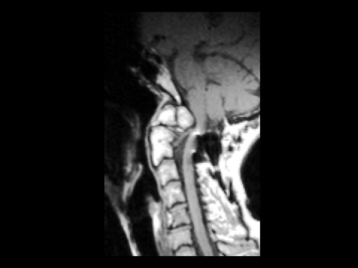
Os odontoidium Findings Complete separation of dens ossification
- Slides: 141
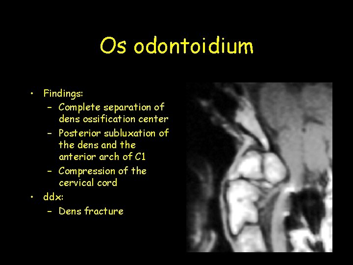
Os odontoidium • Findings: – Complete separation of dens ossification center – Posterior subluxation of the dens and the anterior arch of C 1 – Compression of the cervical cord • ddx: – Dens fracture
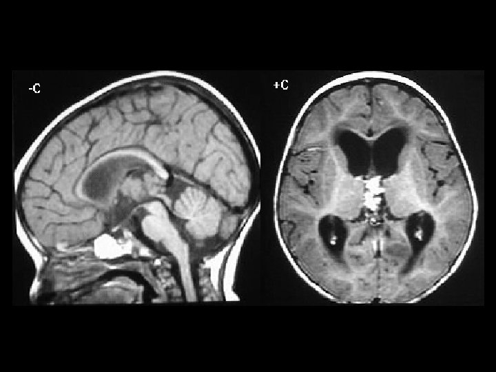
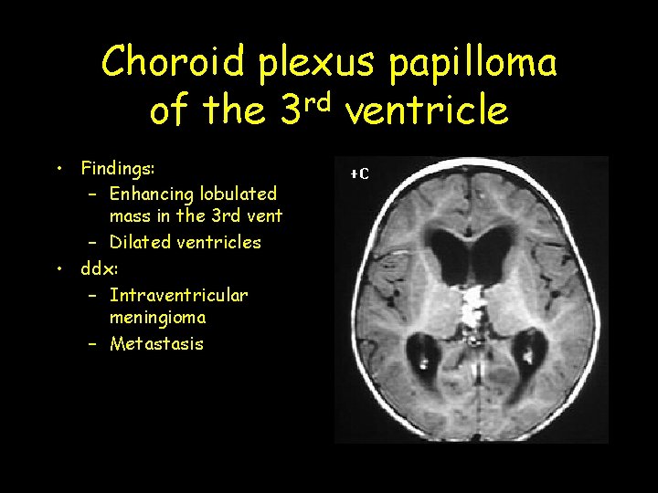
Choroid plexus papilloma of the 3 rd ventricle • Findings: – Enhancing lobulated mass in the 3 rd vent – Dilated ventricles • ddx: – Intraventricular meningioma – Metastasis
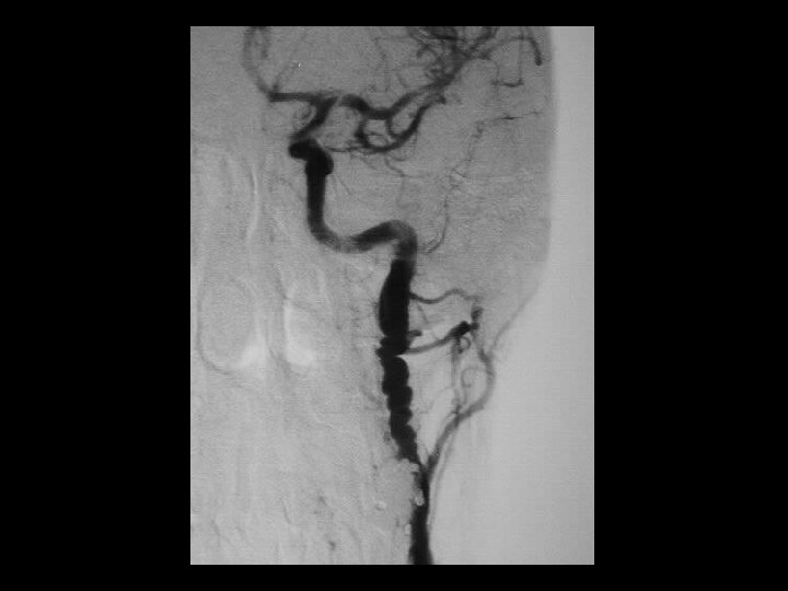
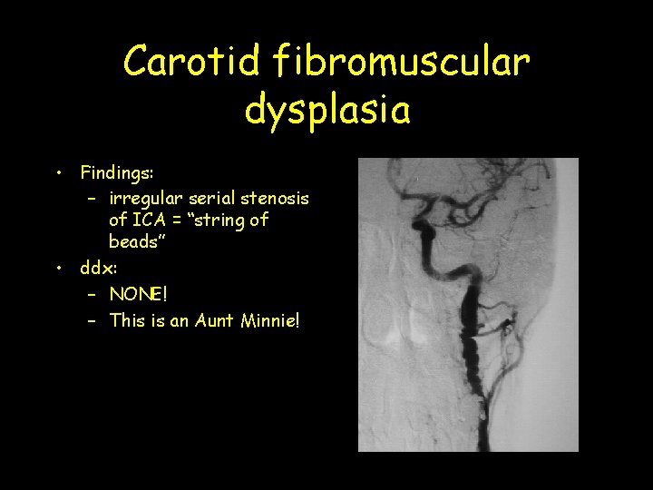
Carotid fibromuscular dysplasia • Findings: – irregular serial stenosis of ICA = “string of beads” • ddx: – NONE! – This is an Aunt Minnie!
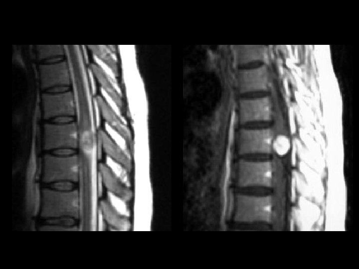
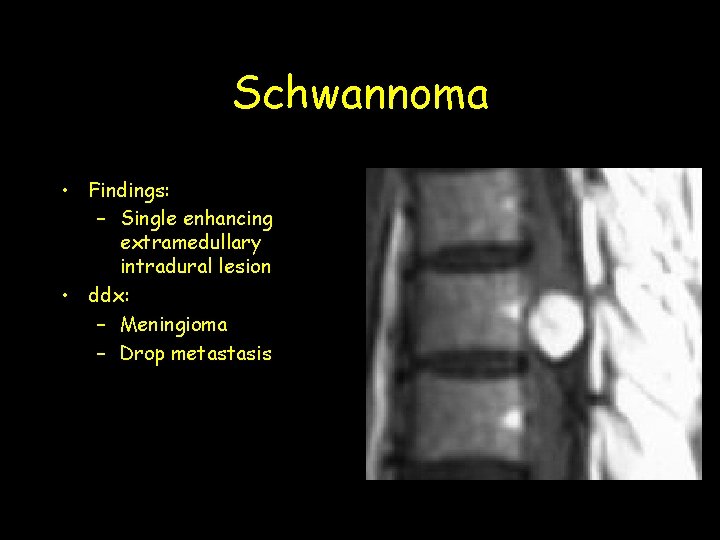
Schwannoma • Findings: – Single enhancing extramedullary intradural lesion • ddx: – Meningioma – Drop metastasis
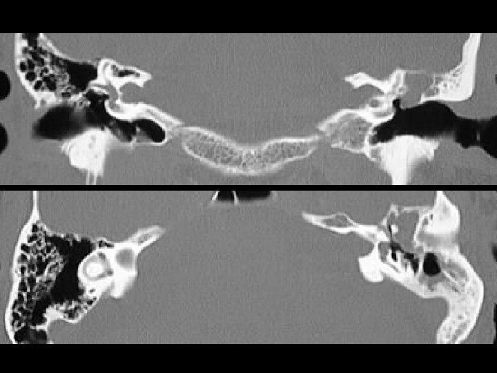
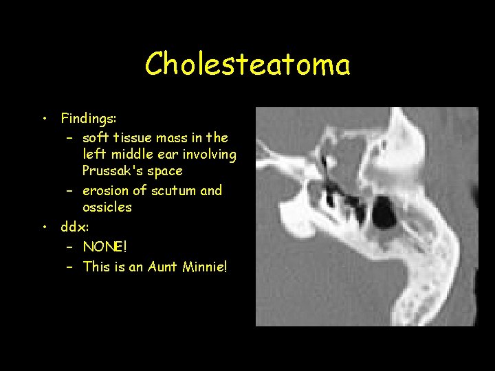
Cholesteatoma • Findings: – soft tissue mass in the left middle ear involving Prussak's space – erosion of scutum and ossicles • ddx: – NONE! – This is an Aunt Minnie!
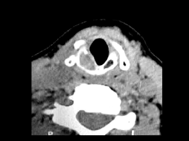
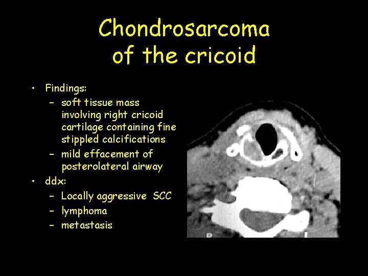
Chondrosarcoma of the cricoid • Findings: – soft tissue mass involving right cricoid cartilage containing fine stippled calcifications – mild effacement of posterolateral airway • ddx: – Locally aggressive SCC – lymphoma – metastasis
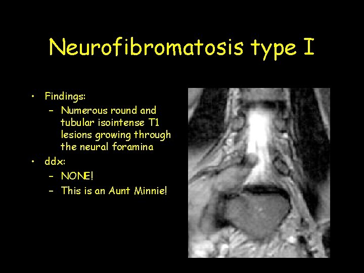
Neurofibromatosis type I • Findings: – Numerous round and tubular isointense T 1 lesions growing through the neural foramina • ddx: – NONE! – This is an Aunt Minnie!
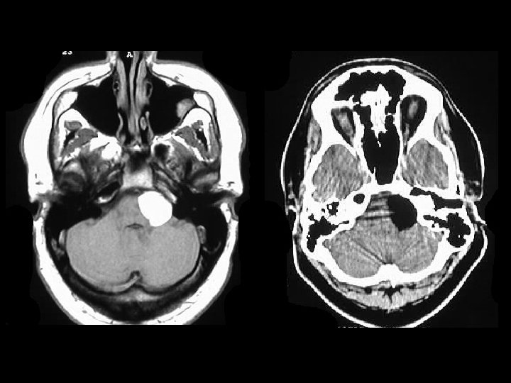
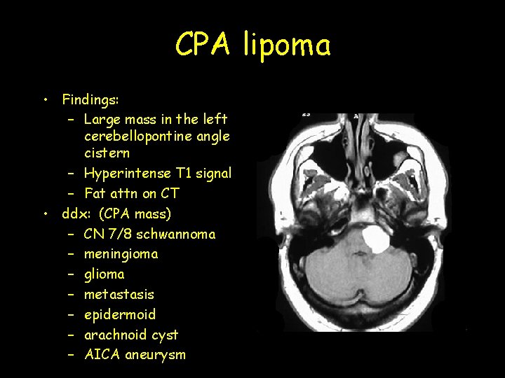
CPA lipoma • Findings: – Large mass in the left cerebellopontine angle cistern – Hyperintense T 1 signal – Fat attn on CT • ddx: (CPA mass) – CN 7/8 schwannoma – meningioma – glioma – metastasis – epidermoid – arachnoid cyst – AICA aneurysm
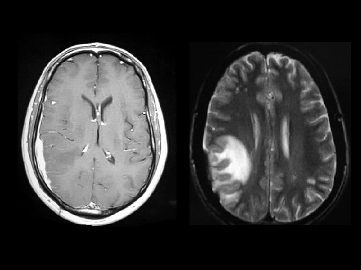
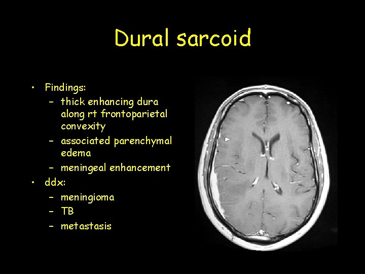
Dural sarcoid • Findings: – thick enhancing dura along rt frontoparietal convexity – associated parenchymal edema – meningeal enhancement • ddx: – meningioma – TB – metastasis
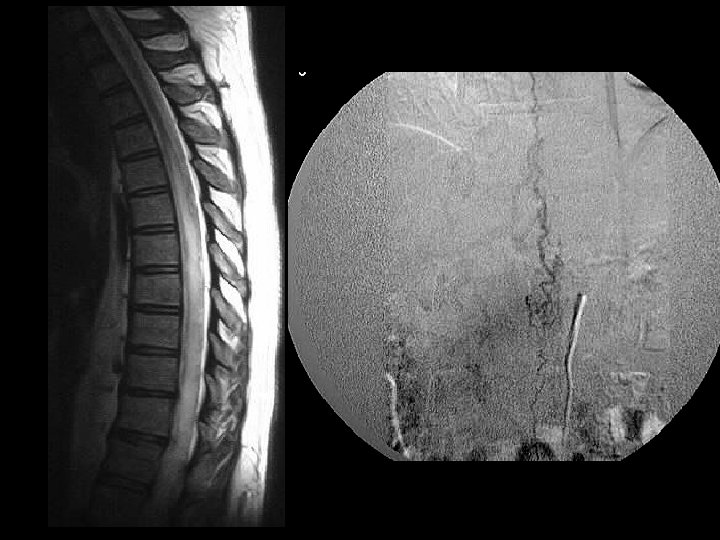
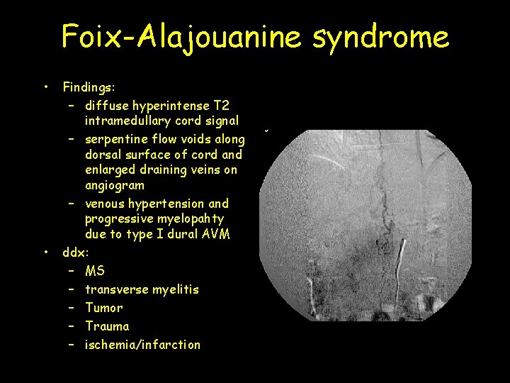
Foix-Alajouanine syndrome • • Findings: – diffuse hyperintense T 2 intramedullary cord signal – serpentine flow voids along dorsal surface of cord and enlarged draining veins on angiogram – venous hypertension and progressive myelopahty due to type I dural AVM ddx: – MS – transverse myelitis – Tumor – Trauma – ischemia/infarction
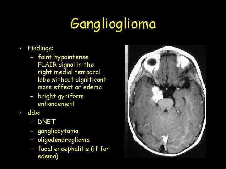
Ganglioma • Findings: – faint hypointense FLAIR signal in the right medial temporal lobe without significant mass effect or edema – bright gyriform enhancement • ddx: – DNET – gangliocytoma – oligodendroglioma – focal encephalitis (if for edema)
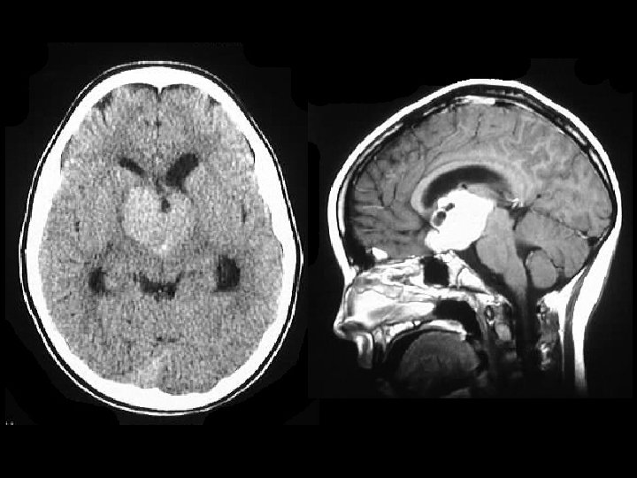
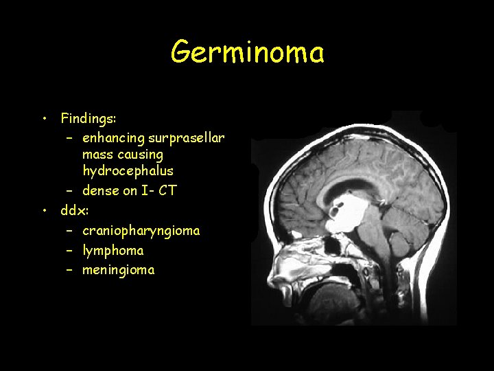
Germinoma • Findings: – enhancing surprasellar mass causing hydrocephalus – dense on I- CT • ddx: – craniopharyngioma – lymphoma – meningioma
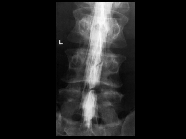
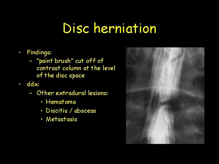
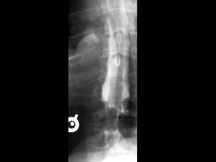
Disc herniation • Findings: – “paint brush” cut off of contrast column at the level of the disc space • ddx: – Other extradural lesions: • Hematoma • Discitis / abscess • Metastasis
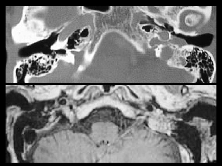
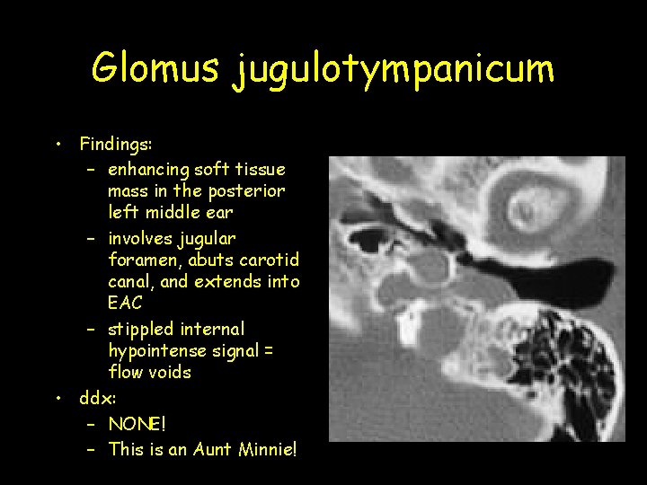
Glomus jugulotympanicum • Findings: – enhancing soft tissue mass in the posterior left middle ear – involves jugular foramen, abuts carotid canal, and extends into EAC – stippled internal hypointense signal = flow voids • ddx: – NONE! – This is an Aunt Minnie!
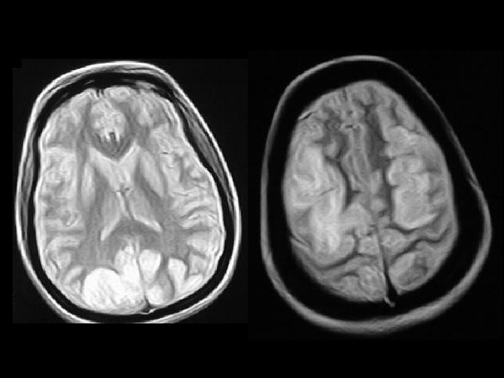
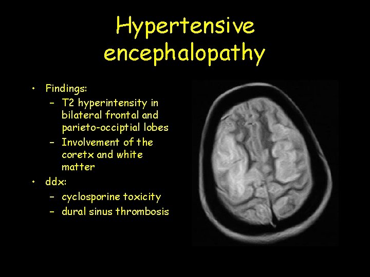
Hypertensive encephalopathy • Findings: – T 2 hyperintensity in bilateral frontal and parieto-occiptial lobes – Involvement of the coretx and white matter • ddx: – cyclosporine toxicity – dural sinus thrombosis
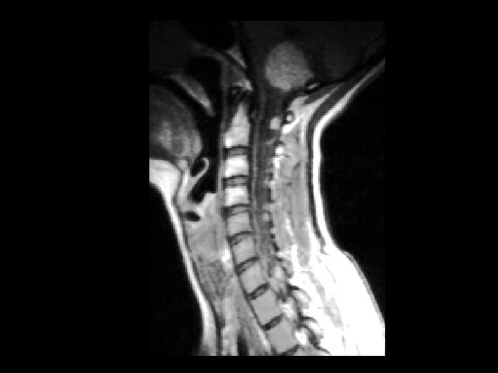
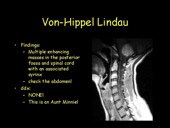
Von-Hippel Lindau • Findings: – Multiple enhancing masses in the posterior fossa and spinal cord with an associated syrinx – check the abdomen! • ddx: – NONE! – This is an Aunt Minnie!
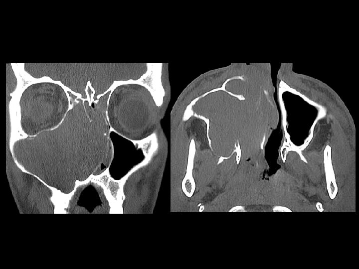
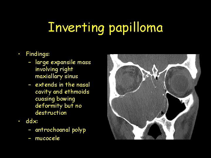
Inverting papilloma • Findings: – large expansile mass involving right maxiallary sinus – extends in the nasal cavity and ethmoids cuasing bowing deformity but no destruction • ddx: – antrochoanal polyp – mucocele
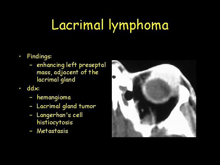
Lacrimal lymphoma • Findings: – enhancing left preseptal mass, adjacent of the lacrimal gland • ddx: – hemangioma – Lacrimal gland tumor – Langerhan's cell histiocytosis – Metastasis
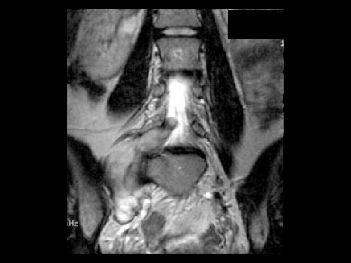
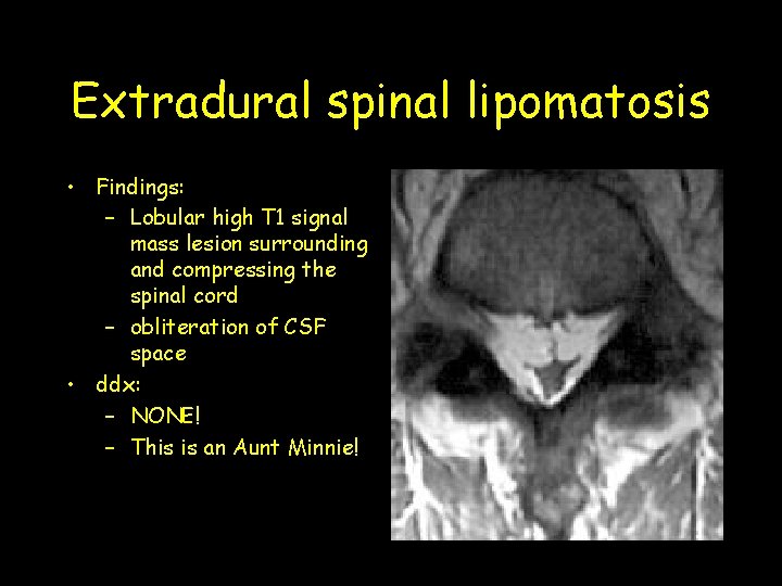
Extradural spinal lipomatosis • Findings: – Lobular high T 1 signal mass lesion surrounding and compressing the spinal cord – obliteration of CSF space • ddx: – NONE! – This is an Aunt Minnie!
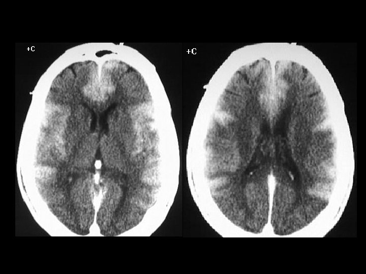
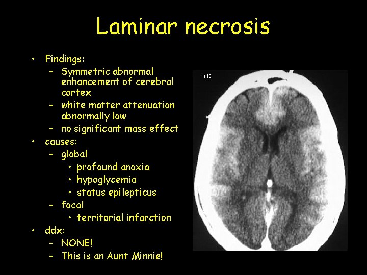
Laminar necrosis • Findings: – Symmetric abnormal enhancement of cerebral cortex – white matter attenuation abnormally low – no significant mass effect • causes: – global • profound anoxia • hypoglycemia • status epilepticus – focal • territorial infarction • ddx: – NONE! – This is an Aunt Minnie!
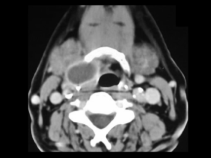
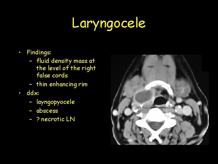
Laryngocele • Findings: – fluid density mass at the level of the right false cords – thin enhancing rim • ddx: – layngopyocele – abscess – ? necrotic LN
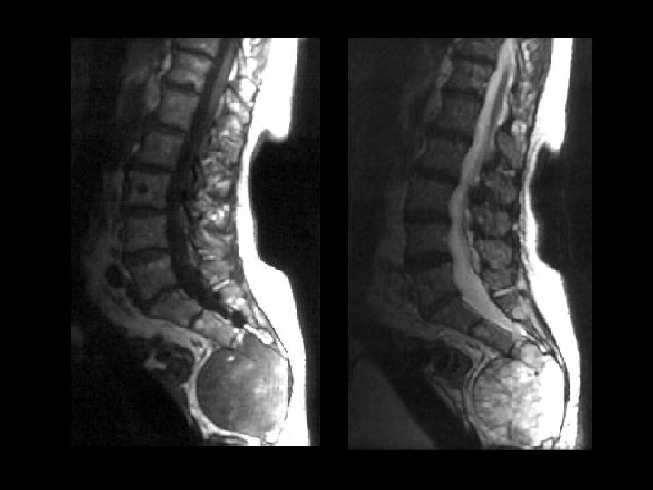
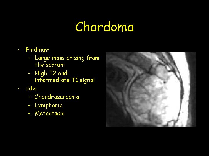
Chordoma • Findings: – Large mass arising from the sacrum – High T 2 and intermediate T 1 signal • ddx: – Chondrosarcoma – Lymphoma – Metastasis
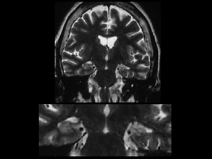
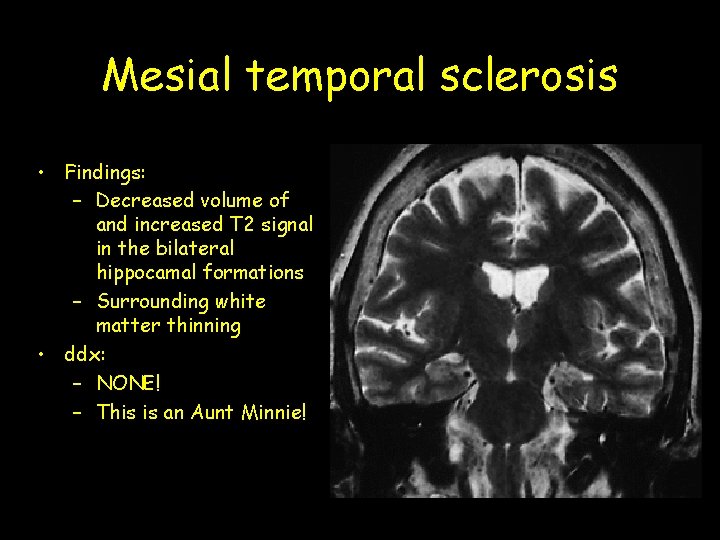
Mesial temporal sclerosis • Findings: – Decreased volume of and increased T 2 signal in the bilateral hippocamal formations – Surrounding white matter thinning • ddx: – NONE! – This is an Aunt Minnie!
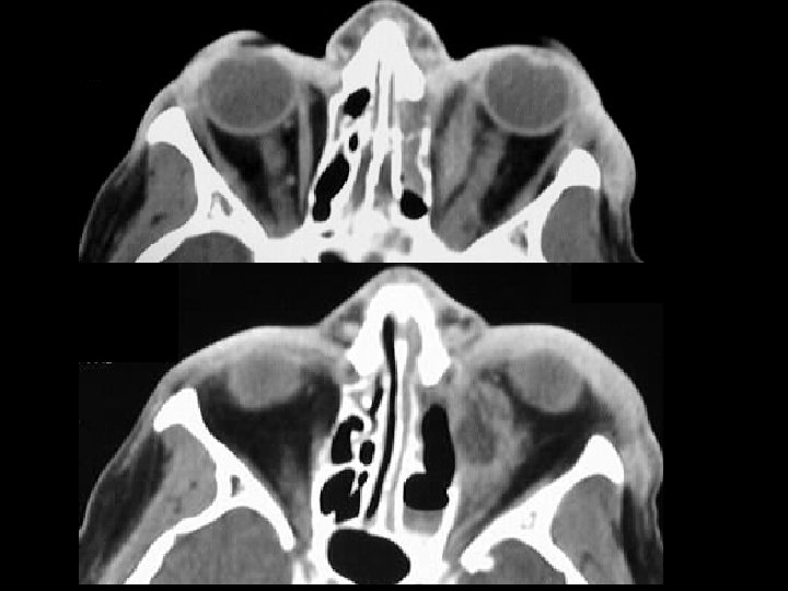
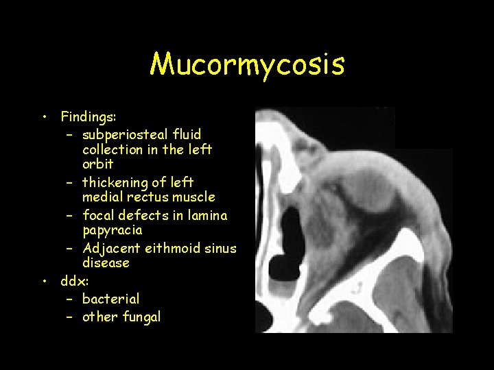
Mucormycosis • Findings: – subperiosteal fluid collection in the left orbit – thickening of left medial rectus muscle – focal defects in lamina papyracia – Adjacent eithmoid sinus disease • ddx: – bacterial – other fungal
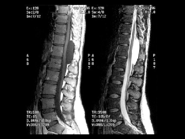
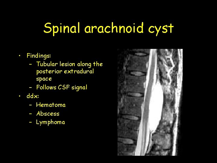
Spinal arachnoid cyst • Findings: – Tubular lesion along the posterior extradural space – Follows CSF signal • ddx: – Hematoma – Abscess – Lymphoma
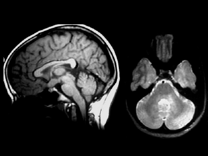
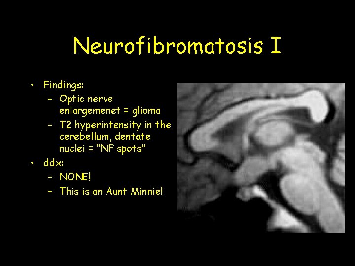
Neurofibromatosis I • Findings: – Optic nerve enlargemenet = glioma – T 2 hyperintensity in the cerebellum, dentate nuclei = “NF spots” • ddx: – NONE! – This is an Aunt Minnie!
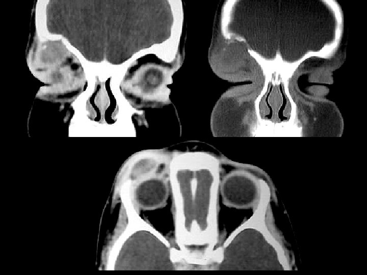
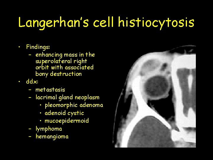
Langerhan’s cell histiocytosis • Findings: – enhancing mass in the superolateral right orbit with associated bony destruction • ddx: – metastasis – lacrimal gland neoplasm • pleomorphic adenoma • adenoid cystic • mucoepidermoid – lymphoma – hemangioma
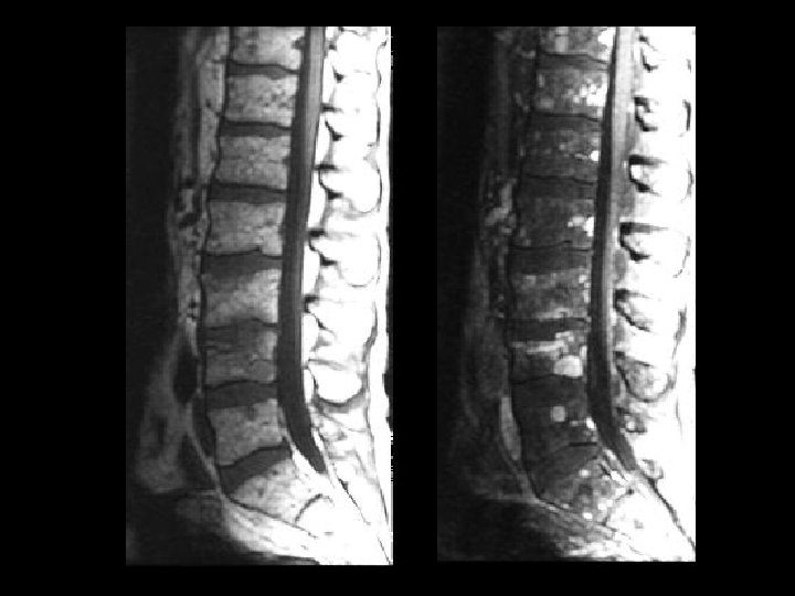
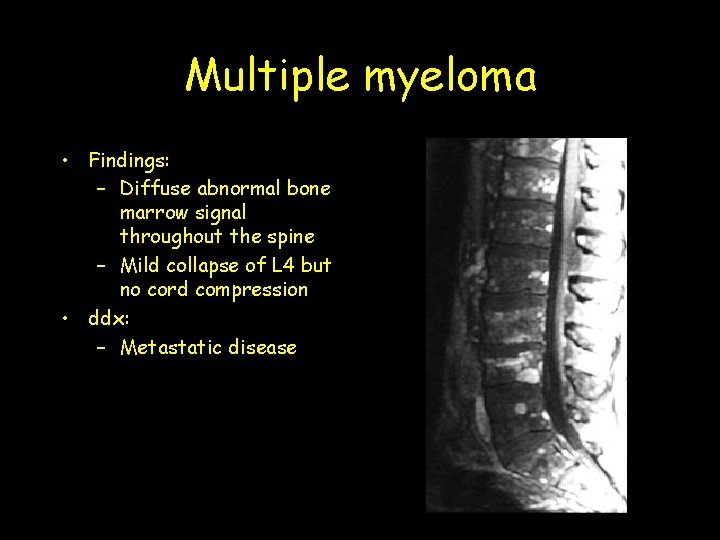
Multiple myeloma • Findings: – Diffuse abnormal bone marrow signal throughout the spine – Mild collapse of L 4 but no cord compression • ddx: – Metastatic disease
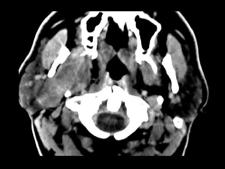
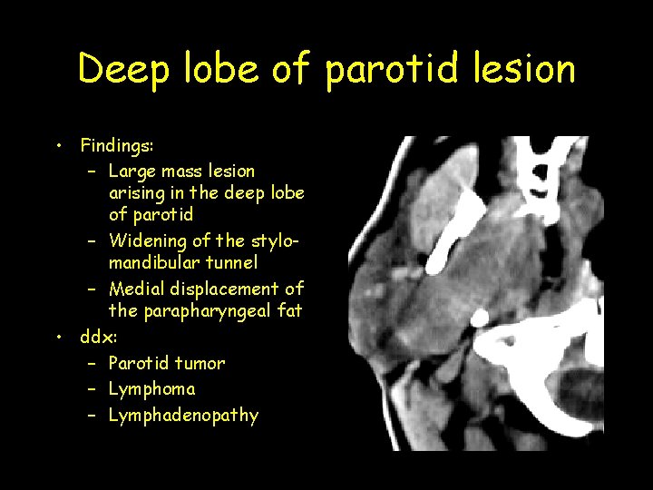
Deep lobe of parotid lesion • Findings: – Large mass lesion arising in the deep lobe of parotid – Widening of the stylomandibular tunnel – Medial displacement of the parapharyngeal fat • ddx: – Parotid tumor – Lymphoma – Lymphadenopathy
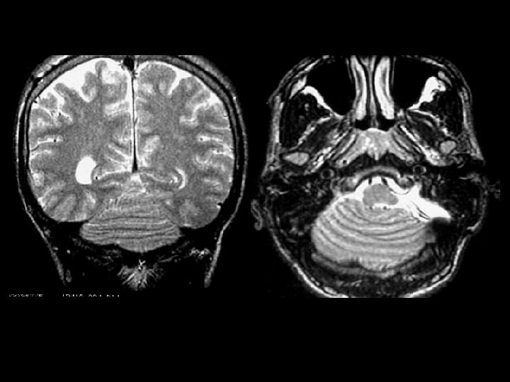
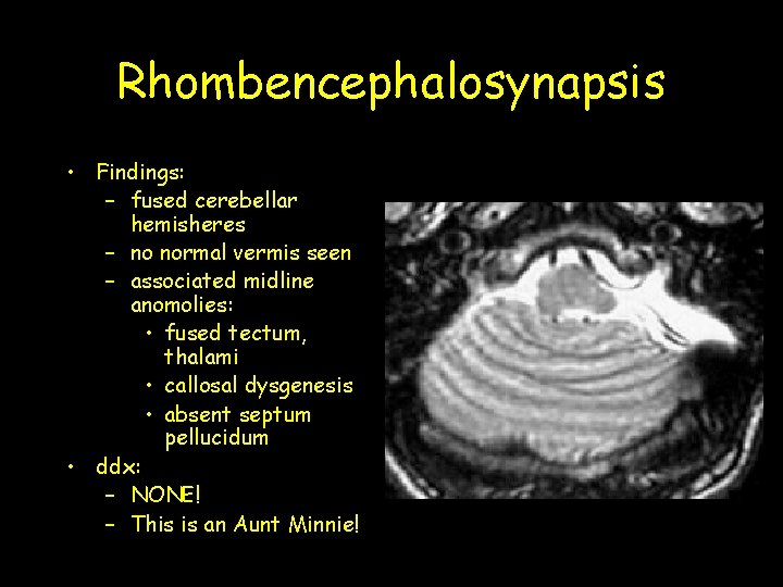
Rhombencephalosynapsis • Findings: – fused cerebellar hemisheres – no normal vermis seen – associated midline anomolies: • fused tectum, thalami • callosal dysgenesis • absent septum pellucidum • ddx: – NONE! – This is an Aunt Minnie!
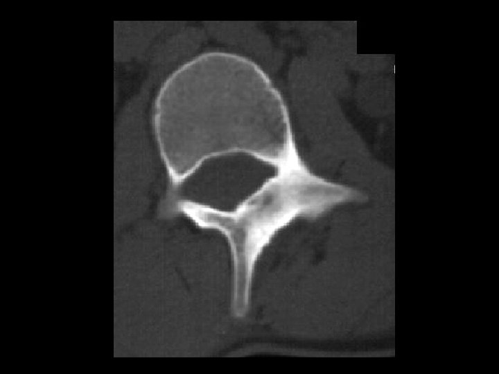
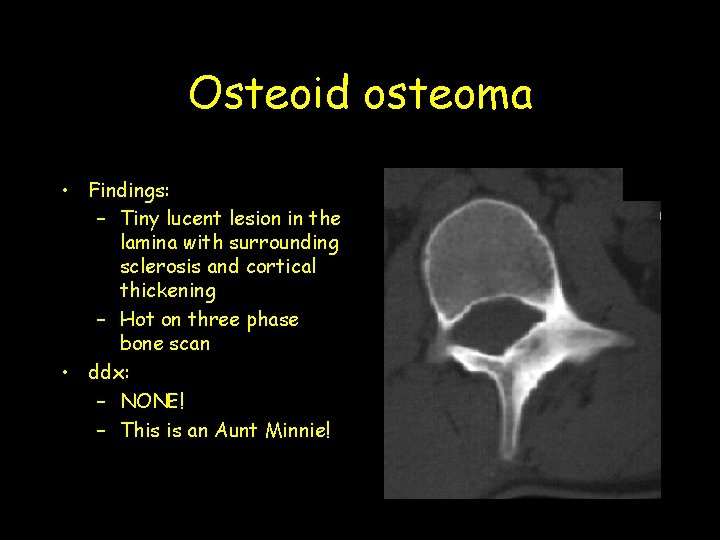
Osteoid osteoma • Findings: – Tiny lucent lesion in the lamina with surrounding sclerosis and cortical thickening – Hot on three phase bone scan • ddx: – NONE! – This is an Aunt Minnie!
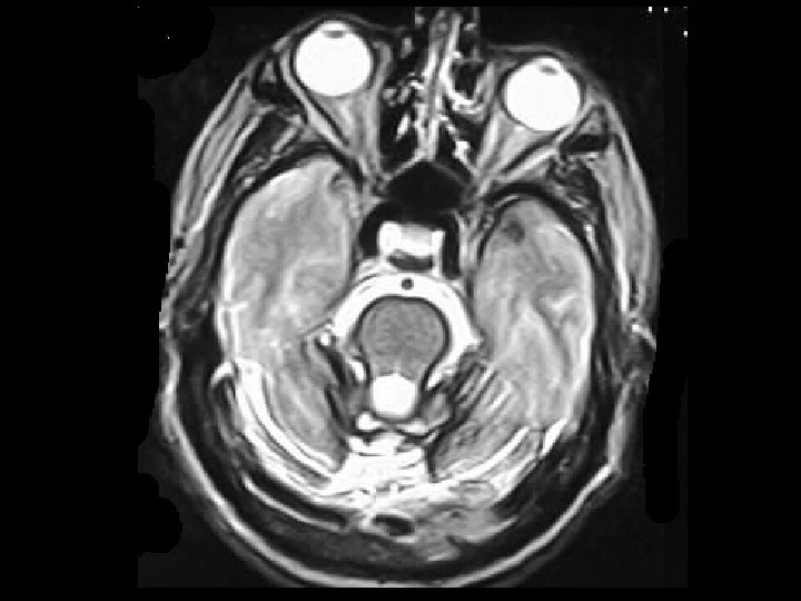
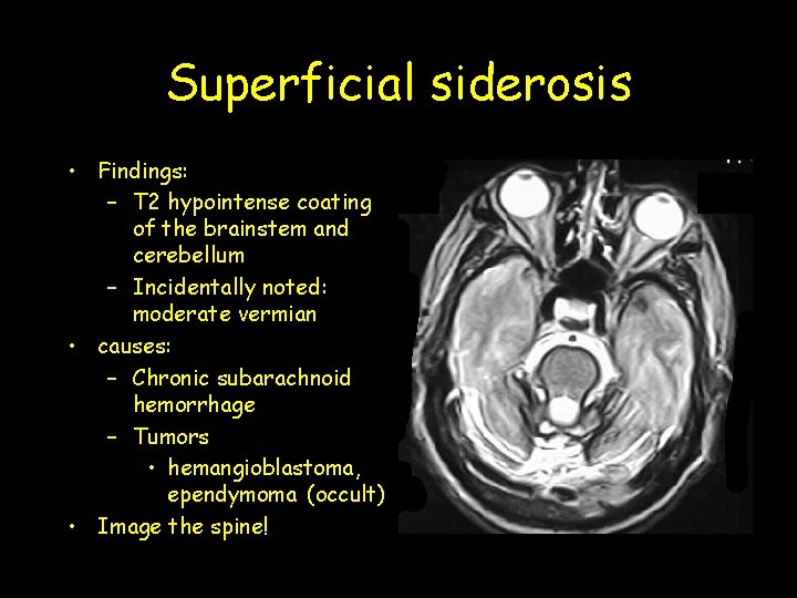
Superficial siderosis • Findings: – T 2 hypointense coating of the brainstem and cerebellum – Incidentally noted: moderate vermian • causes: – Chronic subarachnoid hemorrhage – Tumors • hemangioblastoma, ependymoma (occult) • Image the spine!
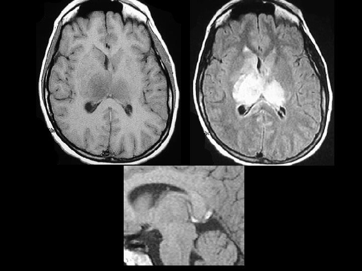
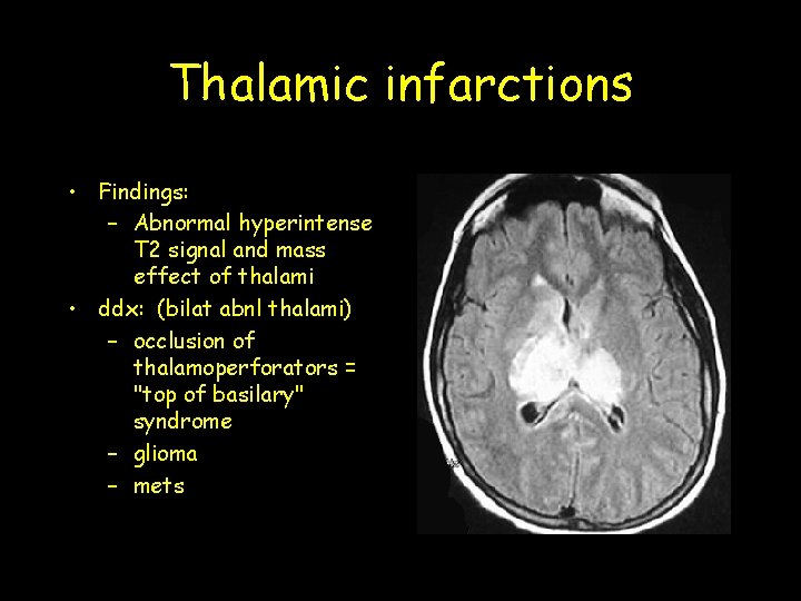
Thalamic infarctions • Findings: – Abnormal hyperintense T 2 signal and mass effect of thalami • ddx: (bilat abnl thalami) – occlusion of thalamoperforators = "top of basilary" syndrome – glioma – mets
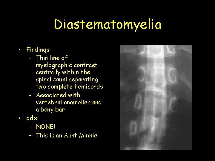
Diastematomyelia • Findings: – Thin line of myelographic contrast centrally within the spinal canal separating two complete hemicords – Associated with vertebral anomolies and a bony bar • ddx: – NONE! – This is an Aunt Minnie!
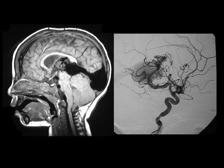
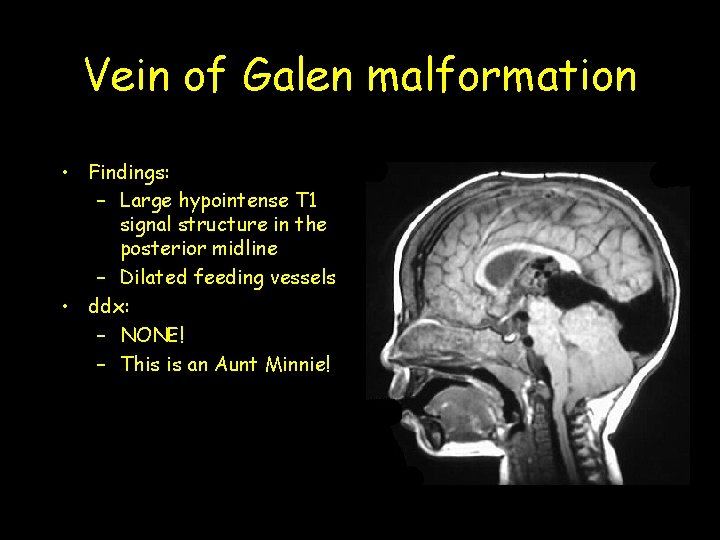
Vein of Galen malformation • Findings: – Large hypointense T 1 signal structure in the posterior midline – Dilated feeding vessels • ddx: – NONE! – This is an Aunt Minnie!
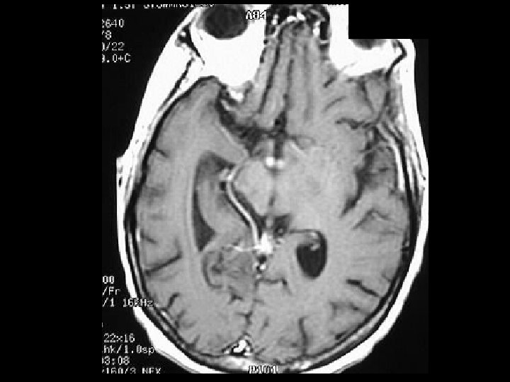
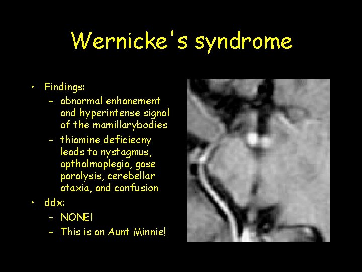
Wernicke's syndrome • Findings: – abnormal enhanement and hyperintense signal of the mamillarybodies – thiamine deficiecny leads to nystagmus, opthalmoplegia, gase paralysis, cerebellar ataxia, and confusion • ddx: – NONE! – This is an Aunt Minnie!
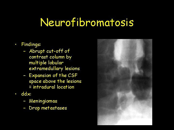
Neurofibromatosis • Findings: – Abrupt cut-off of contrast column by multiple lobular extramedullary lesions – Expansion of the CSF space above the lesions = intradural location • ddx: – Meningiomas – Drop metastases
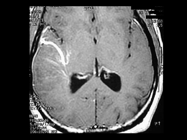
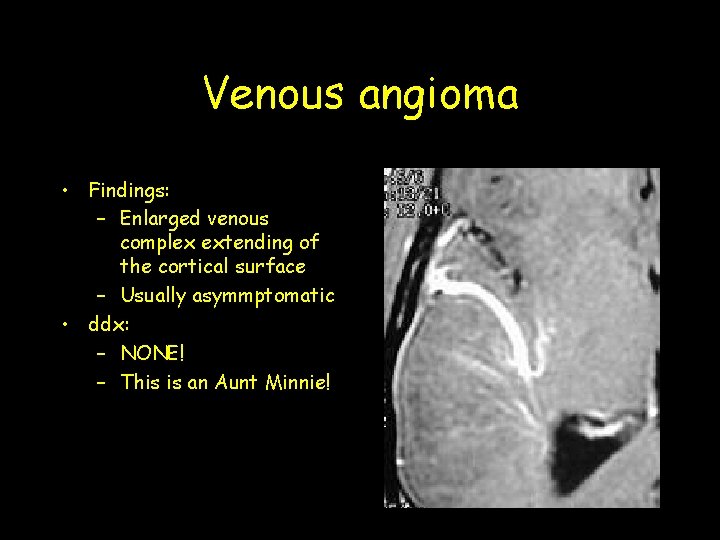
Venous angioma • Findings: – Enlarged venous complex extending of the cortical surface – Usually asymmptomatic • ddx: – NONE! – This is an Aunt Minnie!
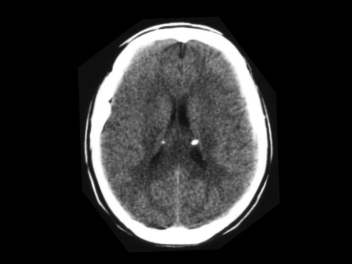
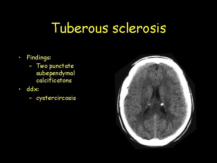
Tuberous sclerosis • Findings: – Two punctate subependymal calcificatons • ddx: – cystercircosis
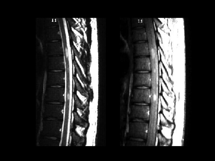
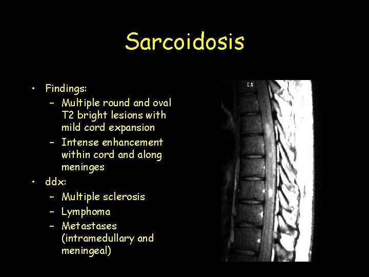
Sarcoidosis • Findings: – Multiple round and oval T 2 bright lesions with mild cord expansion – Intense enhancement within cord and along meninges • ddx: – Multiple sclerosis – Lymphoma – Metastases (intramedullary and meningeal)
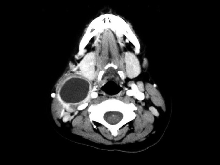
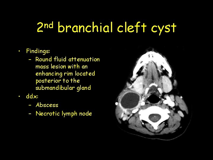
nd 2 branchial cleft cyst • Findings: – Round fluid attenuation mass lesion with an enhancing rim located posterior to the submandibular gland • ddx: – Abscess – Necrotic lymph node
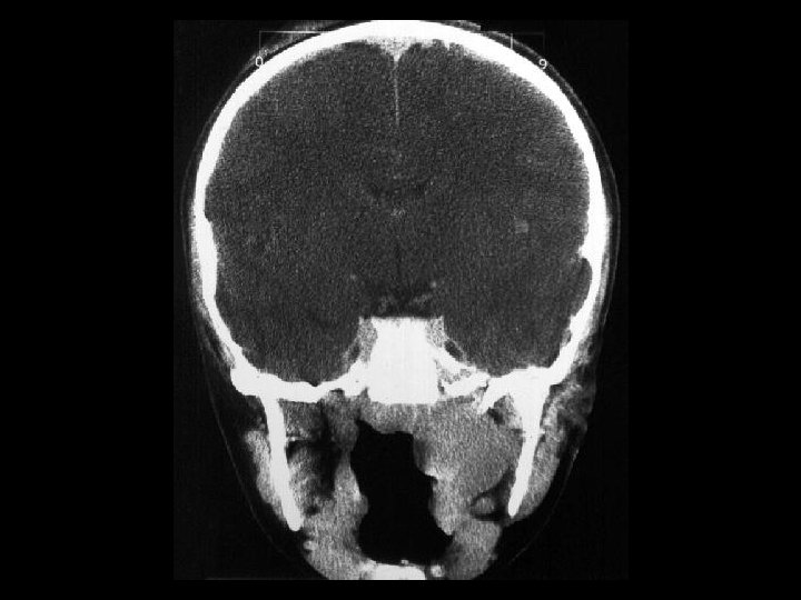
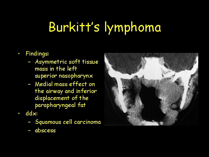
Burkitt’s lymphoma • Findings: – Asymmetric soft tissue mass in the left superior nasopharynx – Medial mass effect on the airway and inferior displacement of the parapharyngeal fat • ddx: – Squamous cell carcinoma – abscess
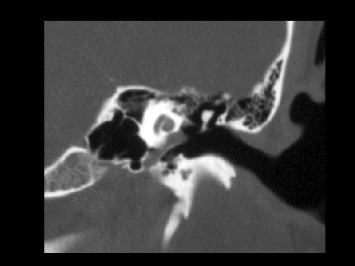
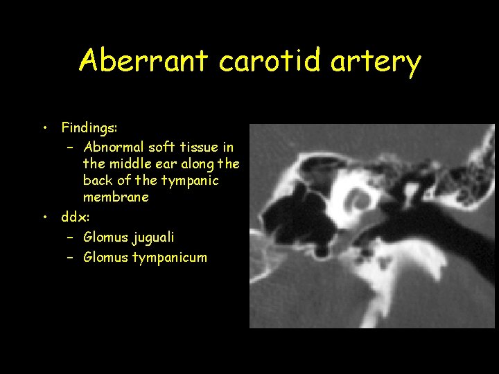
Aberrant carotid artery • Findings: – Abnormal soft tissue in the middle ear along the back of the tympanic membrane • ddx: – Glomus juguali – Glomus tympanicum
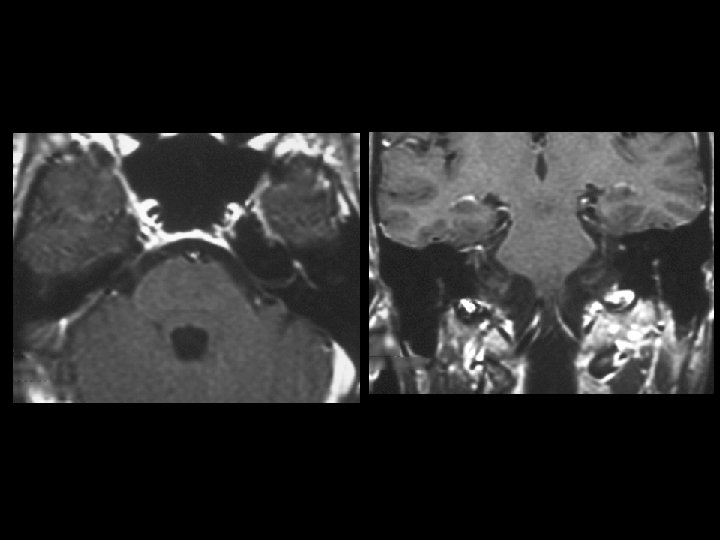
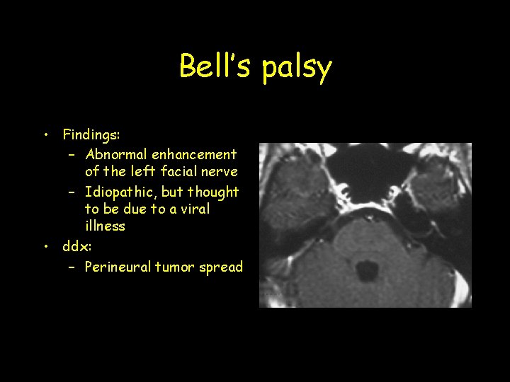
Bell’s palsy • Findings: – Abnormal enhancement of the left facial nerve – Idiopathic, but thought to be due to a viral illness • ddx: – Perineural tumor spread
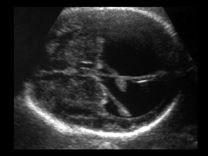
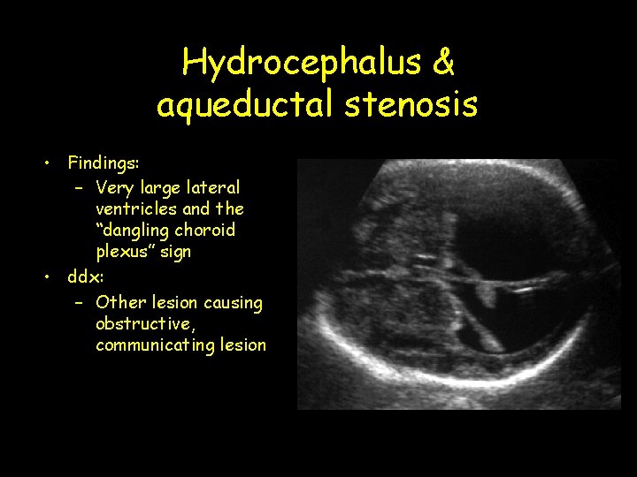
Hydrocephalus & aqueductal stenosis • Findings: – Very large lateral ventricles and the “dangling choroid plexus” sign • ddx: – Other lesion causing obstructive, communicating lesion
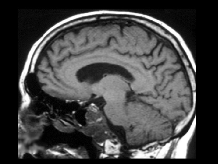
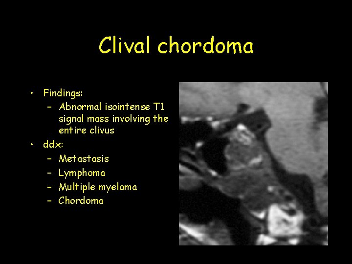
Clival chordoma • Findings: – Abnormal isointense T 1 signal mass involving the entire clivus • ddx: – Metastasis – Lymphoma – Multiple myeloma – Chordoma
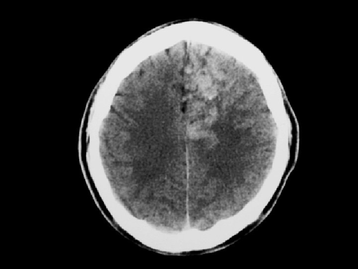
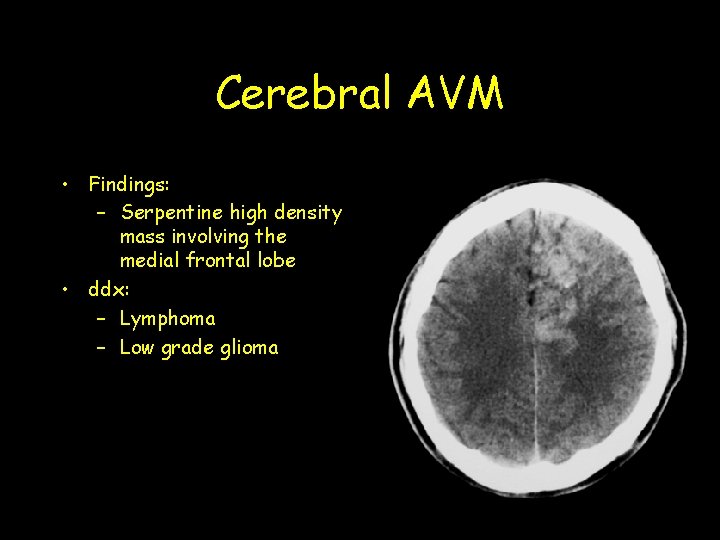
Cerebral AVM • Findings: – Serpentine high density mass involving the medial frontal lobe • ddx: – Lymphoma – Low grade glioma
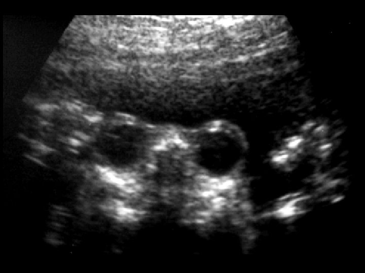
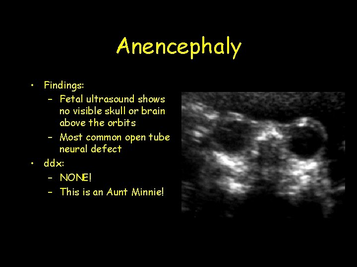
Anencephaly • Findings: – Fetal ultrasound shows no visible skull or brain above the orbits – Most common open tube neural defect • ddx: – NONE! – This is an Aunt Minnie!
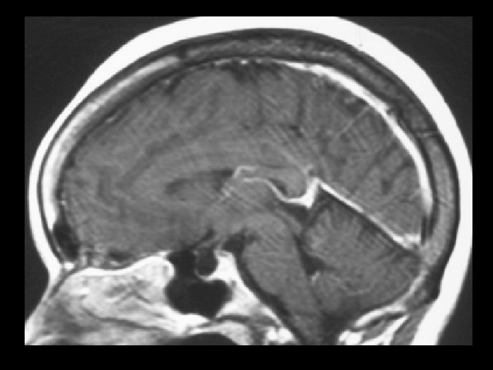
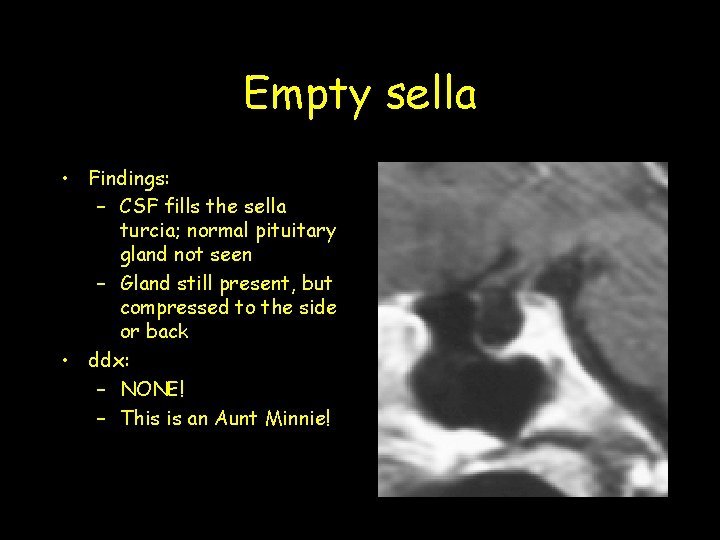
Empty sella • Findings: – CSF fills the sella turcia; normal pituitary gland not seen – Gland still present, but compressed to the side or back • ddx: – NONE! – This is an Aunt Minnie!
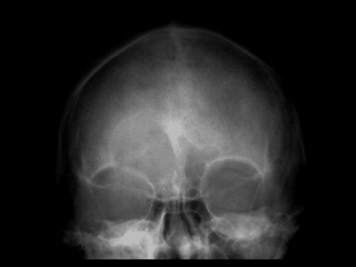
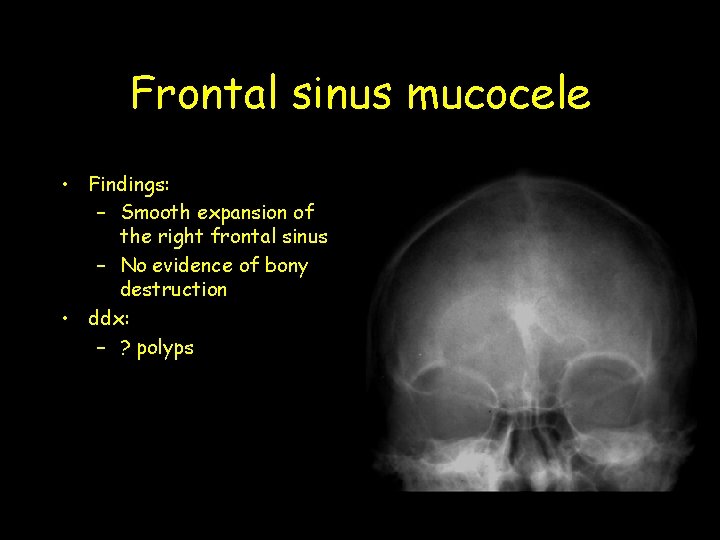
Frontal sinus mucocele • Findings: – Smooth expansion of the right frontal sinus – No evidence of bony destruction • ddx: – ? polyps
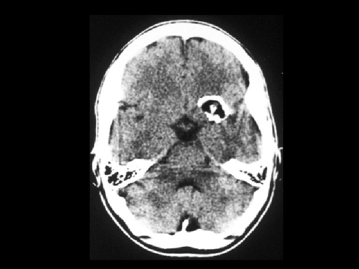
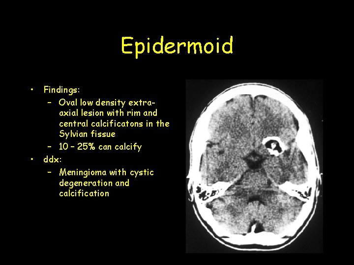
Epidermoid • • Findings: – Oval low density extraaxial lesion with rim and central calcificatons in the Sylvian fissue – 10 – 25% can calcify ddx: – Meningioma with cystic degeneration and calcification
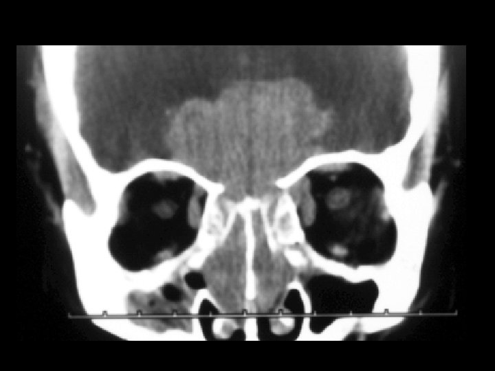
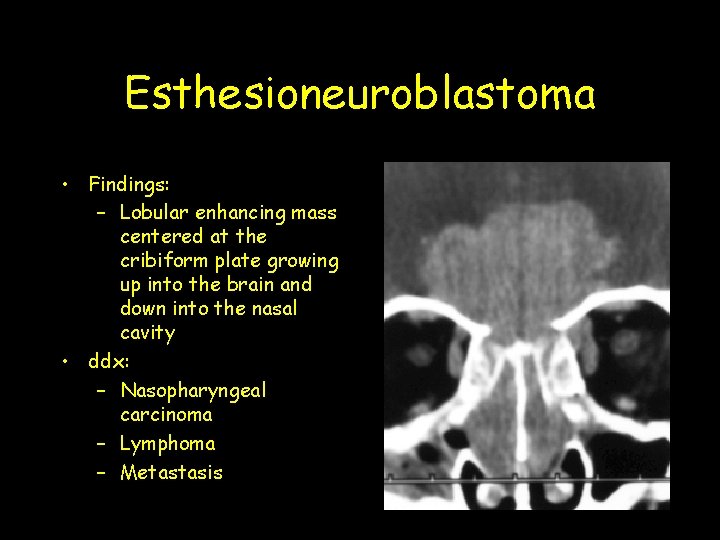
Esthesioneuroblastoma • Findings: – Lobular enhancing mass centered at the cribiform plate growing up into the brain and down into the nasal cavity • ddx: – Nasopharyngeal carcinoma – Lymphoma – Metastasis
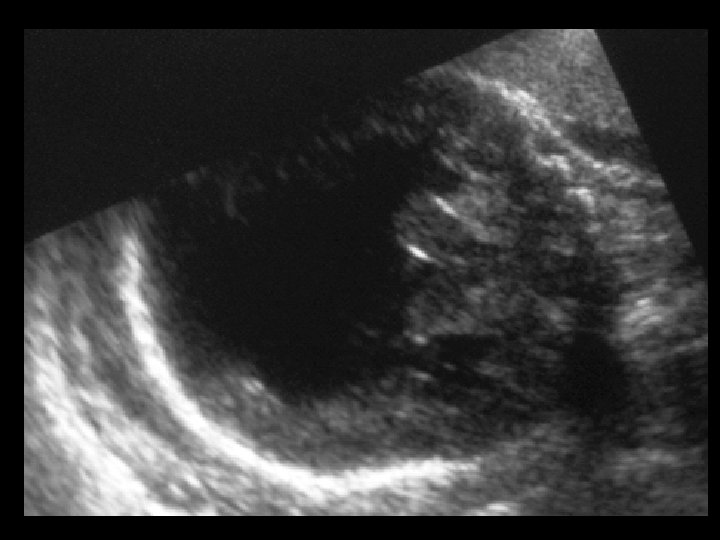
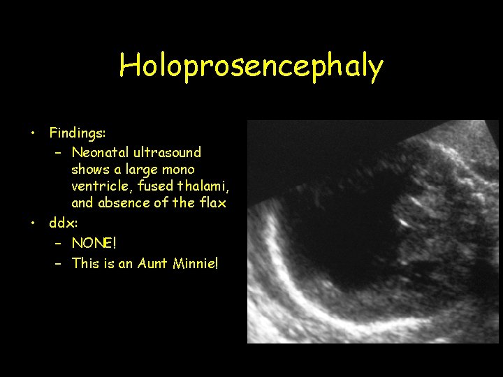
Holoprosencephaly • Findings: – Neonatal ultrasound shows a large mono ventricle, fused thalami, and absence of the flax • ddx: – NONE! – This is an Aunt Minnie!
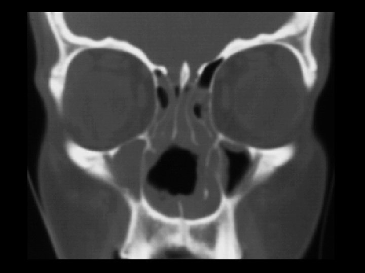
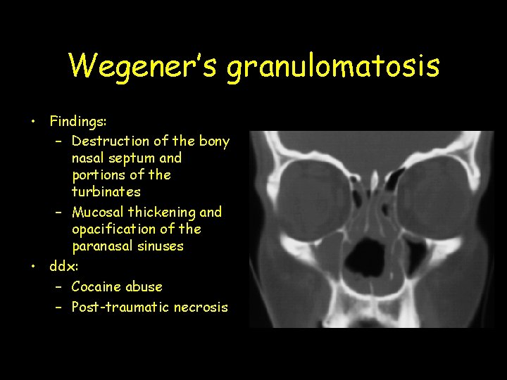
Wegener’s granulomatosis • Findings: – Destruction of the bony nasal septum and portions of the turbinates – Mucosal thickening and opacification of the paranasal sinuses • ddx: – Cocaine abuse – Post-traumatic necrosis
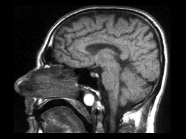
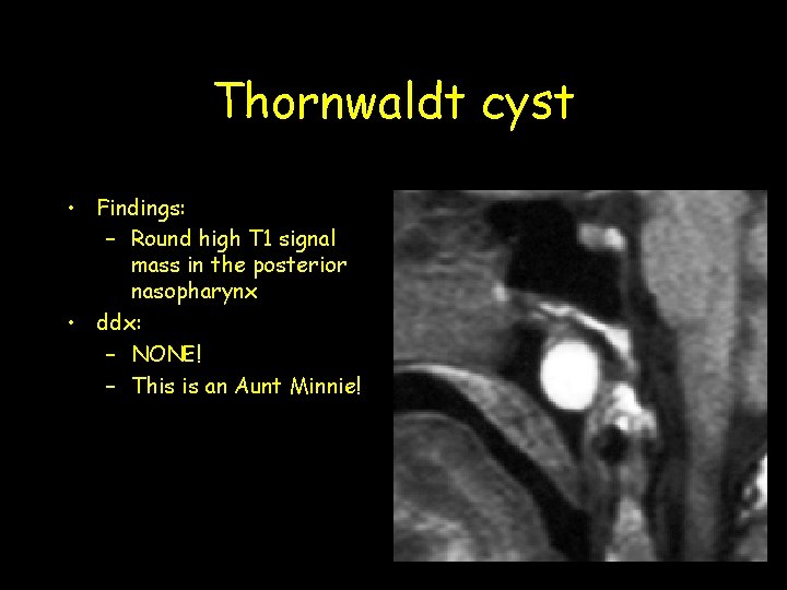
Thornwaldt cyst • Findings: – Round high T 1 signal mass in the posterior nasopharynx • ddx: – NONE! – This is an Aunt Minnie!
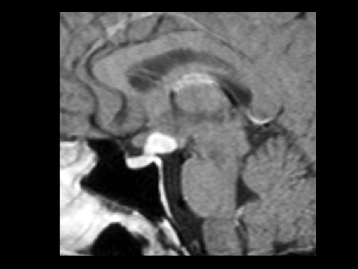
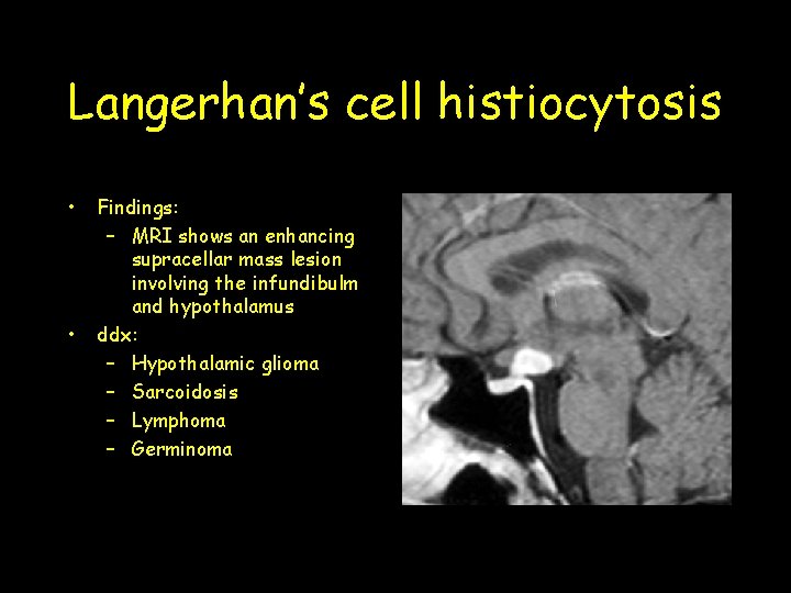
Langerhan’s cell histiocytosis • • Findings: – MRI shows an enhancing supracellar mass lesion involving the infundibulm and hypothalamus ddx: – Hypothalamic glioma – Sarcoidosis – Lymphoma – Germinoma
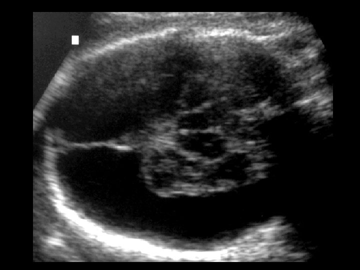
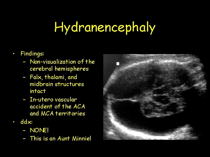
Hydranencephaly • Findings: – Non-visualization of the cerebral hemispheres – Falx, thalami, and midbrain structures intact – In-utero vascular accident of the ACA and MCA territories • ddx: – NONE! – This is an Aunt Minnie!
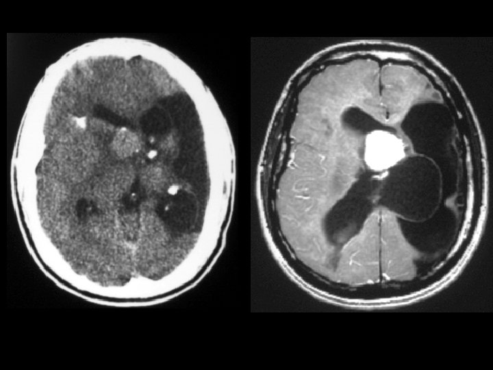
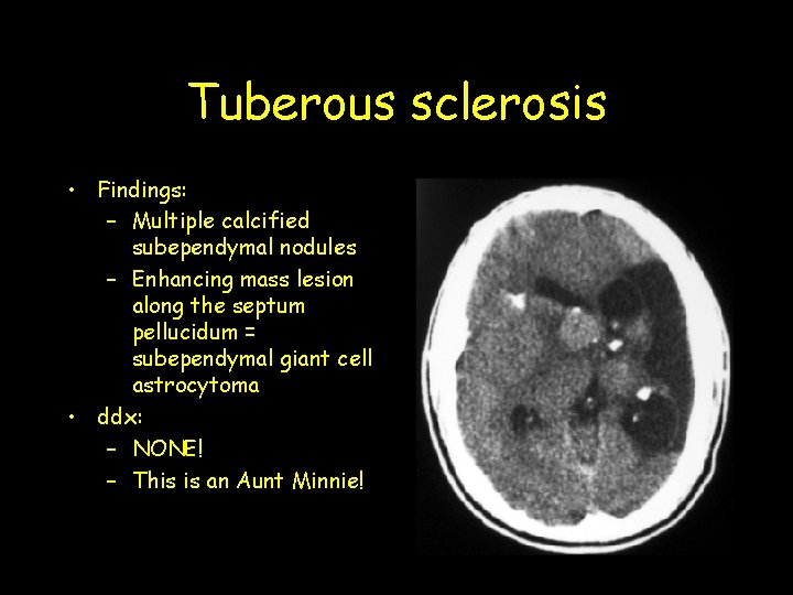
Tuberous sclerosis • Findings: – Multiple calcified subependymal nodules – Enhancing mass lesion along the septum pellucidum = subependymal giant cell astrocytoma • ddx: – NONE! – This is an Aunt Minnie!
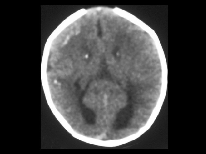
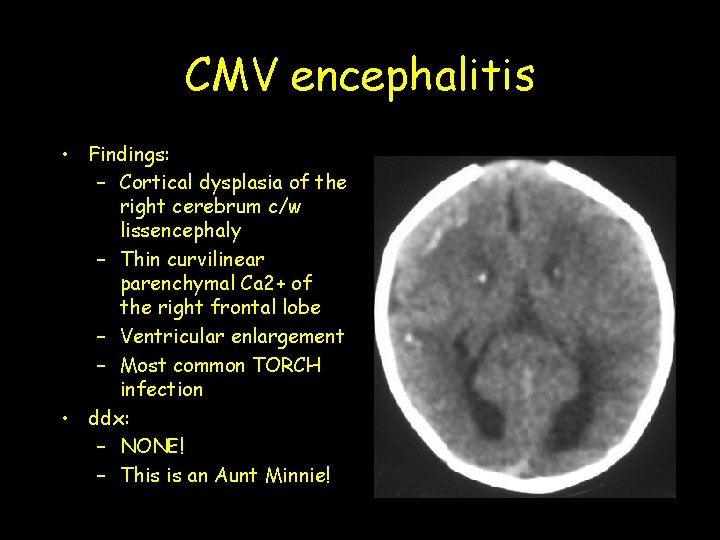
CMV encephalitis • Findings: – Cortical dysplasia of the right cerebrum c/w lissencephaly – Thin curvilinear parenchymal Ca 2+ of the right frontal lobe – Ventricular enlargement – Most common TORCH infection • ddx: – NONE! – This is an Aunt Minnie!
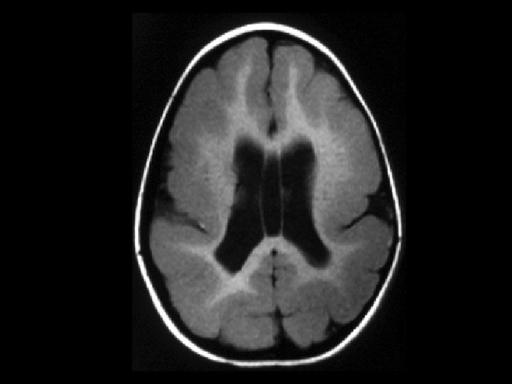
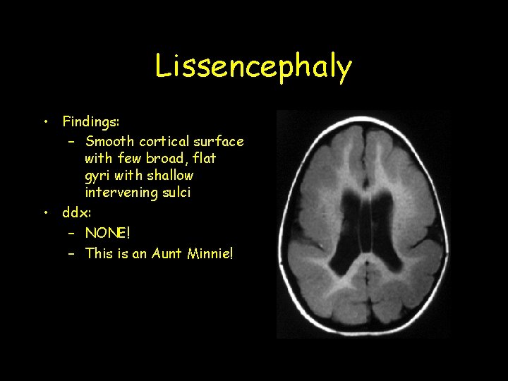
Lissencephaly • Findings: – Smooth cortical surface with few broad, flat gyri with shallow intervening sulci • ddx: – NONE! – This is an Aunt Minnie!
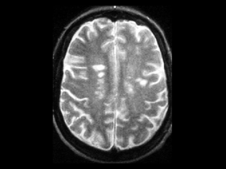
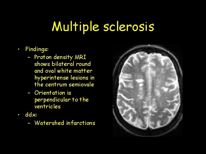
Multiple sclerosis • Findings: – Proton density MRI shows bilateral round and oval white matter hyperintense lesions in the centrum semiovale – Orientation is perpendicular to the ventricles • ddx: – Watershed infarctions
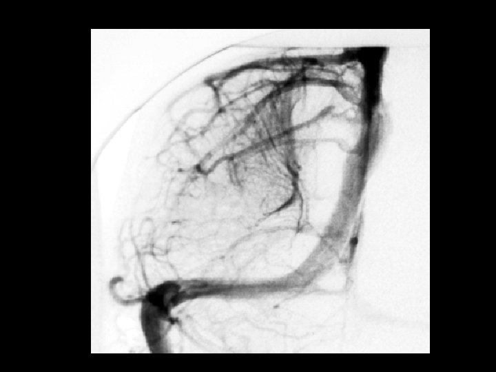
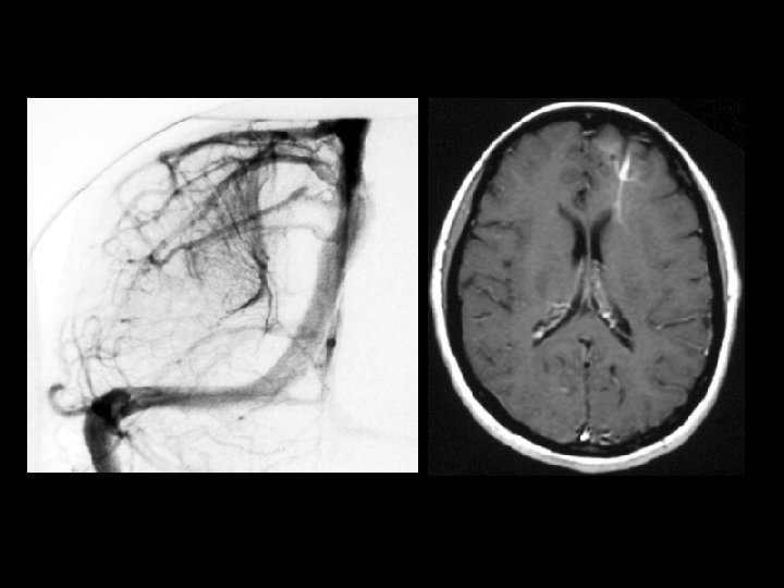
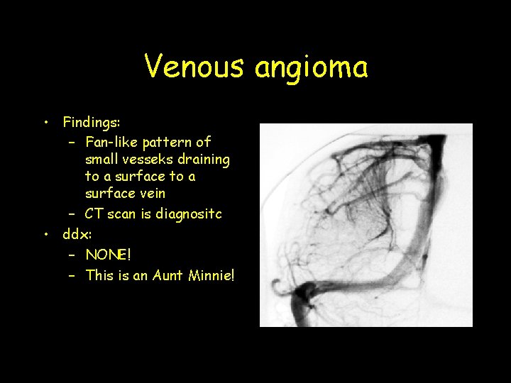
Venous angioma • Findings: – Fan-like pattern of small vesseks draining to a surface vein – CT scan is diagnositc • ddx: – NONE! – This is an Aunt Minnie!
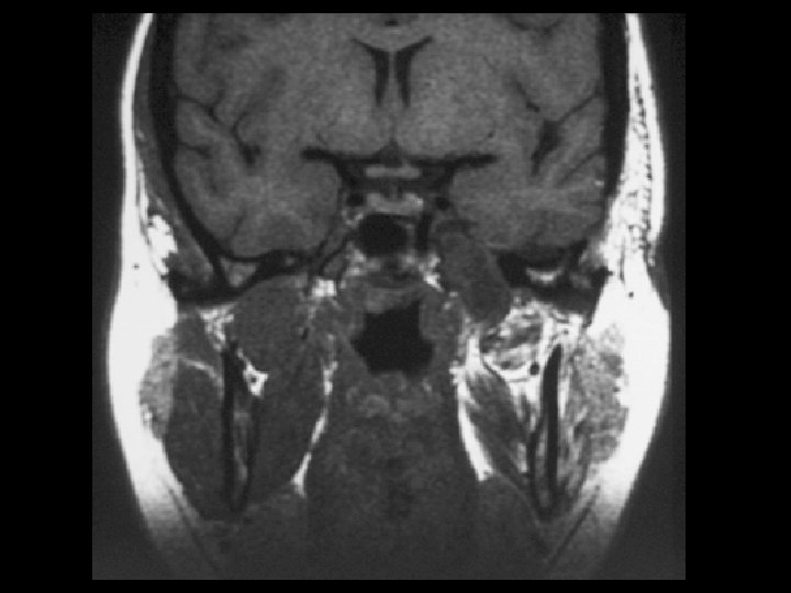
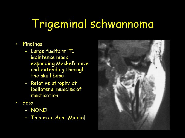
Trigeminal schwannoma • Findings: – Large fusiform T 1 isointense mass expanding Meckel’s cave and extending through the skull base – Relative atrophy of ipsilateral muscles of mastication • ddx: – NONE! – This is an Aunt Minnie!
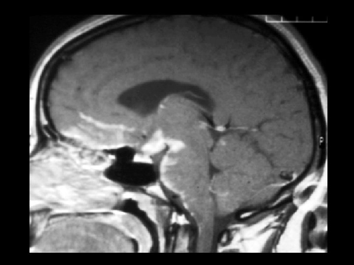
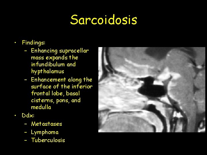
Sarcoidosis • Findings: – Enhancing supracellar mass expands the infundibulum and hypthalamus – Enhancement along the surface of the inferior frontal lobe, basal cisterns, pons, and medulla • Ddx: – Metastases – Lymphoma – Tuberculosis
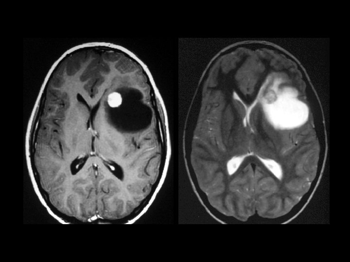
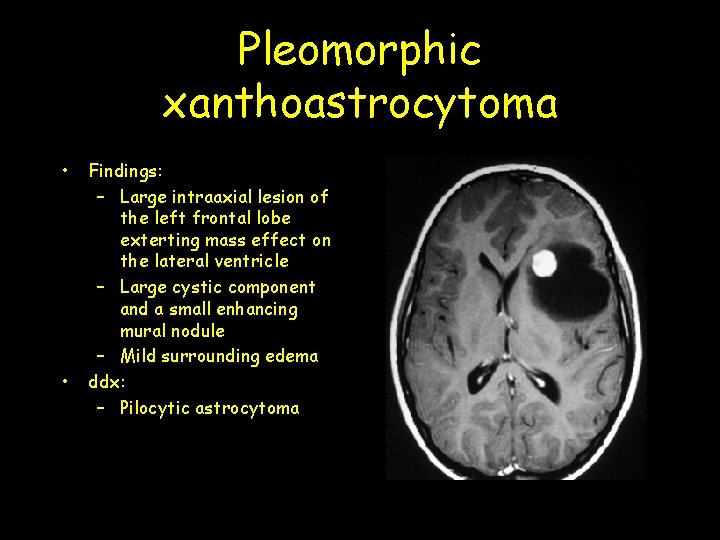
Pleomorphic xanthoastrocytoma • • Findings: – Large intraaxial lesion of the left frontal lobe exterting mass effect on the lateral ventricle – Large cystic component and a small enhancing mural nodule – Mild surrounding edema ddx: – Pilocytic astrocytoma
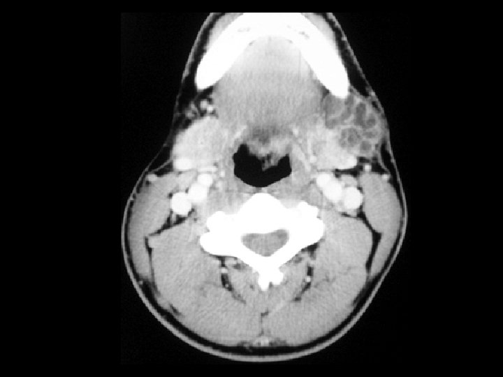
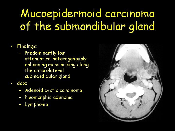
Mucoepidermoid carcinoma of the submandibular gland • Findings: – Predominantly low attenuation heterogenously enhancing mass arising along the anterolateral submandibular gland • ddx: – Adenoid cystic carcinoma – Pleomorphic adenoma – Lymphoma
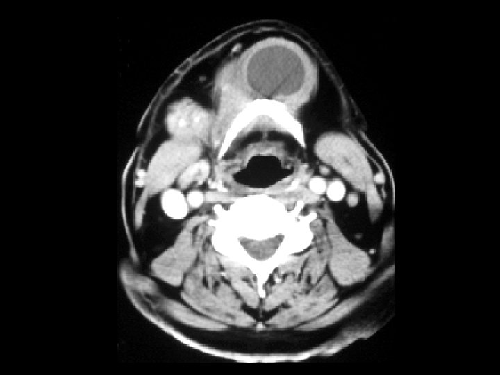
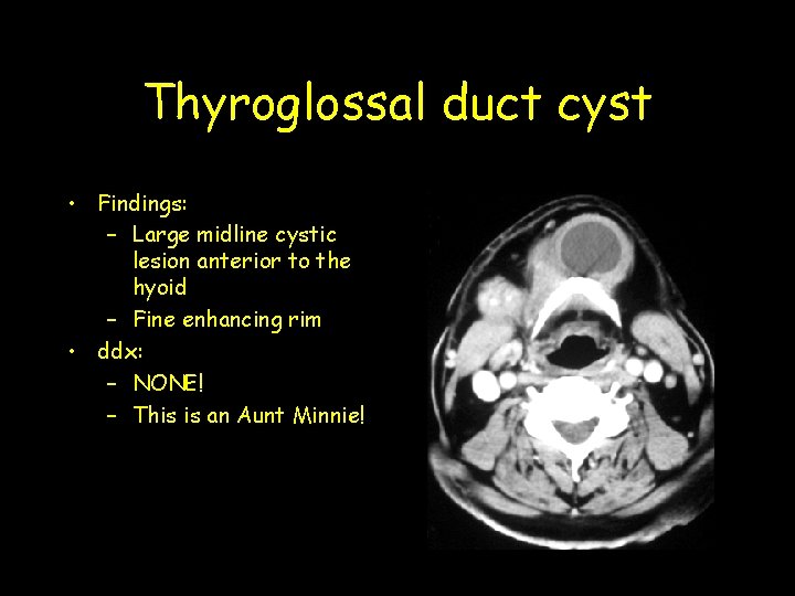
Thyroglossal duct cyst • Findings: – Large midline cystic lesion anterior to the hyoid – Fine enhancing rim • ddx: – NONE! – This is an Aunt Minnie!
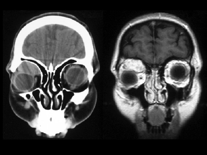
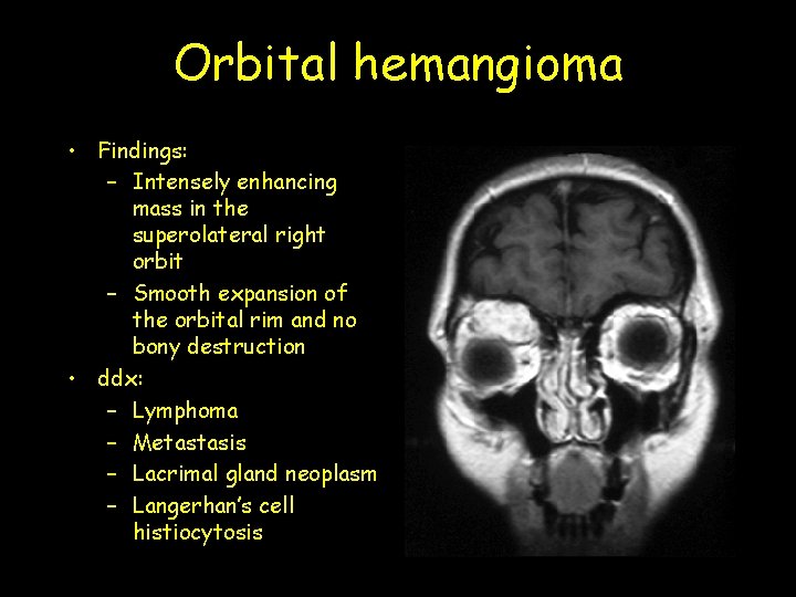
Orbital hemangioma • Findings: – Intensely enhancing mass in the superolateral right orbit – Smooth expansion of the orbital rim and no bony destruction • ddx: – Lymphoma – Metastasis – Lacrimal gland neoplasm – Langerhan’s cell histiocytosis
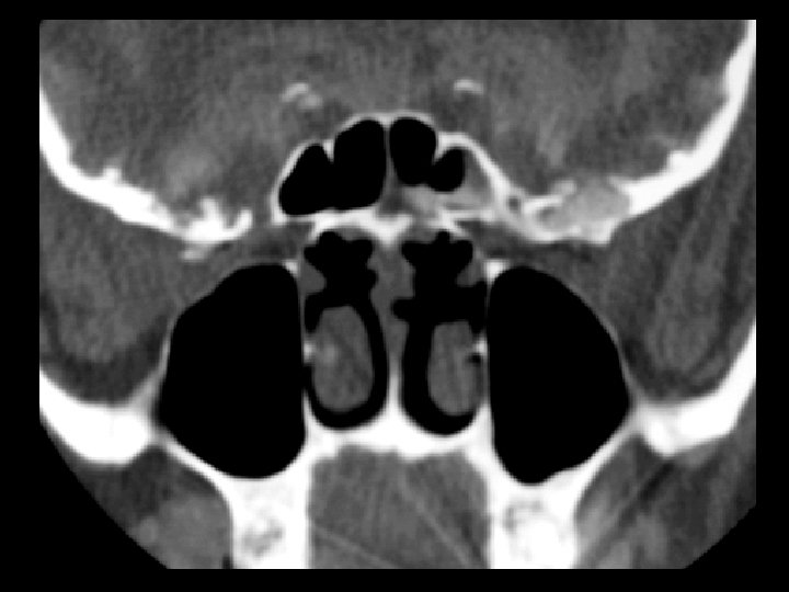
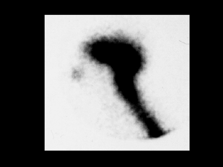
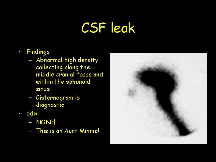
CSF leak • Findings: – Abnormal high density collecting along the middle cranial fossa and within the sphenoid sinus – Cisternogram is diagnostic • ddx: – NONE! – This is an Aunt Minnie!