
Laboratory testing for von Willebrand disease Emmanuel J
Laboratory testing for von Willebrand disease Emmanuel J Favaloro, Haematology, ICPMR, Westmead Hospital October, 2017 emmanuel. favaloro@health. nsw. gov. au

The Basics - von Willebrand factor (VWF) Ø Large and complex molecule Ø Plasma VWF – multimers of core protein (monomer of 2050 AA) ranging in size from 250 k. Da to >20000 k. Da Ø Has binding sites for many ligands – these describe various functions of VWF Ø The larger the size, the more ‘overall functionality’ or adhesiveness. Ø ‘High molecular weight’ (HMW) multimers more adhesive than intermediate or low molecular weight multimers Ø Absent, deficient, or defective VWF = von Willebrand ‘disease’ (or ‘disorder’ [VWD]; congenital) or acquired von Willebrand ‘syndrome’ (a. VWS) 2

VWF structure & binding sites VWF Ligand D Laboratory Assay FVIII VWF: FVIIIB A 1 GPIba VWF: RCo / RIPA GPIb binding assays A 3 Collagen VWF: CB D 3 A 2 D 4 B 1 -3 C 1 C 2 GPIIb. IIIa Modified: From Andreas Hillarp , in: Bolton-Maggs et al. Haemophilia. 2012; 18 (S 4): 66 -72. 3
The Basics - von Willebrand disease (VWD) Ø von Willebrand factor (VWF) disorders: Ø Defect and/or deficiency in VWF Ø congenital - von Willebrand disease (VWD) Ø acquired - von Willebrand syndrome (AVWS) Ø VWD is the most common inherited bleeding disorder Ø reported incidence up to 1% of the general population Ø more realistic incidence (patients seeking support for bleeding related issues) ~0. 1%-0. 01% Ø probably more common than haemophilia A (although severe HA probably more common than ‘severe VWD’) Ø AVWS incidence unknown, but may represent up to ~25% of patients presenting for VWD investigation 4

von Willebrand disease – testing/classification ‘Guidelines’ for VWF testing for VWD diagnosis – ISTH SSC VWD Classification (2006) Ø VWD separated into six types: 1, 2 A, 2 B, 2 N, 2 M, 3 Ø Quantitative deficiencies (type 1 = partial; type 3 = total) Ø Qualitative defects (+/- quantitative deficiency) Ø type 2 A = loss of HMW VWF Ø type 2 B = hyper-adhesive VWF (loss of HMW VWF & mild thrombocytopenia) Ø type 2 N = loss of FVIII binding (loss of FVIII stability and loss of FVIII activity from plasma) Ø type 2 M = ‘the rest’ (dysfunctional VWF not associated with loss of HMW VWF) Sadler et al. JTH. 2006; 4: 2103 -14 5

von Willebrand disease – diagnosis/management ‘Guidelines’ for VWF testing for VWD diagnosis – UKHCDO (2014) Ø Evidence based guideline from United Kingdom Haemophilia Centre Doctors Organization guideline approved by the British Committee for Standards in Haematology Ø Update of their 2004 guidelines Laffan et al. The diagnosis and management of von Willebrand disease: a United Kingdom Haemophilia Centre Doctors Organization guideline approved by the British Committee for Standards in Haematology. Br J Haematol 2014; 167: 453– 65. 6

VWD lab diagnosis – a summary Ø Type 1 – low VWF, but all VWF assays similar (i. e. , ratios ~1 (>0. 7)); VWF: Ag, VWF: CB, VWF: RCo. Ø Type 3 – ‘undetectable’ VWF (LOD issues); VWF <5 U/d. L (VWF: Ag [VWF: CB, VWF: RCo)]). Ø Type 2 A – usually low VWF: Ag, but disproportionally lower VWF: CB & VWF: RCo (both activity/Ag ratios <0. 7) Ø Type 2 B – low dose RIPA; disproportionally lower VWF: CB & VWF: RCo than VWF: Ag (both activity/Ag ratios <0. 7) Ø Type 2 N – VWF: FVIIIB (FVIIIB/VWF: Ag <0. 7) [low FVIII: C/VWF: Ag] Ø Type 2 M – low VWF ‘activity’/Ag ratio 7

VWD investigation - most common test panel Ø Factor VIII (FVIII) activity Ø one stage assay of ‘coagulant’ activity (FVIII: C) (usually automated analyzer); Ø less commonly chromogenic assay (generally only in haemophilia centers). Ø VWF: Ag - measure level of VWF protein: Ø typically by ELISA or LIA (turbimetric); usually automated analyzers. Ø occasionally other techniques (fluorescence or chemiluminescence (VIDAS; Acu. Star); flow cytometry described) Ø VWF Activity by VWF: RCo Ø classically using platelets and an aggregometer (semi-automated) Ø alternatively, using platelets on an automated analyzer Ø alternate techniques now available (e. g. , platelets & flow cytometry; latex with automated instruments [‘VWF: GPIb. R’]; chemiluminescence [Acu. Star]) 8

VWD investigation – new ISTH SSC nomenclature for ‘platelet related activity’ assays Mazurier C, Rodeghiero F. Thromb Haemost 2001; 86: 712. Bodo I, et al. J Thromb Haemost 2015; 13: 1345– 50. Favaloro et al. Pathology 2016; 48: 303 -18. 9

VWF ‘activity assays’ – Australian trends Updated from Favaloro & Mohammed. Thromb Res 2014. 134: 1292– 1300 10

Measurement of VWF protein level - ‘Antigen’ (VWF: Ag) Extent of color reaction proportional to level of VWF ELISA Color reaction Substrate HRP HRP Labeled (Rabbit) a-VWF Favaloro EJ, Mohammed S, Patzke J. Laboratory Testing for von Willebrand Factor Antigen (VWF: Ag). Methods Mol Biol. 2017; 1646: 403 -416. (Rabbit) a-VWF 11

Measurement of VWF protein level - ‘Antigen’ (VWF: Ag) LIA Latex aggregation proportional to level of VWF a-VWF Latex particles Favaloro EJ, Mohammed S, Patzke J. Laboratory Testing for von Willebrand Factor Antigen (VWF: Ag). Methods Mol Biol. 2017; 1646: 403 -416. 12

Measurement of VWF ristocetin cofactor (VWF: RCo) Ø Ristocetin binds to VWF and causes conformational change (‘unfolding’) which leads to exposure of VWF epitopes that then subsequent bind to platelet via platelet GPIb Ø Assay designed to be sensitive to VWF defects (VWF conformational changes; e. g. , 2 A, 2 B, 2 M VWD) and HMW VWF (2 A, 2 B VWD) Ø Can be performed using platelets (‘VWF: RCo’) or other methods including latex and based on r. VWF (‘VWF: GPIb’) ristocetin ‘unfolded’ VWF Gp. Ib Platelet Mohammed S, Favaloro EJ. Laboratory Testing for von Willebrand Factor Ristocetin Cofactor (VWF: RCo). Methods Mol Biol. 2017; 1646: 435 -451. 13 Latex aggregation proportional to level of ‘active’ and HMW VWF
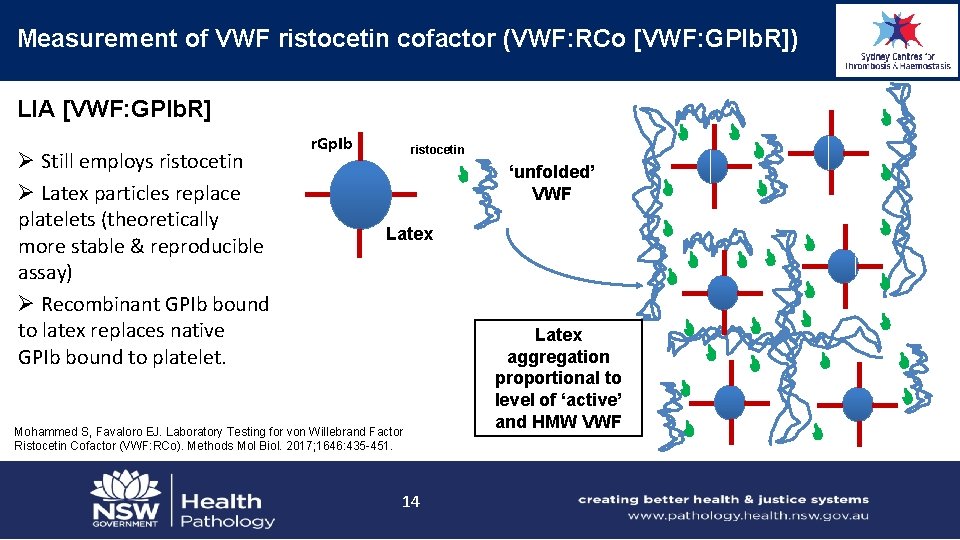
Measurement of VWF ristocetin cofactor (VWF: RCo [VWF: GPIb. R]) LIA [VWF: GPIb. R] Ø Still employs ristocetin Ø Latex particles replace platelets (theoretically more stable & reproducible assay) Ø Recombinant GPIb bound to latex replaces native GPIb bound to platelet. r. Gp. Ib ristocetin ‘unfolded’ VWF Latex Mohammed S, Favaloro EJ. Laboratory Testing for von Willebrand Factor Ristocetin Cofactor (VWF: RCo). Methods Mol Biol. 2017; 1646: 435 -451. 14 Latex aggregation proportional to level of ‘active’ and HMW VWF

Measurement of VWF collagen binding (VWF: CB) - ELISA Ø Another measure of VWF ‘activity’ Ø VWF binding to collagen is a natural in vivo adhesive activity Ø Original description in 1986 ØELISA assay can be designed to be sensitive to specific VWF defects (VWF conformational changes; e. g. , 2 A, 2 B, 2 M VWD) and HMW VWF (2 A, 2 B VWD) Color reaction Extent of color reaction proportional to level of adhesive (HMW) VWF Substrate HRP Labeled (Rabbit) a-VWF *Brown JE, Bosak JO. Thromb Res 1986; 43: 303– 11. VWF Favaloro EJ, Mohammed S. Laboratory Testing for von Willebrand Factor Collagen Binding (VWF: CB). Methods Mol Biol. 2017; 1646: 417 -433. Collagen 15 HRP

Measurement of VWF collagen binding (VWF: CB) - CLIA Werfen IL - ACL Acu. Star Ø Chemiluminescence technology Ø “Superior range and sensitivity compared to ELISA or immunoturbidimetric assays” Ø Fully automated Ø Self-contained, ready-to-use reagent cartridges, stable up to twelve weeks onboard Ø Assays onboard and available 24 hours/day, 7 days/week Ø Assay calibration on lot change only Ø Random access: no batching required Favaloro EJ, Mohammed S. Laboratory Testing for von Willebrand Factor Collagen Binding (VWF: CB). Methods Mol Biol. 2017; 1646: 417 -433. 16

Measurement of VWF collagen binding (VWF: CB) Werfen IL - ACL Acu. Star ‘VWD data set’; n=314 Ø Good correlation Ø Some scatter around line of equivalence Ø Most scatter is likely random noise events (all comparisons) Ø Some outliers circled/arrowed Ø Arrowed = random error event (rpt was OK) Ø Circled = discrepant data – mostly type 2 M VWD Favaloro EJ, Mohammed S. Thromb Res. 2016; 141: 202 -11 17

Measurement of VWF GPIb binding – ‘other’ ØSiemens ‘INNOVANCE’ ‘activity’ assay (VWF Ac; ‘VWF: GPIb. M’) Ø assay employs recombinant (r) GPIb with 2 gain of function mutations in a latex based GPIb-binding assay Ø this r. GPIb spontaneously binds native VWF (like PT-VWD GPIb) Ø this occurs in absence of ristocetin, so is not a VWF: RCo assay Ø however, results of this assay tend to mimic results of VWF: RCo Patzke J, Favaloro EJ. Laboratory Testing for von Willebrand Factor Activity by Glycoprotein Ib Binding Assays (VWF: GPIb). Methods Mol Biol. 2017; 1646: 453 -460. 18

Measurement of VWF GPIb binding – VWF Ac/‘VWF: GPIb. M’ cont 600 samples, comprising nearly 100 VWD, nearly 60 individual normal samples, post DDAVP & Biostate, with the rest mostly non. VWD but for VWF/VWD workups. Favaloro & Mohammed. Thromb Res 2014. 134: 1292– 1300 19

Measurement of VWF GPIb binding – ‘other’ Ø RIPA assay (for identification of 2 B and PT VWD) Ø Ristocetin induced platelet agglutination/aggregation assay Ø Also stemmed from the work of Barry Firkin group Ø Typically performed by platelet aggregation on an aggregometer Ø Different to VWF: RCo Ø VWF: RCo = patient plasma + fixed dose of ristocetin (usually ~1 mg/ml, but probably higher in the Siemen’s assay) + fixed or lyophilised normal platelets; results compared to standard curve using reference plasma = quantitative value (>50 U/d. L = ‘normal’) Ø RIPA = patient platelet rich plasma + variable dose of ristocetin (typically, ‘normal’ dose = ~1 or 1. 2 or 1. 5 mg/m. L; low dose = <0. 7 (usually 0. 5) mg/m. L; high dose + > 1. 5 mg/m. L). Ø Response to low dose = 2 B or PT VWD (can be differentiated by RIPA mixing studies or genetic analysis of VWF and/or GPIb) Ø Response only to high dose (not to normal or low dose) = VWD (severe 1 or type 2 A, 2 M). Ø No response to any dose = type 3 VWD or BSS Ø Response to normal dose but not to low dose – ‘exclude’ severe VWD and 2 A, 2 B, 2 M, PT VWD. Frontroth JP, Favaloro EJ. Ristocetin-Induced Platelet Aggregation (RIPA) and RIPA Mixing Studies. Methods Mol Biol. 2017; 1646: 473 -494 20

Measurement of VWF FVIII binding (2 N VWD identification) Extent of color reaction proportional to level of FVIII bound to VWF Color reaction ØTypically performed by ELISA Ø Patient VWF bound to ELISA plate Ø Separate ELISA wells with calibration standard & controls Ø Sandwich process that removes patient VWF bound FVIII, followed by addition of recombinant (r) FVIII, and assay determines the level of patient VWF bound r. FVIII Substrate HRP Labeled a-FVIII VWF Mohammed S, Favaloro EJ. Laboratory Testing for von Willebrand Factor: Factor VIII Binding (for 2 N VWD). Methods Mol Biol. 2017; 1646: 461 -472. (Rabbit) a-VWF 21 HRP

Measurement of VWF FVIII binding (2 N VWD identification) ØFindings compared to those of parallel VWF: Ag assay Ø Proportion of FVIII bound to VWF (VWF: FVIIIB) compared to amount of VWF bound in VWF: Ag assay Ø Normal VWF: FVIIIB = >0. 7 Ø Values close to 0. 5 suggest heterozygous 2 N VWD Ø Values <0. 2 suggest homozygous or double heterozygous 2 N VWD or heterozygous 2 N VWD with a second ‘null’ allele for VWF Ø Only performed by ~5% of RCPA QAP enrolled VWF test labs Mohammed S, Favaloro EJ. Laboratory Testing for von Willebrand Factor: Factor VIII Binding (for 2 N VWD). Methods Mol Biol. 2017; 1646: 461 -472. 22

Error rates in VWD ‘laboratory diagnosis’ Favaloro et al. Thromb Res 2014. 134(2): 393 -403 Favaloro. STH 2014. 40(5): 551 -70 23

Error rates in VWD ‘laboratory diagnosis’ Favaloro et al. Thromb Res 2014. 134(2): 393 -403 Favaloro. STH 2014. 40(5): 551 -70 24

VWF Laboratory assay limitations Lower limit assay sensitivity Assay variability Favaloro et al. Thromb Res 2014; 134: 393 -403 Favaloro et al. Clin Lab Sci 2008; 21: 178 -185 25

VWF Laboratory assay limitations Discrimination of type 1 vs 2 VWD (RCPAQAP) Normal samples HMW VWF deficient samples (‘type 2 A VWD’) Favaloro et al. Thromb Res 2014; 134: 393 -403 26 VWF deficient samples (‘type 1 VWD’) HMW VWF deficient samples (type 2 B VWD)

What about VWF Multimer analysis? High Molecular Weight VWF Intermediate Molecular Weight VWF Low Molecular Weight VWF Type 2 B Normal Type 2 A Mild Type 1 Normal 27 Ø All multimers present = normal (i. e. , not VWD), or 2 N VWD or 2 M VWD Ø All multimers present but in lower intensity = type 1 VWD (or maybe specimen artefact) Ø Loss of HMW VWF = possible 2 A or 2 B/PT VWD (or maybe specimen artefact) Ø Loss of HMW & IMW VWF = probable 2 A (or maybe specimen artefact)

Multimer analysis – the good, bad & the ugly Ø Multimer analysis is laborious and requires a high level of technical skill Ø Performing multimers on 6 -12 x samples may take several weeks of technical time. Ø Automated testing can perform ~36 x samples for VWF: Ag, VWF: RCo, VWF: CB and FVIII: C in a single day Ø <5% of VWF test labs perform multimer testing (2/56 labs in RCPA QAP) in Australasia Ø Perhaps 15 -25% of labs perform multimer testing in Europe/North America Ø NASCOLA survey (Chandler WL, et al. Am J Clin Pathol. 2011; 135: 862 -869): Ø 15% overall average error rate, Ø 5% of labs reported loss of HMW VWF in normal samples, Ø 18% reported loss of HMW VWF in type 1 VWD samples, Ø 18% reported a normal multimer pattern in type 2 VWD. Ø ECAT survey (Meijer & Haverkate. Semin Thromb Hemost. 2006; 32: 485 -491): Ø 23% error rate for normal samples Ø up to 52% error rate in type 1 VWD 28

What about VWF Multimer analysis? High Molecular Weight VWF: RCo Intermediate Molecular Weight VWF: CB VWF: Ag Assay interrelationships Low Molecular Weight VWF Type 2 B Normal Type 2 A Mild Type 1 Favaloro EJ, Sem Thromb Hemost, 2007; 33: 727 -44 Normal 29 Ø low VWF but concordant activity & antigen (i. e. RCo/Ag & CB/Ag > 0. 7 = type 1 VWD or ‘low VWF’) Ø discordant activity & antigen x 2 (i. e. RCo/Ag & CB/Ag < 0. 5 = probable loss of HMW VWF = type 2 A or 2 B VWD) Ø discordant activity & antigen x 1 (i. e. RCo/Ag or CB/Ag < 0. 5 = probable VWF ‘dysfunction’ = type 2 M VWD)

What about VWF Multimer analysis? Ø recent ‘revalidation’ of expected test patterns and multimer analysis with a semi -automated VWF multimer test system Ø low VWF but concordant activity & antigen (i. e. RCo/Ag & CB/Ag > 0. 7 = type 1 VWD or ‘low VWF’) Ødiscordant activity & antigen x 2 (i. e. RCo/Ag & CB/Ag < 0. 5 = probable loss of HMW VWF = type 2 A or 2 B VWD) Ø discordant activity & antigen x 1 (i. e. RCo/Ag or CB/Ag < 0. 5 = probable VWF ‘dysfunction’ = type 2 M VWD) Favaloro EJ, Oliver S. Haemophilia. 2017; 23: e 373 -e 377. Oliver S, et al. Methods Mol Biol. 2017; 1646: 495 -511. 30

Laboratory ‘Diagnosis’ of VWD Ø Lupus anticoagulant guidelines: Ø Assess by at least two assays using different principles Ø Can exclude LA if both negative Ø Can identify LA if either is positive Ø VWD can be caused by mutations anywhere on VWF Ø Type 2 VWD will only be properly identified if labs perform activity testing using assays employing as many principles as possible Ø At the very least, explore GPIb binding (VWF: RCo [or VWF: GPIb. R] and/or VWF Ac [VWF: GPIb. M]) plus collagen binding (VWF: CB), in addition to VWF: Ag and FVIII Laffan et al. The diagnosis and management of von Willebrand disease: a United Kingdom Haemophilia Centre Doctors Organization guideline approved by the British Committee for Standards in Haematology. Br J Haematol 2014; 167: 453– 65. 31

Laboratory ‘Diagnosis’ of VWD – a summary Ø Type 1 – low VWF, but all VWF assays similar (i. e. , ratios ~1 (>0. 7)); VWF: Ag, VWF: CB, VWF: RCo. Ø Type 3 – ‘undetectable’ VWF (LOD issues); VWF <5 U/d. L (VWF: Ag [VWF: CB, VWF: RCo)]). Ø Type 2 A – usually low VWF: Ag, but disproportionally lower VWF: CB & VWF: RCo (both activity/Ag ratios <0. 7) Ø Type 2 B – low dose RIPA; disproportionally lower VWF: CB & VWF: RCo than VWF: Ag (both activity/Ag ratios <0. 7) Ø Type 2 N – VWF: FVIIIB (FVIIIB/VWF: Ag <0. 7) [low FVIII: C/VWF: Ag] Ø Type 2 M – low VWF ‘activity’/Ag ratio 32

VWD lab diagnosis – an algorithmic approach 33

Acknowledgments • Meeting organisers, especially Joanne Josephs • Westmead clinical colleagues, especially Leonardo Pasalic & Jennifer Curnow • RCPAQAP Haematology, especially Roslyn Bonar • Current/recent past diagnostic Haemostasis laboratory staff – – – Soma Mohammed Jane Mc. Donald Ella Grzechnik Monica Ahuja Shabana Azimulla Yifang Zhang Haemostasis = Love Everybody talks about it, nobody understands it. 34
- Slides: 34