
Pathology Course Haematology 2 Kindly sponsored by What
Pathology Course Haematology 2 Kindly sponsored by:
What will be discussed �Tuesday: Anaemia, coagulation, thrombosis �Thursday: Haematological malignancies Paraproteinaemias Myelodysplastic / myeloproliferative disorders
General tips for written exams �Don’t focus too much on Eponymous syndromes Nitty gritty facts e. g. chromosomal translocations �Do Web. CT questions �Some questions will be hard / strange �Understanding is key
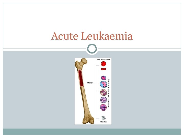
Acute Leukaemia
Acute Leukaemia �Neoplastic process of bone marrow and blood �‘Acute’ therefore rapidly progressive and fatal �Immature blasts > 20% BM cells �Signs/symptoms: BM failure: anaemia, thrombocytopenia, neutropenia Organ infiltration: hepato/spleno-megaly, lymphadenopathy, bone pain, gum hypertrophy
Aetiology �Ionising radiation – radiotherapy �Cytotoxic drugs – chemotherapy �Benzene �Pre-leukaemic disorders e. g. MDS/MPD �Down’s Significnatly increased risk of AML/ALL Also, Down’s neonates often develop transient abnormal myelopoeisis � Resembles AML � Resolves spontaneously after a few weeks
Acute Lymphoblastic Leukaemia �Childhood (ALL) �Symptoms as previously mentioned Lymphadenopathy +++ CNS involvement +++ Testicular enlargment Thymic enlargement �High WCC (accumulation of lymphoblasts) �Ph+ve t(9; 22) – poor prognosis
ALL: Treatment �Chemotherapy: (memory aid: I Can Cure Mankind) Remission Induction Consolidation CNS treatment Maintenance �Supportive Blood products, fluids, electrolyes
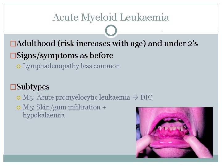
Acute Myeloid Leukaemia �Adulthood (risk increases with age) and under 2’s �Signs/symptoms as before Lymphadenopathy less common �Subtypes M 3: Acute promyelocytic leukaemia DIC M 5: Skin/gum infiltration + hypokalaemia
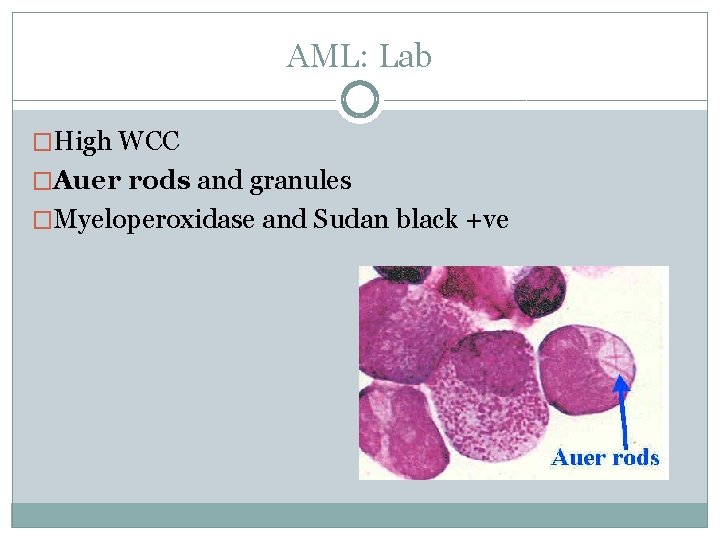
AML: Lab �High WCC �Auer rods and granules �Myeloperoxidase and Sudan black +ve
AML: Treatment �Chemotherapy Similar principles to ALL but… No CNS prophylaxis or maintenance needed �ATRA for M 3 �Consider allo-SCT in young
Chronic Myeloid Leukaemia
Chronic Myeloid Leukaemia �A myeloproliferative disease �Often diagnosed on routine bloods �Now very successfully treated in the majority
CML: Presentation � 40 -60 years peak (middle-aged) �May be asymptomatic �Anaemia �Weight loss, low grade fever, night sweats �Massive splenomegaly (down to RIF) �Gout (raised urate – hypermetabolism)
CML: Lab �Huge WCC > 100 (neutrophilia) �Hypercellular BM with spectrum of mature and immature cells in the blood Malignant cells proliferate excessively but retain ability to differentiate into mature cells �Ph+ve in 80%; t(9; 22) �PCR for BCR-ABL Monitor disease and therapeutic response
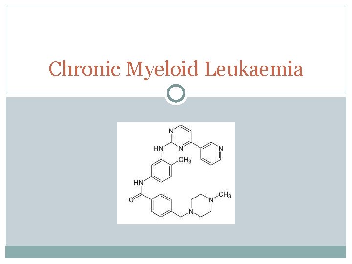
CML: Phases & Treatment Chronic Phase �< 5% blasts in BM/blood �WBC slowly increases �Over 3 -4 years �Rx = Imatinib BCR-ABL tyrosine kinase inhibitor
CML: Phases & Treatment Accelerated phase � > 10% blasts in BM/blood �Increasing manifestations e. g. splenomegaly �Up to a year �Less responsive to therapy
CML: Phases & Treatment Blast phase �> 20% blasts in BM/blood �Resembles an acute leukaemia �Course = months �Treatment similar to AML
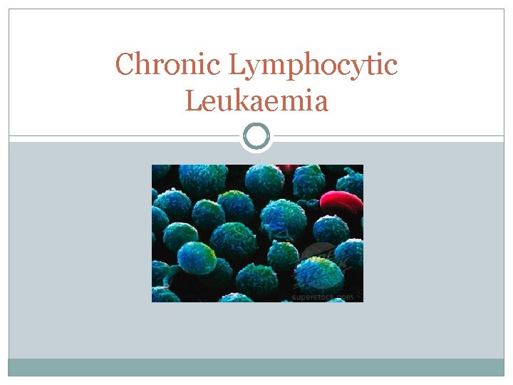
Chronic Lymphocytic Leukaemia
Chronic Lymphocytic Leukaemia �A lymphoproliferative disease Others include ALL, MM and Waldenstrom’s �Also often an incidental finding on routine bloods �Disease of the elderly
CLL & SLL �Chronic lymphocytic leukaemia and small lymphocytic lymphoma �Same underlying disease, different presentation �CLL is primarily located in BM �SLL is primarily located in lymph nodes
CLL: Presentation �> 40 years, elderly �May be asymptomatic �Symmetrical painless lymphadenopathy �Anaemia, thrombocytopenia, neutropenia �Wt loss, low grade fever, night sweats �Hepato/Spleno-megaly (less prominent) �Ass w/ autoimmunity AIHA (Coomb’s +ve) + ITP = Evan’s syndrome
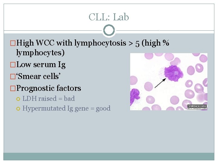
CLL: Lab �High WCC with lymphocytosis > 5 (high % lymphocytes) �Low serum Ig �‘Smear cells’ �Prognostic factors LDH raised = bad Hypermutated Ig gene = good
CLL: Stages & Treatment �Binet Staging A, B & C �Stage A < 3 groups of enlarged LN No treatment required �Stage B & C > 3 groups of LN, patient becomes more symptomatic Rx: 1 st line is chlorambucil
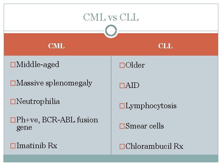
CML vs CLL CML �Middle-aged �Older �Massive splenomegaly �AID �Neutrophilia �Ph+ve, BCR-ABL fusion gene �Imatinib Rx �Lymphocytosis �Smear cells �Chlorambucil Rx
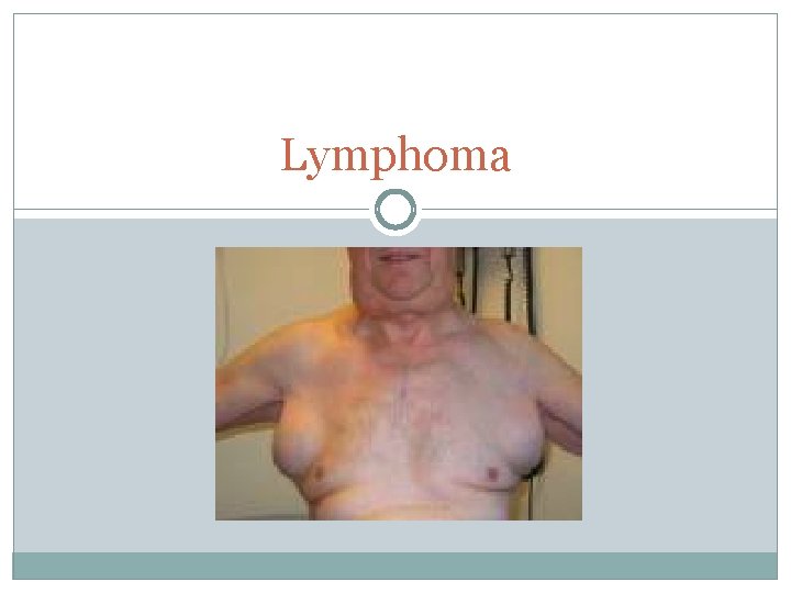
Lymphoma
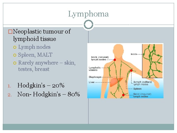
Lymphoma �Neoplastic tumour of lymphoid tissue Lymph nodes Spleen, MALT Rarely anywhere – skin, testes, breast Hodgkin’s – 20% 2. Non- Hodgkin’s – 80% 1.
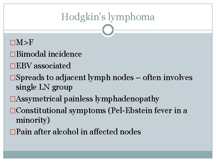
Hodgkin’s lymphoma �M>F �Bimodal incidence �EBV associated �Spreads to adjacent lymph nodes – often involves single LN group �Assymetrical painless lymphadenopathy �Constitutional symptoms (Pel-Ebstein fever in a minority) �Pain after alcohol in affected nodes
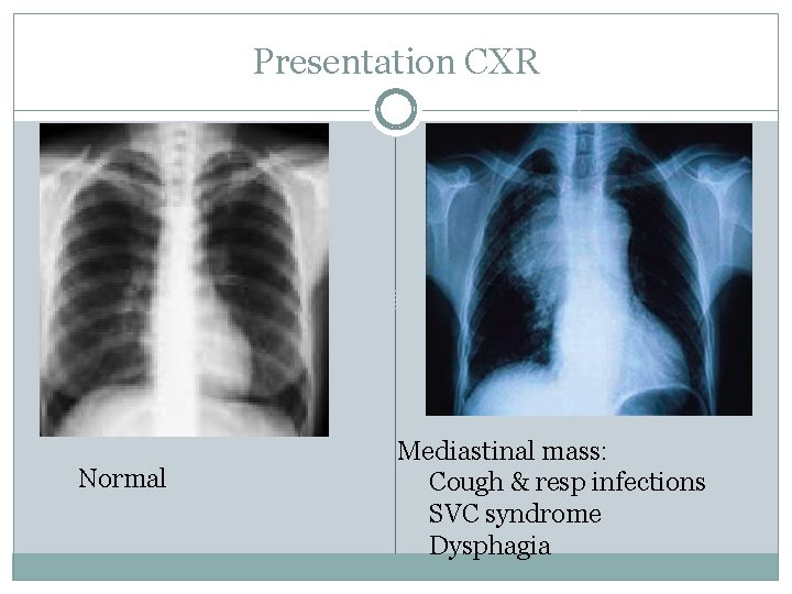
Presentation CXR �nk Normal Mediastinal mass: Cough & resp infections SVC syndrome Dysphagia
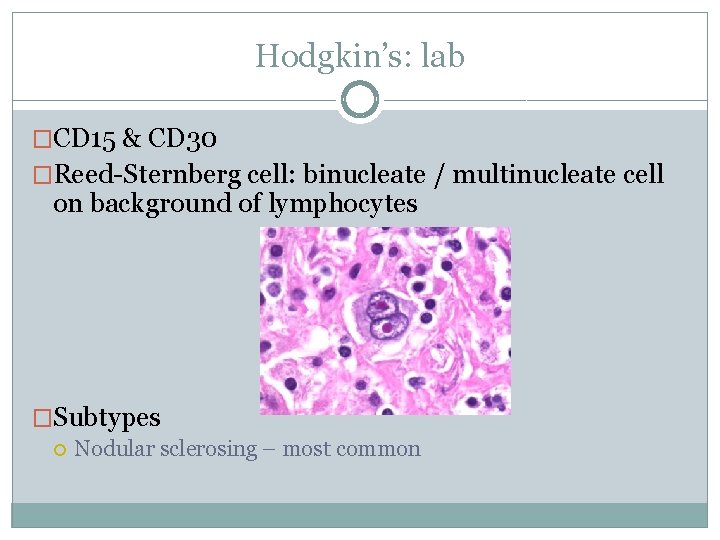
Hodgkin’s: lab �CD 15 & CD 30 �Reed-Sternberg cell: binucleate / multinucleate cell on background of lymphocytes �Subtypes Nodular sclerosing – most common
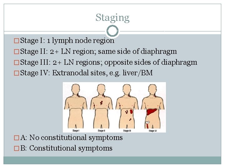
Staging � Stage I: 1 lymph node region � Stage II: 2+ LN region; same side of diaphragm � Stage III: 2+ LN regions; opposite sides of diaphragm � Stage IV: Extranodal sites, e. g. liver/BM � A: No constitutional symptoms � B: Constitutional symptoms
Treatment �Combination chemotherapy ABVD – adriamycin, bleomycin, vinblastine, dacarbazine 2 -4 cycles in I/II, 6 -8 cycles in III/IV �Radiotherapy Sometimes used alongside chemo against bulky areas �Intensive chemo + autologous SCT Relapsed patients �ESR used for progress and monitoring
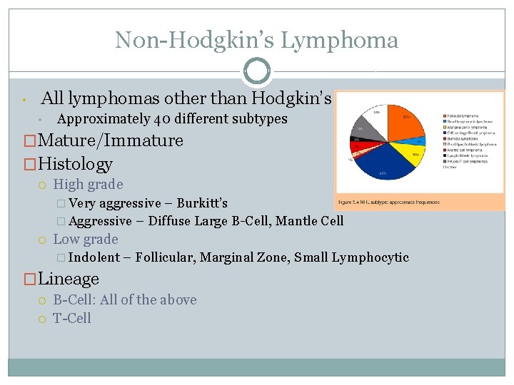
Non-Hodgkin’s Lymphoma • All lymphomas other than Hodgkin’s • Approximately 40 different subtypes �Mature/Immature �Histology High grade � Very aggressive – Burkitt’s � Aggressive – Diffuse Large B-Cell, Mantle Cell Low grade � Indolent – Follicular, Marginal Zone, Small Lymphocytic �Lineage B-Cell: All of the above T-Cell
Non-Hodgkin’s Lymphoma �Presentation varies significantly from subtype to subtype Painless lymphadenopathy Often involves multiple sites Constitutional symptoms No pain after alcohol �Staging as per Hodgkin’s
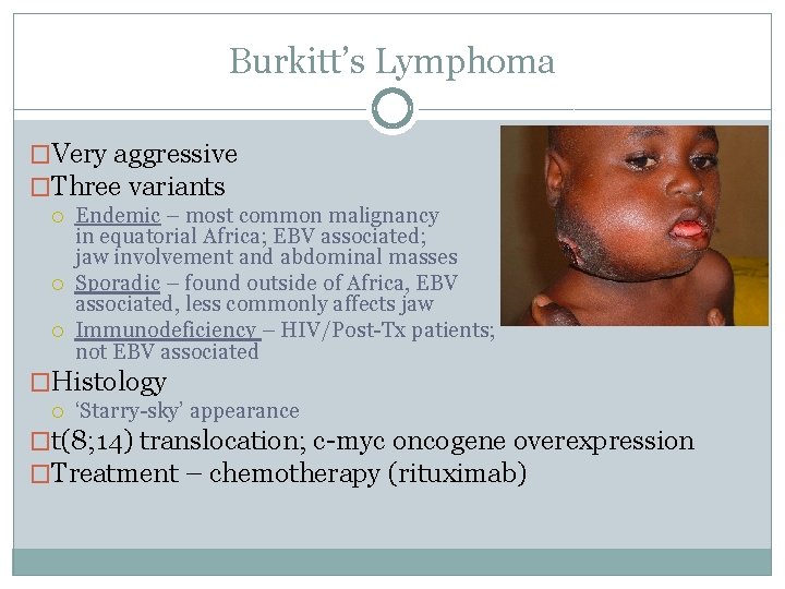
Burkitt’s Lymphoma �Very aggressive �Three variants Endemic – most common malignancy in equatorial Africa; EBV associated; jaw involvement and abdominal masses Sporadic – found outside of Africa, EBV associated, less commonly affects jaw Immunodeficiency – HIV/Post-Tx patients; not EBV associated �Histology ‘Starry-sky’ appearance �t(8; 14) translocation; c-myc oncogene overexpression �Treatment – chemotherapy (rituximab)
Diffuse Large B-Cell Lymphoma (DLBCL) �Middle-aged and elderly �Aggressive Richter’s transformation: leukaemias DLBCL �Histology • Sheets of large lymphoid cells �Treatment Rituximab-CHOP
Mantle Cell Lymphoma �Middle-aged �Aggressive Disseminated at presentation Median survival 3 -5 years �Histology ‘Angular nuclei’ �t(11; 14) translocation �Dysregulation of cyclin D 1 �Tx similar to DLBCL
Follicular Lymphoma �Elderly �Indolent Median life expectancy 12 -15 years Mostly incurable �Histology ‘Follicular pattern’, ‘Nodular appearance’ �t(14; 18) translocation �Treatment Watch and wait Rituximab-CVP
MALT lymphomas �Mucosal Associated Lymphoid Tissue Marginal Zone NHL �Middle aged �Chronic antigen stimulation H. Pylori gastric MALT lymphoma Sjogren’s syndrome parotid lymphoma �Treatment Remove antigenic stimulus e. g. H pylori triple therapy Chemotherapy
T-Cell Lymphomas � Adult T-Cell leukaemia/lymphoma HTLV-1 infection � Enteropathy associated T-cell lymphoma Long standing coeliac disease � Cutaneous T-cell lymphoma � Anaplastic large cell lymphoma Children and young adults Aggressive Large ‘epitheloid’ lymphocytes � Peripheral T-Cell Lymphoma Middle aged and elderly Aggressive Large T cells
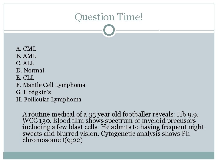
Question Time! A. CML B. AML C. ALL D. Normal E. CLL F. Mantle Cell Lymphoma G. Hodgkin’s H. Follicular Lymphoma A routine medical of a 33 year old footballer reveals: Hb 9. 9, WCC 130. Blood film shows spectrum of myeloid precusors including a few blast cells. He admits to having frequent night sweats and blurred vision. Cytogenetic analysis shows Ph chromosome t(9; 22)
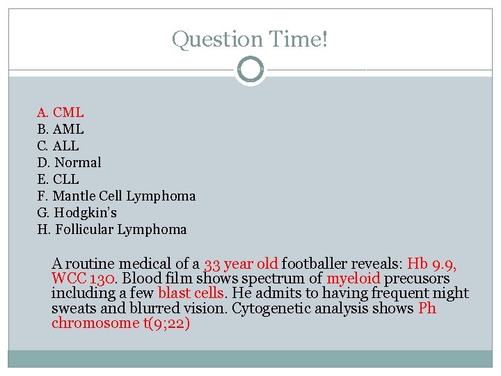
Question Time! A. CML B. AML C. ALL D. Normal E. CLL F. Mantle Cell Lymphoma G. Hodgkin’s H. Follicular Lymphoma A routine medical of a 33 year old footballer reveals: Hb 9. 9, WCC 130. Blood film shows spectrum of myeloid precusors including a few blast cells. He admits to having frequent night sweats and blurred vision. Cytogenetic analysis shows Ph chromosome t(9; 22)
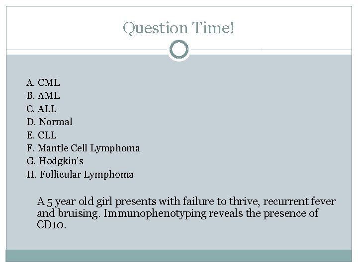
Question Time! A. CML B. AML C. ALL D. Normal E. CLL F. Mantle Cell Lymphoma G. Hodgkin’s H. Follicular Lymphoma A 5 year old girl presents with failure to thrive, recurrent fever and bruising. Immunophenotyping reveals the presence of CD 10.
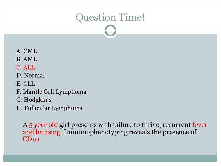
Question Time! A. CML B. AML C. ALL D. Normal E. CLL F. Mantle Cell Lymphoma G. Hodgkin’s H. Follicular Lymphoma A 5 year old girl presents with failure to thrive, recurrent fever and bruising. Immunophenotyping reveals the presence of CD 10.
Question Time! A. CML B. AML C. ALL D. Normal E. CLL F. Mantle Cell Lymphoma G. Hodgkin’s H. Follicular Lymphoma A 64 year old woman receiving long-term chemotherapy for lymphoma presents with worsening bone pain, recurrent fever and night sweats. Blood film shows blast cells and Aeur rods.
Question Time! A. CML B. AML C. ALL D. Normal E. CLL F. Mantle Cell Lymphoma G. Hodgkin’s H. Follicular Lymphoma A 64 year old woman receiving long-term chemotherapy for lymphoma presents with worsening bone pain, recurrent fever and night sweats. Blood film shows blast cells and Aeur rods.
Question Time! A. CML B. AML C. ALL D. Normal E. CLL F. Mantle Cell Lymphoma G. Hodgkin’s H. Follicular Lymphoma An 85 year old lady with a painless swelling in her neck. After undergoing several investigation, she is told that there is cancer present but it is very slow growing. She is advised that she will be followed up regularly but no intervention is necessary at present.
Question Time! A. CML B. AML C. ALL D. Normal E. CLL F. Mantle Cell Lymphoma G. Hodgkin’s H. Follicular Lymphoma An 85 year old lady with a painless swelling in her neck. After undergoing several investigation, she is told that there is cancer present but it is very slow growing. She is advised that she will be followed up regularly but no intervention is necessary at present.
Multiple Myeloma
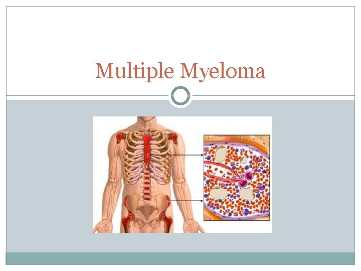
Multiple Myeloma �Neoplasia of plasma cells of BM �Production of monocloncal Ig (Ig. G most common) = paraproteinaemia �Middle aged – elderly �Increased incidence in Afro-Caribbeans
Multiple Myeloma: Presentation
Multiple Myeloma: Presentation �Calcium high Thirst, abdo pain, constipation, stones, confusion �Renal failure Light chain obstruction of tubules Chains in urine = Bence Jones protein �Anaemia Infiltrate of plasma cells in BM Tiredness (also easy bleeding and infection) �Bone pain Osteoperosis, osteolytic lesions, wedge compression fractures
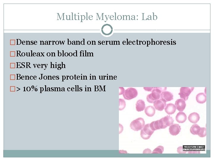
Multiple Myeloma: Lab �Dense narrow band on serum electrophoresis �Rouleax on blood film �ESR very high �Bence Jones protein in urine �> 10% plasma cells in BM
Multiple Myeloma: Treatment �Supportive for CRAB symptoms Bisphonates EPO �Chemotherapy Melphalan �Proteosome inhibitor: Bortezomib is 2 nd line �Lenalidomide 3 rd line
MGUS & Smouldering Myeloma Monoclonal gammaglobinopathy of unknown significance �<10% plasma cells in BM (>10 % = MM) �Incidental finding �Progresses to MM at rate of 1 -2%/year Smouldering Myeloma �>10% plasma cells in BM but no CRAB
Waldenstrom’s Macroglobinaemia �Older men �Lymphoplasmacytoid cells in BM �Ig. M paraproteinaemia �Unlike myeloma, similar to NHL – lymphadenopathy, wt loss, fatigue �Hyperviscosity syndrome – blurred vision, headaches �Plasmapheresis for hyperviscosity
MDS, MPD & ‘The Rest’
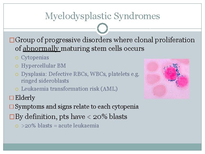
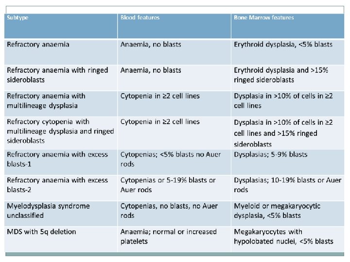
Myelodysplastic Syndromes �Group of progressive disorders where clonal proliferation of abnormally maturing stem cells occurs Cytopenias Hypercellular BM Dysplasia: Defective RBCs, WBCs, platelets e. g. ringed sideroblasts Leukaemia transformation risk (AML) � Elderly � Symptoms and signs relate to each cytopenia �By definition, pts have < 20% blasts >20% blasts = acute leukaemia
MDS: Treatment �Supportive – transfusions, EPO �Biological Modifiers �Chemotherapy: hydroxyurea �Allogenic SCT Prognosis �Depends on International Prognostic Scoring System �Mortality rule of 1/3: infection, bleeding, acute leukaemia
Aplastic Anaemia �Inability of BM to produce adequate blood cells AA typically refers to anaemia i. e. just RBCs, but these pts have a pancytopenia �Symptoms and signs relate to each cytopenia Patients typically present with bleeding problems �AA closely linked to Leukaemia Paroxysmal nocturnal haemoglobinuria
Aplastic Anaemia �Classification Primary � Idiopathic (70%) � Inherited (10%) – see next slide Secondary (10 -20%) � Radiation, drugs, viruses, immune
Inherited AA / BM Failure Syndromes �Fanconi Anaemia AR Presents at 5 -10 years Skeletal abnormalities, short stature, renal malformations, microopthalmia, skin pigmentation AML risk �Schwachman-Diamond Syndrome AR Skeletal abnormalities, short stature, endocrine panreactic dysfynction, hepatic impairment AML risk
Inherited AA / BM Failure Syndromes �Dyskeratososis Congenita X linked Triad: skin pigmentation, nail dystrophy, leukoplakia �Diamond-Blackfan Syndrome Pure red cell aplasia; normal WCC and platelets Presents at 1 yr Dysmorphology
Myeloproliferative disorders �Group of conditions characterised by clonal proliferation of one or more haemopoietic component Polycythaemia vera – RBCs 2. Essential thrombocythaemia – platelets 3. Idoiopathic myelofibrosis – WBCs 1. Associated with JAK 2 mutations (PRV)
Polycythaemia �Absolute polycthaemia Red cell mass raised Primary � PRV, familial Secondary � Disease EPO states (renal Ca, hypoxia - COPD), high altitude raised �Relative (pseudo) polycythaemia Red cell mass normal – plasma volume reduced Dehydration, burns, vomiting, diarrhoea Cigarette smoking, alcohol, HTN, obesity
Polycythaemia rubra vera (PRV) �MPD where erythroid precursors dominate BM JAK 2 point mutations �Symptoms/signs Hyper-viscosity/volaemia/metabolism plethoric (red nose), gout, splenomegaly Histamine release aquagenice pruritus (e. g. hot shower) and peptic ulcers �Lab Raised Hb, HCT Low serum EPO (cf secondary polycythaemia) Platelets, WCC may also be raised �Tx: venesection, hydroxycarbamide, aspirin
Idiopathic myelofibrosis �MPD where abnormal stem cells fibrosis of BM JAK 2 mutation in 50% �Symptoms/signs Related to cytopenias Hepatomegaly, massive splenomegaly Weight loss, fever �Lab findings Tear drop poikilocytes, leucoerythroblasts Abnormally shaped RBCs, nucleated RBCs, immature myeloid cells BM: fibrosis, dry tap appearance �Treatment Supportive with blood products, splenectomy Cytoreductive therapy if platelet count is high: hydroxycarbamide
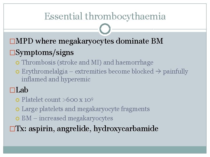
Essential thrombocythaemia �MPD where megakaryocytes dominate BM �Symptoms/signs Thrombosis (stroke and MI) and haemorrhage Erythromelalgia – extremities become blocked painfully inflamed and hyperemic �Lab Platelet count >600 x 109 Large platelets and megakaryocyte fragments BM – increased megakaryocytes �Tx: aspirin, angrelide, hydroxycarbamide
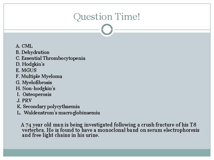
Question Time! A. CML B. Dehydration C. Essential Thrombocytopenia D. Hodgkin’s E. MGUS F. Multiple Myeloma G. Myelofibrosis H. Non-hodgkin’s I. Osteoperosis J. PRV K. Secondary polycythaemia L. Waldenstrom’s macroglobinaemia A 74 year old man is being investigated following a crush fracture of his T 8 verterbra. He is found to have a monoclonal band on serum electrophoresis and free light chains in his urine.
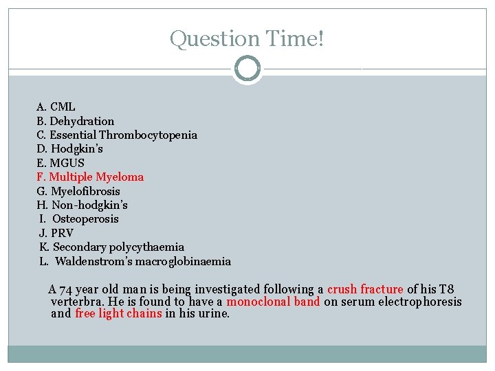
Question Time! A. CML B. Dehydration C. Essential Thrombocytopenia D. Hodgkin’s E. MGUS F. Multiple Myeloma G. Myelofibrosis H. Non-hodgkin’s I. Osteoperosis J. PRV K. Secondary polycythaemia L. Waldenstrom’s macroglobinaemia A 74 year old man is being investigated following a crush fracture of his T 8 verterbra. He is found to have a monoclonal band on serum electrophoresis and free light chains in his urine.
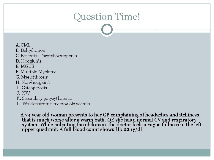
Question Time! A. CML B. Dehydration C. Essential Thrombocytopenia D. Hodgkin’s E. MGUS F. Multiple Myeloma G. Myelofibrosis H. Non-hodgkin’s I. Osteoperosis J. PRV K. Secondary polycythaemia L. Waldenstrom’s macroglobinaemia A 74 year old woman presents to her GP complaining of headaches and itchiness that is much worse after a warm bath. OE she has a normal CV and respiratory system. While palpating the abdomen, the doctor feels a vague fullness in the left upper quadrant. A full blood count shows Hb 22. 1 g/dl
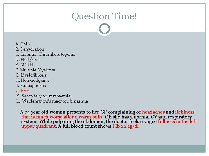
Question Time! A. CML B. Dehydration C. Essential Thrombocytopenia D. Hodgkin’s E. MGUS F. Multiple Myeloma G. Myelofibrosis H. Non-hodgkin’s I. Osteoperosis J. PRV K. Secondary polycythaemia L. Waldenstrom’s macroglobinaemia A 74 year old woman presents to her GP complaining of headaches and itchiness that is much worse after a warm bath. OE she has a normal CV and respiratory system. While palpating the abdomen, the doctor feels a vague fullness in the left upper quadrant. A full blood count shows Hb 22. 1 g/dl
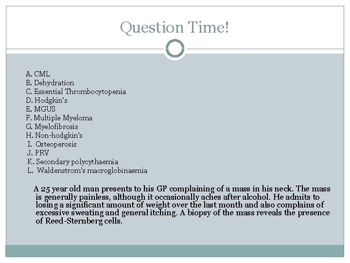
Question Time! A. CML B. Dehydration C. Essential Thrombocytopenia D. Hodgkin’s E. MGUS F. Multiple Myeloma G. Myelofibrosis H. Non-hodgkin’s I. Osteoperosis J. PRV K. Secondary polycythaemia L. Waldenstrom’s macroglobinaemia A 25 year old man presents to his GP complaining of a mass in his neck. The mass is generally painless, although it occasionally aches after alcohol. He admits to losing a significant amount of weight over the last month and also complains of excessive sweating and general itching. A biopsy of the mass reveals the presence of Reed-Sternberg cells.
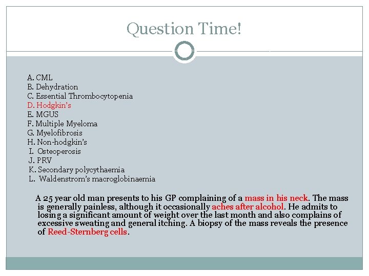
Question Time! A. CML B. Dehydration C. Essential Thrombocytopenia D. Hodgkin’s E. MGUS F. Multiple Myeloma G. Myelofibrosis H. Non-hodgkin’s I. Osteoperosis J. PRV K. Secondary polycythaemia L. Waldenstrom’s macroglobinaemia A 25 year old man presents to his GP complaining of a mass in his neck. The mass is generally painless, although it occasionally aches after alcohol. He admits to losing a significant amount of weight over the last month and also complains of excessive sweating and general itching. A biopsy of the mass reveals the presence of Reed-Sternberg cells.
Question Time! A. CML B. Dehydration C. Essential Thrombocytopenia D. Hodgkin’s E. MGUS F. Multiple Myeloma G. Myelofibrosis H. Non-hodgkin’s I. Osteoperosis J. PRV K. Secondary polycythaemia L. Waldenstrom’s macroglobinaemia A 65 year old man on long term oxygen therapy for COPD is found to have a Hb of 18. 4 g/d. L
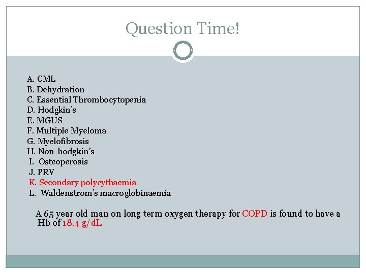
Question Time! A. CML B. Dehydration C. Essential Thrombocytopenia D. Hodgkin’s E. MGUS F. Multiple Myeloma G. Myelofibrosis H. Non-hodgkin’s I. Osteoperosis J. PRV K. Secondary polycythaemia L. Waldenstrom’s macroglobinaemia A 65 year old man on long term oxygen therapy for COPD is found to have a Hb of 18. 4 g/d. L
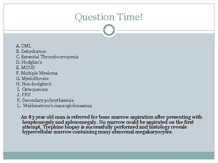
Question Time! A. CML B. Dehydration C. Essential Thrombocytopenia D. Hodgkin’s E. MGUS F. Multiple Myeloma G. Myelofibrosis H. Non-hodgkin’s I. Osteoperosis J. PRV K. Secondary polycythaemia L. Waldenstrom’s macroglobinaemia An 83 year old man is referred for bone marrow aspiration after presenting with heaptomegaly and splenomegaly. No marrow could be aspirated on the first attempt. Trephine biopsy is successfully performed and histology reveals hypercellular marrow containing many abnormal megakaryocytes.
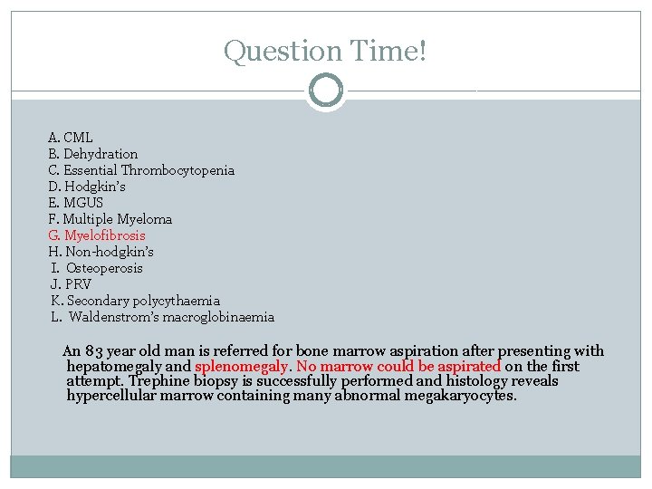
Question Time! A. CML B. Dehydration C. Essential Thrombocytopenia D. Hodgkin’s E. MGUS F. Multiple Myeloma G. Myelofibrosis H. Non-hodgkin’s I. Osteoperosis J. PRV K. Secondary polycythaemia L. Waldenstrom’s macroglobinaemia An 83 year old man is referred for bone marrow aspiration after presenting with hepatomegaly and splenomegaly. No marrow could be aspirated on the first attempt. Trephine biopsy is successfully performed and histology reveals hypercellular marrow containing many abnormal megakaryocytes.
The END! Go through the Blood Transfusion lecture Understanding is key Any questions email: �anika. kaura 09@imperial. ac. uk �sarneet. singh 09@imperial. ac. uk
- Slides: 80