Oleh Slamet Sumarno 251207 Pengantar Suction adalah satu


. n Untuk membersihkan sekret pada pasien yang")

 menimbulkan aktif sympatik dan menyebabkan")


 dan motoriknya")


 Mucouse membrane of nose : n. cranial. V")
 Glossopharyngeal n (IX) : pharynx roof, tonsil soft,")

 Sup. laryngeal n. (ext. laryngeal n. motor n. 로 cricothyroid")
 n – Diaphragm and intercostal muscles contract,")
 n Diaphragm and intercostal muscles relax decreasing the size")


 n Penyulit pemakaian : n – Masuk lewat")
 – Used to suction mouth or nose")

. n n n Bila Secret tidak dapat terhisap dianjurkan dibantu")

: 91 -5 Kozak")

 Used to maintain a patent airway only on deeply unresponsive patients")

 n n n Nose hose, nasal trumpet Used on patients who")


 >20")


 n n EMT-B can feel the lung compliance Consists of self-inflating bag,")


 n Avoid contact with petroleum")


 A breathing tube may be present n If obstructed, suction it n")

 that blocks the airway will cause some")


- Slides: 110
Oleh: Slamet Sumarno 251207
Pengantar Suction adalah satu cara untuk membersihkan jalan nafas yang mengalami hambatan karena sputum, mukus atau skret sehingga jalan nafas menjadi bersih dan kebutuhan gas dapat terpenuhi. n Suction harus dilakukan secara tepat, benar dan aman sehingga dilakukan dengan proses dan dianalisa tepat n
TUJUAN: Mempertahankan jalan nafas yang bebas (hegienis). n Untuk membersihkan sekret pada pasien yang tidak mampu batuk. n
Indikasi 1. 2. Pasien tidak mampu batuk : neonatus, tracheatomi, indotracheal tube Pasien tidak mampu batuk efektif: Retensi skret, neonatus, gagal nafas. Membersihkan berfungsi tube.
Komplikasi Parengeal suction akan merangsang syaraf sympatis (N. vagus) menimbulkan aktif sympatik dan menyebabkan bradicardi dan henti jantung dan henti nafas ( vagal reflek).
Keterangan umum/Perhatian 1. 2. 3. Sekret yang menggangu jalan nafas harus segera dikeluarkan krn dapat menyebabkan gagal nafas. Digunakan tehnik aseptik dan alat steril. Penghisapan sekret harus dilakukan dengan prosedur yang tepat untuk mencegah infeksi, luka, spasme, udema serta perdarahan jalan nafas.
Keterangan umum/Perhatian 4. 5. 6. Lama penghisapan lendir tidak boleh lebih dari: 5 -10 dt untuk bayi dan anak. 10 -15 dt untuk dewasa, Vacum presure: 8 -13, 6 k. Pa (60 -100 mm. Hg) untuk bayi. 13 – 20 (100 -120 mm. Hg) untuk anak. 20 - 27 k. Pa ( 120 -200 mm. Hg untuk dewasa. Botol penampung sekret harus diisi dengan cairan aseptik kira-kira ¼ bagian dicatat selama 24 jam dan diganti.
Keterangan umum/Perhatian 7. 8. Untuk menjegah bradikardi selama suction harus dilakukan pencegahan dengan pre suction pemberian oksigen pada pasien, Gunakan kateter suction seperti indotrache cube. Suction dapat menstimulasi batuk bila tidak ada gangguann Vagus dengan disertai batuk maka mobilisasi scret lebih mudah.
Suction pada bayi dan anak n Sebelum memberikan suction sebaiknya dibberikan Oksigenasi untuk memperbaiki Hypoksia, Inspirasi ooksigen pada gangguan nafas hanya meningkat k/l 10% pada bayi, hypoksia jangka pendek dapat menyebabkan Retinopathy (bayi prematur) (Robutan 1997)
Intervensi n n n Vacum presure diberikan tidak terlalu tinggi tetapi cukup kuat untuk menarik mukus ke luar dengan intensitas antara 8 -20 k. Pa (60150 mm. Hg). Kateter yg digunakan antara 6 -8(french gange) FG. Ukuran 5 FG atau dibawahnya tidak efektif, ukuran >10 untuk anak dan dewasa >10 Th Diameter kateter tidak digunakan 50% dari diameter jalan nafas.
Prosedur 1. 2. 3. 4. 5. 6. Terangkan prosedur yang akan dilakukan pada pasien. Letakkan alat-alat disamping tempat tidur pasien. Jika mungkin buat posisi semi fowler. Cuci tangan anda dengan aseptik. Hidupkan sumber penghisap dengan tekanan sesuai kebutuhan. Ukur kateter sepanjang ujung hidung sampai telinga.
Membersihkan melalui mulut. n n n Hubungkan sumber penghisap dinding dengan penghisap logam. Masukkan penghisap logam kedalam mulut tanpa memberi tekanan penghisap, kemudian lakukan penghisapan sekret dengan hati-hati. Hindarkan mata pasien dari percikan sekret jalan nafas. Bersihkan kateter logam dengan larutan steril. Berikan oksigen pada pasien dan lakukan penghisapan lagi bila perlu.
Membersihkan memalui hidung. n n n Hubungkan sumber penghisap dengan kateter penghisap. Berikan pelicin pada ujung kateter penghisap. Masukkan kateter penghisap melalui lubang hidung atau dapat juga melalui mulut dengan hati-hati tanpa memberikan tekanan pada penghisap, kemudian lubang penghisap ditutup dan sekret dihisap sambil kateter ditarik perlahan-lahan. Bersihkan kateter penghisap dengan larutan steril. Berikan oksigen pada pasien dan lakukan pengisapan lagi bila perlu.
Untuk pasien dengan pipa entratrakea terpasang n n n Perlu 2 orang untuk penghisapan bila mungkin, terutama pada anak yang aktif. Satu memberikan oksigenasi. Tekan tombal alrm ventilator jika perlu. Lepaskan hubungan dengan sirkuit ventilator atau pipa humidifer jika pasien mempergunakan alat tersebut. Gunakan ballon pemompa dan beri oksigen 3 -5 kali inflasi dengan konsentrasi F 1 O 2: ………
= 10% lebih besar dari konsentrasi O 2 yang digunakan untuk neonatus dengan berat badan kurang dari 3 kg. =100% untuk pasien lain kecuali ada ketentuan lain. Selama prosedur, jaga agar gerakan dada tetap adekuat dan jika diberikan ventilator jaga agar tetap dalam tekanan positif, Hindari tekanan yang berlebihan. Bila berat badan bayi kurang dari 2 kg harus dipasang pengukur tekanan pada sirkuit.
Orang ke dua. melakukan penghisapan n n Hidupkan penghisap dinding dengan tekanan sesuai kebutuhan. Pasang sarung tangan dan letakkan pengalas diatas dada pasien. Hubungkan penghisap dengan kateter penghisap dengan tangan kanan. Jaga agar kateter tetap steril. Masukkan kateter kedalam lumen pipa trakea dengan cepat sejauh mungkin tanpa dipaksa, dengan lubang kateter terbuka dalam keadaan tidak menghisap.
n n n Lakukan penghisapan sekret dengan menutup lubang penghisap yang ada disamping. Kateter penghisap ditarik perlahan-lahan untuk beberapa cm pertama, kemudian ditarik secara cepat sambil diputar (rotasi). Bila kateter sulit ditarik mungkin menempel pada dinding bronkus, buka lubang penghisap dan ulangi penghisapan dengan tekanan lebih rendah. Selama penghisapan pipa endotrakea dipegang dengan tangan kiri untuk mempertahankan posisi. Hubungkan pipa endotrakea dengan balon pemompa dan berikan oksigen.
n n n n Pada neonatus dengan berat badan kurang dari 3 kg konsentrasi O 2 10% lebih tinggi dari yang sedang digunakan. Pada pasien yang lain berikan oksigen 100% kecuali ada intruksi khusus. Ulangi seluruh prosedur dengan tehnik yang sama sampai jalan nafas relatif bersih dari sekret. Selama penghisapan perhatikan warna kulit dan denyut nadi pasien. Bila terjadi kelainan hentikan penghisapan dan berikan ventilasi dengan segera. Kateter penghisap dari endotrakea boleh digunakan untuk dari mulutatau hidung sebaiknya tidak boleh. Kembalikan pasien ke sirkuit ventilator atau pipa humidifer bila sebelumnya tidak digunakan. Matikan sumber penghisap, slang dalam keadaan bersih Peralatan dikembalikan dalam keadaan bersih dan pasien dalam posisi semula.
Frekuensi penghisapan n 1. 2. 3. 4. 5. Dilakukan tiap 2 jam bila perlu. Atau setelah dilakukan chest fisioterapi. Segera laporkan bila terdapat: Kesulitan memasukkan kateter penghisap. Bila skret sudah tidak bisa dihisap. Sekret yang pekat dan banyak. Sekret campur darah, berbusa dan atau berbau. Penderita sianotik, keadaan menurun, apnnu dll
Hal-hal yg perlu dicatat Waktu penghisapan. 2. Keadaan skret: jumlah, warna, bau dan konsentrasi 3. Hal-hal yang terjadi selama penghisapan: Posisi, keadaan selama penghisapan: Hipoksia, bradikardi 1.
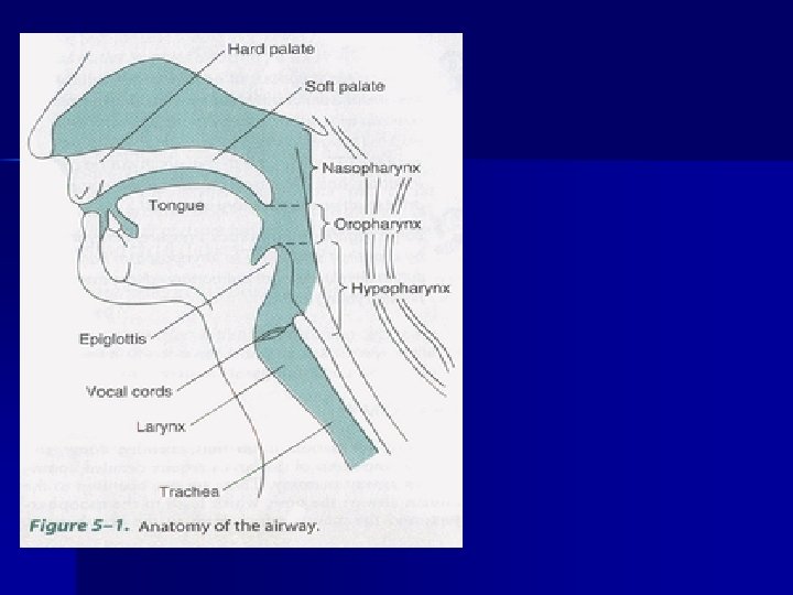
Respiratory Anatomy n n n Nose and mouth : Lungs (Menghangatkan, Visceral pleura (surface of melembabkan dan menyaring lungs) udara). Parietal pleura (internal Pharynx chest wall) – Oropharynx – Nasopharynx Interpleural space Epiglottis (potential space) Trachea (windpipe Cricoid cartilage Larynx (voice box). Bronchi
Larynx
Upper Airway Epiglottis Tongue Glottis
Lower Airway
Sistem persarafan pada jalan nafas
V - Trigeminal Nerve Sensoriknya ke daerah wajah (touch, pain and temperature) dan motoriknya ke Temporal, massester Bila terganggu n. V sensasi & motorik terganggu or dapat menimbulkan nyeri = trigeminal
Trigemin al neuralgi n a
VII - Facial Nerve n n Ekpresi wajah, sensasi lidah anterior 2/3’s, salivary glands and tear, nasal & palatine glands Kerusakan otot-otot fasial & penyampaian sensasi(missing sweet & salty) called Bell’s Palsy
IX - Glossopharyngeal Nerve n n Provides control over swallowing, salivation, gagging, sensations from posterior 1/3 of tongue, control of BP and respiration Damage results in loss of bitter & sour taste & impaired swallowing.
X - Vagus Nerve The wonderer n Provides swallowing, speech, regulation of 2/3 of GI tract n Damage causes impaired voice, n
Sensory supply of upper airway 1) Mucouse membrane of nose : n. cranial. V (trigeminal. ) ophthalmic div. V 1 (ant. Ethmoidal n. ) : ant. part of nose maxillary div. V 2 (sphenopaltine n. ) : post. part of nose 2) Soft and hard palate : palatine n. 3) Tongue br. of mandibular div V 3 (lingual n. ) : ant. 2/3 general sensation dan 1/3 oleh n. IX (glossopharyngeal n. ) branch of VII ( facial n. ) & IX : sensation of taste
Sensory supply of upper airway 4) Glossopharyngeal n (IX) : pharynx roof, tonsil soft, palate under-surface innervation 5) Vagus n. (X) : epiglottis , airway , sensation Sup. Laryngeal br. : ext. br (motor) int. br (sensory) epiglottis, vocal cord , sensory supply Recurrent laryngeal n. : vocal cord , trachea , sensory supply
ANATOMY
Laryngeal n. injury 1) Sup. laryngeal n. (ext. laryngeal n. motor n. 로 cricothyroid m) unilat. : minimal effect bilat. : hoaresness, tiring of voice (but airway effect voice) 2) Recurrent laryngeal n. unilat. : ipsilat. Vocal cord paralysis (voice quality ) bilat. : acute : stridor, respiratory distress chronic : aphonia 3) Vagus n. unilat. : hoarseness bilat. : aphonia
Respiratory physiology Diaphragm n Inhalation (active process) n – Diaphragm and intercostal muscles contract, increasing the size of the thoracic cavity. – Diaphragm moves slightly downward, ribs move upward and outward. n The negative pressure in the chest cavity causes air flow into the lungs.
Respiratory physiology Exhalation (passive process) n Diaphragm and intercostal muscles relax decreasing the size of the thoracic cavity. n – Diaphragm moves upward, ribs move downward and inward. n The positive pressure inside the chest cavity causes air flow out of the lungs.
Respiratory Physiology n n n Oxygenation - blood and the cells become saturated with oxygen Hypoxia - inadequate oxygen levels in the blood Signs of Hypoxia – – – Increased or decreased heart rate Altered mental status (early sign) Agitation Initial elevation of B. P. followed by a decrease Cyanosis (often a late sign)
Alveolar Gas Exchange Oxygen-rich air enters the alveoli during each inspiration. n Oxygen enters the blood in the capillaries as carbon dioxide enters the alveoli for exhalation. n
Infant and Child Considerations n n n Mouth and nose - generally all structures are smaller and more easily obstructed than in adults. Pharynx - infant’s and children’s tongues take up proportionally more space in the mouth than adults. Trachea - (windpipe) – Infants and children have narrower tracheas that are obstructed more easily by swelling. – Trachea is softer and more flexible in infants and children.
Infant and Child Considerations n n Cricoid cartilage - like other cartilage in the infant and child, the cricoid cartilage is less developed and less rigid. It is the narrowest part of the infant’s or child’s airway. Diaphragm - chest wall is softer, infants and children tend to depend more heavily on the diaphragm for breathing.
Opening the Mouth Crossed-finger technique n Inspect the mouth n – Vomit – Blood – Secretions – Foreign bodies n Be extremely cautious – Fingers – Gag or vomit
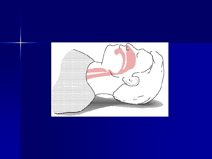
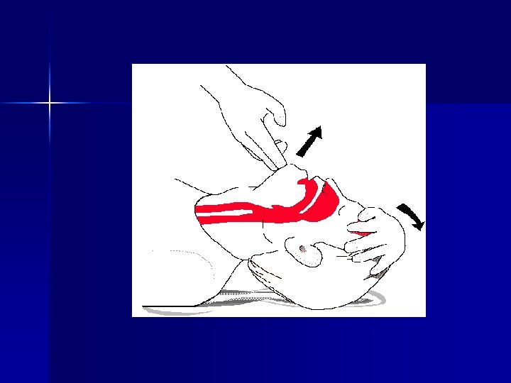
Opening the Airway n Head-tilt, chin lift maneuver – Adults vs. . Infants and Children n Jaw thrust maneuver
Techniques of Suctioning Dasar intervensi suction: precautions n Maksud/ tujuan n – Mengalirkan benda asing dlm saluran nafas spt: blood, liquids, and food particles. – Patient needs to be suctioned dengan segera saat pasien ada suara meneguk
Types of Suction Units 1. Mounted Suction Devices – Fixed on-board the ambulance – 300 mm. Hg pull on gauge when tubing is clamped – Should be adjustable (disesuaikan) for infants and children
2. Portable Suction Devices Electric - battery powered n Oxygen - powered n Hand - powered n Each device must have n – Wide-bore, thick walled, nonkink tubing – Plastic collection bottle, supply of water – Enough vacuum to clear the throat
3. Suction Catheters Catheter keras (yankaeur) n Penyulit pemakaian : n – Masuk lewat mulut. – Terhambat Tonsil dan lidah. – Used to suction mouth and oropharynx – Inserted a limited depth – Use caution on infants and children n Soft tissue damage
Suction Catheters n Soft catheter (French catheter) – Used to suction mouth or nose and nasopharynx – Measured from tip of the nose to the tip of the ear. – Not inserted beyond the base of the tongue
Techniques of Suctioning n Posisikan pasienn yg baik terlentang atau miring kan kepala dan badan bila bayi/anak. (Best positioned at patient’s head) n n Perhatikan perubahan suction unit Pilih catheter yg pas ukurannya, diameter catheter 50% Diameter jalan nafas. Ukur/periksa dan berapa dalam chateter dapat masuk Suction from side to side – Dewasa tidak lebih 15 seconds – Infants & children Kurang dari 15 seconds (5 -10) n Bilas catheter dengan air desinfectance.
Special Considerations (pertimbangan khusus). n n n Bila Secret tidak dapat terhisap dianjurkan dibantu dengan dibersihkan dengan jari. Patient producing frothy secretions as rapidly as suctioning can remove them – Suction 15 seconds – Positive pressure with supplemental oxygen for 2 minutes then suction again and repeat the process Residual air removed from lungs, monitor pulse and heart rate
Suction n The importance of readiness can not be overstated.
Study of suction equipment utilization. Prehosp Emerg Care 1997 Apr-Jun; 1(2): 91 -5 Kozak RJ, Ginther BE, Bean Study of suction equipment utilization. The paramedics reported: carrying suction equipment to the scene of medical aid calls less than 25% of the time. suction equipment is utilized during 50% of advanced airway procedures. WS.
Suction - Key Points Diingatkan agar memahami dasar -dasar pemberian suction. n Suctions are limited in what they remove n Immediate action is needed n Have a secondary device n
Oropharyngeal Airway (OPA) Used to maintain a patent airway only on deeply unresponsive patients n No gag reflex n Designed to allow suctioning while in place n Must have the proper size n If patient becomes responsive and starts to fight the OPA remove it. . . n
Inserting the OPA n n n Select the proper size (corner of the mouth to tip of the ear) Open the patient’s mouth Insert the OPA with the tip facing the roof of the mouth Advance while rotating 180° Continue until flange rests on the teeth Infants and children insertion
Nasopharyngeal Airway (NPA) n n n Nose hose, nasal trumpet Used on patients who are unable to tolerate an OPA or is not fully responsive Do not use on suspected basilar skull fracture Still need to maintain head-tilt chin lift or jaw thrust when inserted Must select the proper size Made to go into right nare or nostril
Inserting the NPA Select the proper size in length and diameter n Lubricate n Insert into right nostril with bevel always toward the septum n Continue inserting until flange rests against the nostril n Insertion into left nostril n
Assessment of Breathing After establishing an airway your next step should be to assess breathing n Look n – Breathing pattern regular or irregular – Nasal flaring – Adequate expansion, retractions
Assessment of Breathing n Listen – Shortness of breath when speaking – Unresponsive place ear next to patients mouth – Is there any movement of air?
Assessment of Breathing n Feel – Check the volume of breathing by placing you ear and cheek next to the patient’s mouth
Assessment of Breathing n Auscultate – Stethoscope n Mid clavicular about the second intercostal space and the fourth or fifth anterior midaxillary line or next to sternum – Check both sides n Present and equal bilaterally n Diminished or absent
Adequate Breathing n Normal rate – Adult 12 - 20/min – Child 15 - 30/min – Infant 25 - 50/min n Rhythm – Regular – Irregular
Ventilation Volume Tidal volume-air inspired in each breath n Minute volume-tidal volume multiplied by the respiratory rate n
Adequate Breathing n Quality – Breath sounds present and equal – Chest expansion adequate and equal – Effort of breathing n use of accessory muscles predominately in infants and children n Depth (tidal volume) – Adequate chest rise and fall – Full breath sounds heard
Inadequate Breathing n Rate – Outside the normal limits n Tachypnea (rapid breathing) >20 n Badypnea (slow breathing) <12 n Rhythm – Irregular breathing pattern
Inadequate Breathing n Quality – – – n Breath sounds diminished, noisy or absent Excessive use of accessory muscles, retractions Reduced air flow at nose/mouth Inadequate chest expansion Nostril flaring (infants & children) Depth – Shallow (impaired depth) breathing – Agonal respirations - occasional gasping respirations
Inadequate Breathing Skin Color n Retractions n “Seesaw” breathing (abd & chest move in opposite directions) n n Any of these signs is by itself may be reason to ventilate a patient without delay
Positive Pressure ventilation n The practice of artificially ventilating, or forcing air into a patient who is breathing inadequately or not breathing at all
Techniques of Artificial Ventilation n In order of preference – Mouth to mask – Two-person bag-valve-mask – Flow-restricted oxygen-powered ventilation device – One-person bag-valve-mask
Considerations When Using Artificial Ventilation Maintain a good mask seal n Device must deliver adequate volume of air to sufficiently inflate the lungs n Supplemental oxygen must be used n
Adequate Artificial Ventilations Chest rises and falls with each ventilation n Rate of ventilations are sufficient n Heart rate returns to normal n Color improves n
Inadequate Artificial Ventilations Chest does not rise and fall n Ventilation rate is too fast or slow n Heart rate does not return to normal n Color is not improved n
Mouth-to-Mouth Ventilation Air we breath contains 21% oxygen n 5% used by the body n 16% is exhaled n Danger of infectious disease n
Mouth-to-Mask Eliminates direct contact with patient n One-way valve system n Can provide adequate or greater volume than a BVM n Oxygen port (should be connected to 15 lpm) n
Bag-Valve-Mask (BVM) n n EMT-B can feel the lung compliance Consists of self-inflating bag, one-way valve, face mask, intake/oxygen reservoir valve, and an oxygen reservoir. By adding oxygen and a reservoir close to 100% oxygen can be delivered to the patient When using a BVM an OPA/NPA should be used if possible
Bag-Valve-Mask Cont. . . n n n Volume of approximately 1, 600 milliliters Provides less volume than mouth-to-mask Single EMT may have trouble maintaining seal Two EMT’s more effective Pop-off valve must be disabled Available in infant, child, and adult sizes
Bag-Valve-Mask Cont. . . Breaths should be 1. 5 to 2 seconds n Guard against overinflation n Monitor the seal n Bring the jaw to the mask n
Bag-Valve-Mask Cont. . . n Assisted ventilations for hyper or hypoventilating patients – Explain procedure – Place the mask – Squeeze bag on inhalation – Over next 5 to 10 breaths slowly adjust rate and tidal volume to desired rate and volume
Sellick Maneuver
Sellick Maneuver
Mask ventilation will be made difficult by: n n n n n poor mask seal -- beards facial burns facial scarring/cuts facial dressings edentulous patients any evidence of airway obstruction neck instability penetrating neck trauma repeated failed direct laryngoscopy obesity/bull neck
Other ventilation techniques will be made difficult by: • lack of knowledge and experience • lower airway obstruction • neck instability • penetrating neck injury
Flow-Restricted, Oxygen. Powered Ventilation Device n n n n Known as a demand-valve device Can be operated by patient or EMT Unable to feel lung compliance With proper seal will deliver 100% oxygen Designed for use on adult patients Gastric distension Rupture of the lungs A trigger positioned to allow EMT to keep both hands on the mask
Automatic Transport Ventilators n n Deliver 100% oxygen Provide and maintain a constant rate and tidal volume during ventilation Advantages – Frees both hands – Rate, & tidal volume can be set – Alarm for low oxygen tank Disadvantages – Oxygen powered – not used in children under 5 – Cannot feel increase in airway resistance
Oxygen Therapy Oxygen is a drug that can be given by the EMT-B n “Generally speaking”, a patient who is breathing less than 12 and more than 24 times a minute needs oxygen n
Oxygen Dangers Oxygen supports combustion, (it is not flammable) n Avoid contact with petroleum products n Smoking n Handle carefully since contents are under pressure n
Oxygen Cylinders All of the cylinders when full are the same pressure of 2, 000 psi. n Usually green or aluminum grey n D cylinder - 350 liters n E cylinders - 625 liters n M cylinders - 3, 000 liters n G cylinders - 5, 300 liters n H cylinders - 6, 900 liters n
High-Pressure Regulator Provides 50 psi to an oxygen-powered, ventilation device. n Flow rate cannot be controlled n
Low Pressure/Therapy Regulator Permit oxygen delivery to the patient at a desired rate in liters per minute n Flow rate can go from 1 to 25 liters/min. n
Oxygen Humidifiers Dry oxygen is not harmful in the short term n Generally not needed in prehospital care n Transport time of an hour or more humidifier should be considered n
Changing Oxygen Bottle n n n Check cylinder for oxygen remove protective seal Quickly open and shut tank to remove debris Place regulator over yoke and align pins. Make sure new O ring is in place Hand tighten the T screw Open to check for leaks
Nonrebreather Mask n n n n Preferred method of giving oxygen to prehospital patients Up to 90% oxygen can be delivered Bag should be filled before placing on patient Flow rate should be adjusted to 15 liters/min. Patients who are cyanotic, cool, clammy or short of breath need oxygen Concerns of too much oxygen Different size masks
Nasal Cannula n n n Provides limited oxygen concentration Used when patients cannot tolerate mask Prongs and other uses Concentration of 24 to 44% Flow rate set between 1 to 6 liters For every liter per minute of flow delivered, the oxygen concentration the patient inhales increases by 4%
Nasal Cannula Flow Rates 1 liters/min. =24% n 2 liters/min. = 28% n 3 liters/min. = 32% n 4 liters/min. = 36% n 5 liters/min. = 40% n 6 liters/min. = 44% n
Simple Face Mask No reservoir n Can deliver up to 60% concentration n Rate 6 to 10 liters/min. n Not recommended for prehospital use n
Partial Rebreather Mask Similar to nonrebreather except it has a two-way valve allowing patient to rebreath his exhaled air. n Flow rate 6 to 10 liters/min. n Oxygen concentration between 35 to 60% n
Venturi Mask Provides precise concentrations of oxygen n Entrainment valve to adjust oxygen delivery n Mostly used in the hospital setting for COPD patients n
Special Situations
Inhaler Therapy History n Medical Direction n Review of specific bronchodilator medication n
Laryngectomies (Stomas) A breathing tube may be present n If obstructed, suction it n Some patients may have partial laryngectomies n Be sure to close the mouth and nose to prevent air escaping n
Infants and Child Patients n n n Neutral position infant Just a little past neutral for child Avoid hyperextension of head Avoid excessive BVM pressure Gastric distension more common in children Oral or nasal airway may be considered when other procedures fail to clear the airway
Obstruction Anything (food, blood, swollen tissue, vomit) that blocks the airway will cause some level of decrease of available oxygen to the body.
Obstruction The size of obstruction affects the available air exchange. For example, snoring will reduce air Exchange while a food bolus can actually stop air exchange.
Obstruction n When obstruction persists, repeat FBAO procedures three times and transport as soon as possible.
Facial Injuries Rich blood supply to the face n Blunt injuries and burns to the face result in severe swelling n Bleeding into the airway can be a challenge to manage n
Jaw Thrust
Technique Suction. n n n n Gunakan cateter steril untuk mencegah nasocomial (infeksi dirumah sakit). Sebelum suction informasikan ke keluarga/pasien apa maksud anda melakukan suction. Siapkan alat siap pakai. Sebelum dan sesudah suction dapat diberikan terapi oksigen untuk mencegah vagal reflek. Lakukan dengan waktu suction 3 -5 detik untuk anak dan 5 -10 atau 10 -15 detik untuk dewasa. Vacum presure =60 -150 mm. Hg untuk dewasa dan 40 -80 mm. Hg untuk anak ( 8 - 20 k. Pa dewasa) 6 -12 k. Pa anak (Tergantung kepekatan mukus). Dengan nafas dalam akan merangsang batuk pada sebagian pasien.