OLEH SLAMET SUMARNO Disampaikan pada seminar sehari DISELENGGARAKAN


 is the proses by which air is moved into and out")


 2. O 2 &")

 1. Lymphatic structures 1. Capillaries with valves 2.")


- Slides: 48
OLEH: SLAMET SUMARNO Disampaikan pada seminar sehari DISELENGGARAKAN : AKADEMI FISIOTERAPI UKI TGL: 15 SEPTEMBER 2007.
Kurikulum vite • Slamet Sumarno, Drs, SMPh • Wonogiri : 15 -09 -1950 • Dik. S 1 1992 Unis Syeh Yusuf Tangerang : Administrasi Negara • Akademi Fisioterapi Surakarta 1975 • Kardia Respirasi : Revalidasi Centrum Ultreh Nedherland 1983
Pekerjaan. • Fisioterapi Rumkit Polpus RS Sukanto Kramat jati Jak Tim 1976 – Sekarang. • Dosen Akfis UPN 1983 – Sekarang • Dosen Akfis Uki 1986 – Sekarang • Dosen Fakultas Fisioterapi Indonusa Esainggil 1998 • Dosen Fakultas Fisioterapi STIKES Binawan 2004 • Dosen Akbid Sukawangi 2004
Agar peserta dapat memahami: • Mekanisme pernafasan dan sirkulasi normal. • Fungsi kardiovakuler dan respirasi. • Kapasitas pernafasan dan tranportasi oksigen normal. • Mampu mengaplikasikan kordio vaskuler respirasi dalam penilaian klien/ pasien dengan baik dan benar. • Mampu menerapkan dalam gangguan kordivaskulerrespirasi khususnya dan Neuromuskular dan muskuloskeletal maupun integumen.
• We cannot live without oxygen. • Anda tidak bisa hidup tanpa oksigen • All living cells require to get oxygen from the fluid around them and to get rid of corbon dioxide to it.
1. Exchange O 2 1. Air to blood 2. Blood to cells 2. Exchange CO 2 1. Cells to blood 2. Blood to air 3. Regulate blood p. H 4. Vocalizations 5. Protect alveoli External and cellular respiration
Oksigen = 20, 93 % Carbon diaksida= 0, 03 % Nitrogen = 79, 04 % TEKANAN OKSIGEN Di Daerah SEA LEVEL
Pulmonary ventilation (breathing) is the proses by which air is moved into and out of the lungs. It has two phases: inspiration and expiration.
Proses pernafasan. 1. External Respirasi a. Inspirasi. b. Ekspirasi. 2. Internal Respirasi. a. Tranportasi (sirkulasi). b. Buffer
Inspirasi is an active process in which the diaphragm and the external intercostal muscles increase the dimensions, and thus the volume, of the thoracic cage. This decreases the pressure in the lungs and draws air in. Tekanan alveoli minus 3 s/d 10 mm. Hg < dari 760 mm. Hg.
Normal expiration is the passive process. The inspiratory muscle relax and the elastic tissue of the lungs recoils, returning the thoracic cage to its smaller, normal dimensions. This increases the presure in the lungs and forces air out.
q Forced or labored inspiration and expiration are active proceses, dependent on muscle actions. q Aktif inspirasi dibantu otot: Scalani, Sterno cledo mastoideus, pektoralis, membantu angkat sternum dan costae ke depan atas. Tekanan alveoli turun sampai 100 -80 mm. Hg. q Aktif ekspirasi dibantu otot : Abdominalis, Latisimus dorsi Inspirasi. Ekspirasi
1. Tranportasinya Exchange O 2 in cells a. Air to blood b. Blood to cells 2. Exchange CO 2 1. Cells to blood 2. Blood to air 3. Buffer
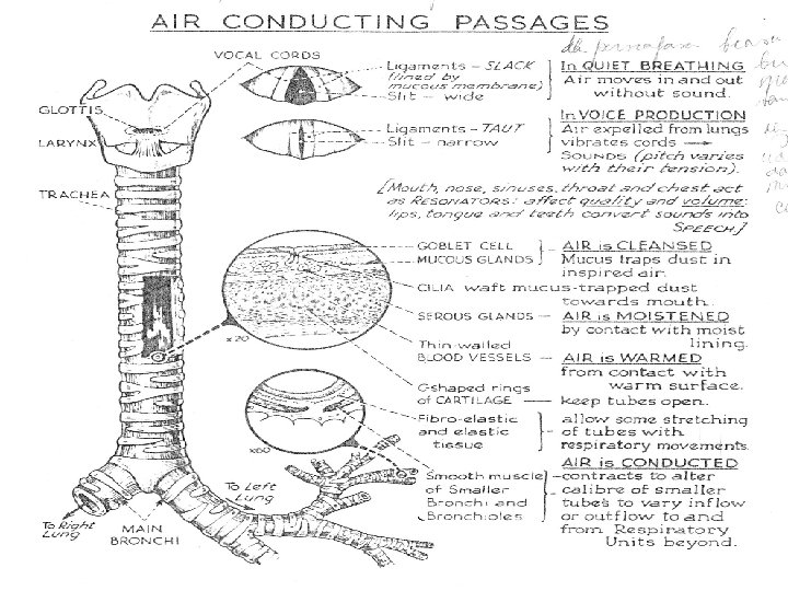
Ruang rugi anatomi dan fisiologi Ruang rugi Anatomi: Gas yanga Ada pada Jalan nafas 150 CC Ruang rugi Fisiologi: Keterbatasan Ventilasi Paru atau Raung residu Yang tinggi
DAERAH PERTUKARAN O 2 Dan CO 2: 1. Brokus respiratoris 2. Ducctus 3. Succus dan 4. Alveoli
1 Pertahanan Respiratory system Inhalasi melalui hidung Mempunyai keuntungan 1. Udara disaring. 2. Udara dilembabkan. 3. Udara dihangatkan. 4. Cegah peradangan jalan nafas bag atas.
2. Pembersih jalan nafas oleh cillia dan fungsi menelan
Perfusi eksterna Eksteranal respirasi: Dalam paru Dipengaruhi: respirasi 1. Cairan surfaktan 2. Tebal membran. 3. Volume darah 4. Hb darah 5. Kecepatan sirkulasi. 6. Vicositas darah. 7. p. H darah
Tranportasi O 2 dan CO 2 dalam darah • Kemampuan jantung (COP= HR X Stroke volume). • Tahanan perifer. • Fleksibilitas pembuluh darah. • Lumen. • Vikositas. • Hb. • Saturasi darah dll.
Ventilation: The Pumps 1. Inspiration 2. Expiration 3. Diaphragm 1. Low energy pump 2. Concavity – flattens 4. Thorax: ribs & muscles 5. Pleura: double membrane 1. Vacuum seal 2. Fluid-lubrication
Ventilation: The Pumps Pada tekanan rendah bila dua gelembung udara mempunyai tension Permukaan yg sama , Gelembung kecil mempunyai tekanan besar Surfactant reduces surface tension
Respiratory Damage & Diseases Pada tekanan rendah bila dua gelembung udara mempunyai tension Permukaan yg sama , Gelembung kecil mempunyai tekanan besar Surfactant reduces surface tension
Pengaruhi internal terhadap kemampuan tubuh suply O 2 1. Jalan nafas yang adeguat. 2. Fleksibilitan otot, fasia, kulit sangkar thorax. 3. Perfusi O 2 dan CO 2 4. Volume, Hb, Ph, Plasma darah 5. Keadan pleurae 6. Pusat kontrol fungsi pernafasan 7. Fungsi bufer.
Pengaruh eksternal terhadap kemampuan tubuh suply O 2 1. Kadar Oksigen udara. 2. Posisi tubuh 3. Usia jenis kelamin.
Factors Affecting Ventilation 1. 2. 3. 4. 5. 6. Airway Resistance Diameter Mucous blockage Bronchoconstriction Bronchodilation Alveolar compliance 1. Surfactants 2. Surface tension 7. Alveolar elasticity
1. Lung volumes , Volume paru a. RV b. ERV c. TV d. IRV 2. Capasitas paru a. Capasitas inspirasi b. Capasitas Ekspirasi c. Capasitas vital d. Capasitas total paru. e. Kapasitas residual 1200 cc 1000 500 1900 -3300 PRIA WANITA VCI 3, 3 1, 9 TV 0, 5 VCE 1, 0 0, 7 VR 1, 2 1, 1 V TOTAL 6, 0 4, 2
Clinical Cardiovascular Anatomy & Physiology Concepts, Definitions, & Principles
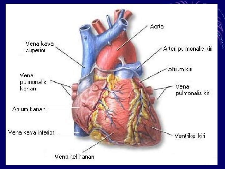
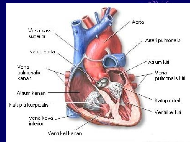
Jantung A. LETAK. B. UKURAN. C. LAPISAN JANTUNG. D. RONGGA. E. KATUP DAN SEKAT. F. SYARAF JANTUNG
Pumpa jantung Agar darah sampai pada sel Tubuh harus dipompa jantung Menuju jaringan, termasuk Jaringan jantung sendiri. Jumlah darah yang dipompa jantung dipengaruhi: Strooke vulume dan frekuensi Denyut jantung per menit. Makin tinggi strooke vulumee Dan frekuensi makin tingg COP Tetapi makin tinggi frekuensi Jantung makin sedikit aliran Koroner yang memberi nutresi Jantung.
Efisiensi kerja jantung. Makin kuat dan fleksibel otot Jantung , strooke volume Makin besar dan makin hemat energi kerja jantung Sehingga frekuensi jantung Permenit makin kecil. Termasuk tahanan perifer Sistem sirkulasi Tahanan perifer meliputi: 1. Vulume pembuluh darah 2. Vikositas darah 3. Trammister 4. Jenis Kerja otot.
MICRO CIRKULASI ANATOMI
Pusat kontrol cardio vaskuler respirasi
Conduction System of the Heart
Tranportasi darah dan O 2 CO 2 1. Kemampuan jantung (COP= HR X Stroke volume). 2. Tahanan perifer. 3. Fleksibilitas pembuluh darah. 4. Lumen. 5. Vikositas. 6. Hb. 7. Saturasi darah. 8. Sistem konduksi
Blood Components: Plasma Transports Solutes 1. Water, ions, trace elements 2. Gasses: O 2 & CO 2 3. Organic Molecules 1. 2. 3. 4. 5. Glucose N–wastes Proteins Antibodies Hormones
Blood Components: Plasma Transports Solutes
Blood Compon ents: "Blood Count" – % of Each Compon ent
Blood Components: Cells 1. Erythrocytes 1. Red Blood Cells (RBC) 2. O 2 & CO 2 transport 2. White Blood Cells (WBC) 1. Immune defense 2. Phagocytosis 3. Platelets: clotting
Vicositas darah
Lymphatic System: Structure and Roles (overview) 1. Lymphatic structures 1. Capillaries with valves 2. Lymph vessels 3. Lymph nodes & organs 2. Immune defense: lymphocytes 3. Transport of fats 4. Collects excess ECF 1. Returns to plasma 2. Edema
System lymphe
System Aliran Lymphe Net Out Flow Into ECF Hubungan antara kapiler dan pembuluh lymphe Aliran air dan cairan difilter keluar oleh kapiler Ke atas oleh pembuluh lymphe dan masuk sirkulasi • Net filtration – net absorption = net out flow • About 2 L/day collected by lymph vessels Figure 15 -18 b: Fluid exchange at the capillary