Neck Space Infections Dr Vishal Sharma Fascial layers


 & covered")


: 1. Poor dental hygiene (caries & abscess)")


: due")

 Anterior: Buccopharyngeal fascia")


 1. Widened pre-vertebral soft tissue shadow a. > 7")


: homogenous opacity")


")


 to a dehydrated parotid gland")
 2. Medications that decrease salivary")


- Slides: 99
Neck Space Infections Dr. Vishal Sharma
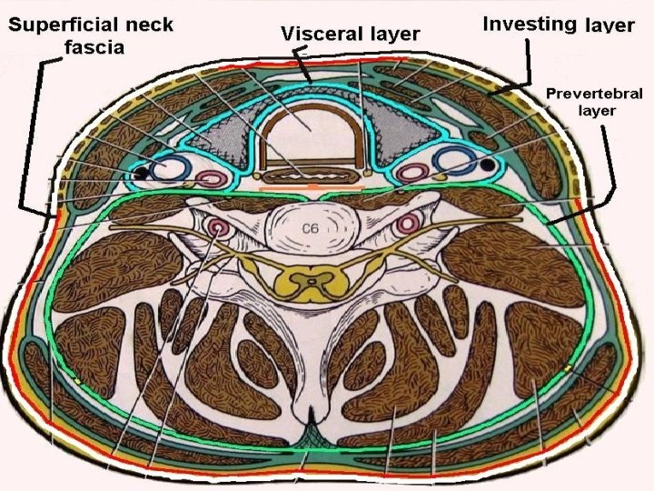
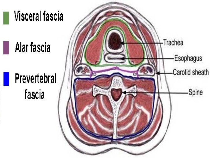
Fascial layers of neck A. Superficial cervical fascia: encloses platysma B. Deep cervical fascia 1. Superficial or Investing layer 2. Middle layer 3. Deep layer a. Muscular division a. Alar fascia b. Visceral division b. Pre-vertebral fascia
Deep Cervical Fascia Investing layer: Encloses trapezius & SCM; parotid, submandibular gland & carotid sheath Visceral layer: Surrounds strap muscles, pharynx, larynx, esophagus, trachea, thyroid Deep layer: Covers deep neck muscles, cervical plexus, phrenic nerve & brachial plexus. Cervical sympathetic chain lies superficial to this fascia.
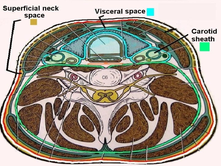
Classification of neck spaces
A. Involves entire neck B. Spaces above hyoid 1. Superficial neck space 1. Submental 2. Deep neck spaces 2. Submandibular a. Carotid sheath a. Sublingual b. Retro-pharyngeal b. Submaxillary c. Danger space 3. Masticator d. Pre-vertebral 4. Parotid C. Below Hyoid 5. Parapharyngeal 1. Pre-tracheal space 6. Peri-tonsillar
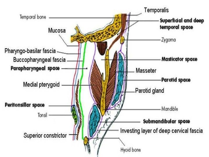
Masticator spaces Formed around muscles of mastication (masseter, pterygoids, insertion of temporalis) & covered by investing layer of deep cervical fascia
Classification of neck space infections
A. Involves entire neck B. Supra-hyoid abscess 1. Superficial space Sub-mental Necrotizing fascitis Masticator 2. Deep space abscess Parotid Carotid sheath Ludwig’s angina Retro-pharyngeal Para-pharyngeal Danger space Peri-tonsillar (quinsy) Pre-vertebral C. Infra-hyoid abscess Pre-tracheal
Necrotizing fasciitis
p Rare infection of superficial neck space causing necrosis of fascia + subcutaneous tissue, initially sparing skin & muscle p Term coined in 1952 by Wilson p Etiology: Dental infections, skin trauma, quinsy & parapharyngeal abscess p Bacteriology: β-hemolytic streptococcus, Staphylococcus aureus, anaerobes
Clinical Presentation p Outer zone of erythema, intermediate zone of tender ecchymosis & central zone of vesiculation + black necrosis + ulceration p Fascial necrosis extends beyond skin necrosis p Skin anesthesia (damage of cutaneous nerves) p Soft tissue crepitus due to gas formation p Hypocalcemia, hyponatremia & dehydration
Necrotizing fasciitis of chest
CT scan showing gas formation
Treatment p Early correction of fluid & electrolyte imbalance p I. V. Ampicillin + Gentamicin + Clindamycin p Immediate radical debridement of necrotic tissue (in presence of subcutaneous air, progressive infection despite 48 hours of medical therapy, obvious fluctuation or skin necrosis) p Skin grafting after debridement
Wound debridement
Skin grafting
Healed wound
p Poor prognostic factors: Diabetes mellitus, atherosclerosis, chronic renal failure, obesity, immuno-suppression, malnutrition p Complications: necrosis of chest wall fascia, mediastinitis, pleural effusion, pericardial effusion, empyema, airway obstruction, arterial erosion, jugular vein thrombophlebitis, septic shock, lung abscess, carotid artery thrombosis
Ludwig’s Angina
Rapidly progressing poly-microbial cellulitis of sublingual & submaxillary spaces with potentially life-threatening airway compromise
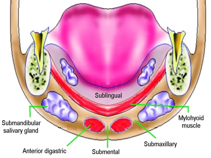
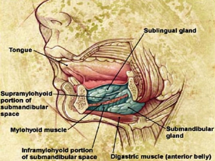
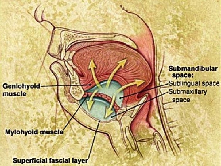
Submandibular space Boundaries: Anterior & lateral: mandible Medial: anterior belly of digastric Posterior: submandibular gland Inferior: level of hyoid bone Subdivisions: 1. Sublingual space: above mylohyoid muscle 2. Submaxillary space: below mylohyoid muscle Contents: Submandibular salivary gland, lymph nodes
Etiology of Ludwig’s angina
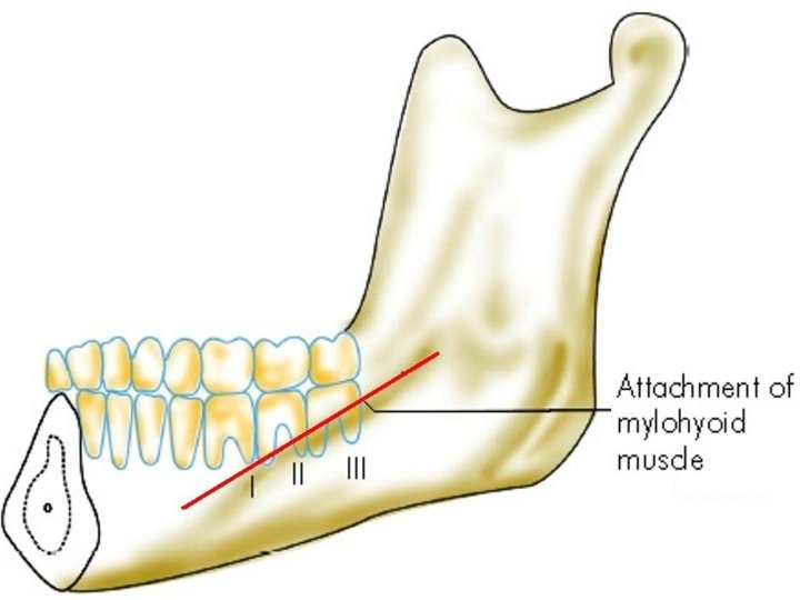
A. Lower dental or periodontal infection (80%): 1. Poor dental hygiene (caries & abscess) 2. Tooth extraction (lower molars & premolars) Roots of premolars & 1 st molar lie above mylohyoid sublingual space infection Roots of 2 nd & 3 rd molars lie below mylohyoid submaxillary space infection B. Others (20%): submandibular sialadenitis, floor of mouth trauma, mandibular fractures
Causative organisms Mixed aerobic & anaerobic infection p Streptococcus pyogenes p Streptococcus viridans p Streptococcus pneumoniae p Staphylococcus p Fusobacterium p Bacteroides p Peptostreptococcus
Clinical Features p Toothache, fever, odynophagia, drooling p Floor of mouth swelling + tongue elevation in sublingual space infection p Brawny / woody tender swelling below chin in submaxillary space infection p Trismus p Stridor: falling back of tongue, laryngeal edema p Initial cellulitis delayed pus formation
Elevation of tongue
Submandibular swelling
Submandibular swelling
X-ray soft tissue neck lateral assess degree of soft tissue swelling & airway obstruction
C. T. scan
Treatment of Ludwig’s angina
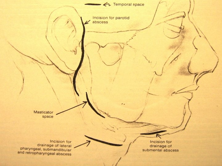
1. I. V. antibiotics: Cefuroxime / Ceftriaxone + Metronidazole / Clindamycin 2. Airway: endotracheal intubation / tracheostomy 3. Incision & drainage of serous fluid / pus a. Intra-oral: for sublingual space infection b. Extra-oral: for submaxillary space infection Transverse incision from one angle of mandible to opposite angle of mandible 4. IV fluid for adequate hydration 5. Periodic assessment for disease progression & airway compromise
Incision drainage + Tracheostomy
Incision drainage + Tracheostomy
Complications p Parapharyngeal abscess p Retropharyngeal abscess p Acute airway obstruction (within hours): due to pushing back of tongue, laryngeal edema p Aspiration pneumonia p Septicemia p Death
Retropharyngeal abscess
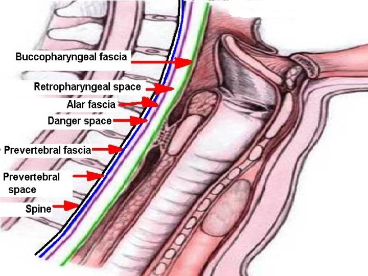
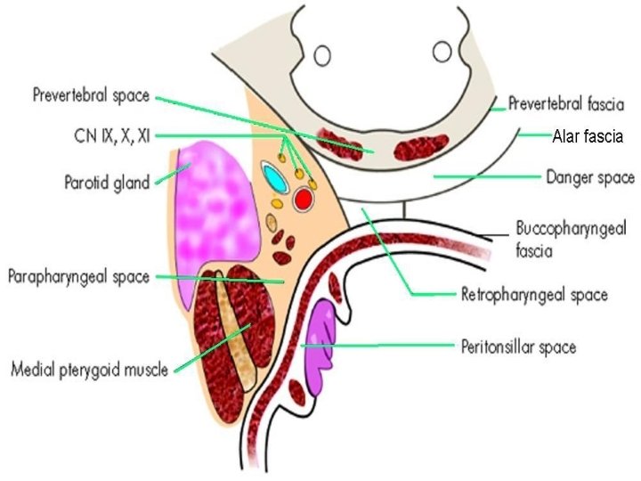
Retropharyngeal Space Superior: Base of skull Inferior: Mediastinum (till tracheal bifurcation) Anterior: Buccopharyngeal fascia Posterior: Alar fascia Lateral: Parapharyngeal spaces Divided into two lateral compartments (space of Gillette) by midline fibrous raphe
Retropharyngeal abscess Collection of pus in retropharyngeal space Classification: 1. Acute 2. Chronic Acute abscess is common in children below 3 -5 yrs as retropharyngeal nodes of Rouviere regress later
Acute Retropharyngeal Abscess
Etiology p Suppuration of retropharyngeal lymph node of Rouviere from upper respiratory tract infection p Penetrating injury of posterior pharyngeal wall (e. g. . fish bone, vertebral fracture) p Following endoscopic trauma to pharynx p Acute mastoitis: pus tracking under petrous bone
Symptoms p H/o upper respiratory tract infection p Dysphagia / odynophagia p Difficulty in breathing p Croupy cough p Hot potato voice p Neck stiffness
Signs p Febrile, ill-looking, child with drooling p Tender neck swelling + fistula p Torticollis (twisted neck) on side of abscess followed by hyperextension of neck p U/L bulge on posterior pharyngeal wall
Posterior pharyngeal wall swelling on left side
Endoscopic view of posterior pharyngeal wall bulge
X-ray soft tissue neck (lateral) 1. Widened pre-vertebral soft tissue shadow a. > 7 mm at C 2 vertebra b. > 14 mm at C 6 vertebra below 14 years c. > 22 mm at C 6 vertebra above 14 years 2. Presence of air-fluid level & / gas (acute cases) 3. Homogenous pre-vertebral shadow (chronic) 4. Straightening of cervical spine curve due to spasm of pre-vertebral muscles
High retropharyngeal abscess
Air-fluid level & gas shadow
CT scan axial cuts
Treatment 1. IV antibiotics: Ceftriaxone + Metronidazole 2. Incision & drainage: p No anesthesia (as it may rupture abscess) or very careful endotracheal intubation p Supine with head hanging low from table p Vertical or horizontal incision on fluctuant area p Incision + immediate suction of pus 3. Tracheostomy for airway obstruction
Chronic Retropharyngeal Abscess
Etiology p Caries of cervical spine: presents as central posterior pharyngeal wall swelling p Tubercular infection of retropharyngeal lymph nodes from infected deep cervical nodes: presents as lateral posterior pharyngeal wall swelling true retropharyngeal abscess p Post traumatic: vertebral fracture p Spread from parapharyngeal abscess
Clinical Features p Chronic mild dysphagia p Pain is absent due to cold abscess p Bulge of posterior pharyngeal wall with fluctuant swelling (central or lateral) Investigations p As in acute retropharyngeal abscess p Ziehl Neelsen stain of pus after aspiration
X-ray soft tissue neck (lateral): homogenous opacity
Tuberculosis of cervical spine with chronic retropharyngeal abscess
Treatment 1. I. V. antibiotics: Ceftriaxone + Metronidazole 2. Incision & drainage: p Low abscess: along anterior border of sternocleidomastoid muscle p High abscess: along posterior border of sternocleidomastoid muscle 3. Anti-tubercular therapy for 9 - 12 months
Complications 1. Airway obstruction: mechanical obstruction laryngeal edema 2. Spread of abscess to other neck spaces 3. Spontaneous rupture of abscess 4. Septicemia 5. Death
Parapharyngeal abscess
Parapharyngeal space Base & superior limit: Skull Base Apex: Lesser cornu of hyoid Lateral: Mandible ramus, Medial Pterygoid, Parotid Medial: Bucco-pharyngeal fascia, superior constrictor Anterior: Pterygo-mandibular raphe Posterior: Pre-vertebral fascia Inferior: Deep cervical fascia lateral to mandible angle
Contents Pre-styloid Post-styloid Deep lobe of parotid Internal carotid artery Internal maxillary artery Internal jugular vein Inferior alveolar nerve Last 4 cranial nerves Lingual nerve Sympathetic chain Auriculo-temporal nerve Glomus system Lymph nodes Styloid: Styloid process, its 3 muscles + 2 ligaments
Etiology p Pharynx: acute tonsillitis, peritonsillar abscess p Teeth: dental infection (esp. lower last molar) p Ear: Bezold’s abscess p Spread from other neck abscess: parotid, retropharyngeal, submandibular p Penetrating neck injuries
Clinical Features 1. Fever, sore throat, odynophagia, torticollis 2. Anterior compartment involvement: a. Tonsils pushed medially b. Trismus c. Neck swelling behind angle of mandible 3. Posterior compartment involvement: a. Medial bulge behind posterior pillar of tonsil b. Paralysis of IX, X, XII & sympathetic chain
CT scan neck: axial cuts
Treatment 1. IV antibiotics: Ceftriaxone + Metronidazole 2. Incision & drainage: p Under GA with endotracheal intubation p Horizontal incision made 3 cm below angle of mandible p Trans-oral drainage avoided to prevent injury to carotid artery & internal jugular vein 3. Tracheostomy for airway obstruction / trismus
Peritonsillar abscess (Quinsy)
Etio-pathogenesis Pus present between tonsillar capsule & superior constrictor muscle Pathology: aerobic + anaerobic organisms 1. Acute tonsillitis blockage of crypts intra tonsillar abscess peritonsillitis quinsy 2. Abscess of Weber's salivary gland in supra tonsillar fossa quinsy
Clinical features Symptoms: Young adult with severe odynophagia, fever, halitosis & muffled voice Signs: 1. Para-tonsil area swollen & congested 2. U/L tonsil ed, pushed medially, congested 3. Jugulo-digastric lymph node tender, enlarged 4. Trismus 5. Torticollis
Peri-tonsillitis & Quinsy
Management Diagnosis: Needle aspiration reveals pus Medical treatment: 1. Urgent admission, I. V. fluids 2. I. V. Cefotaxime + Metronidazole 3. Antihistamine - decongestant + analgesic 4. Betadine gargle
Needle aspiration
Incision
Incision line & quinsy forceps
Alternate incision site at maximum bulge
Abscess drainage
Incision & drainage p Incision made with # 11 blade or Thilenius peritonsillar abscess drainage forceps p Nick made above & lateral to junction of 2 imaginary lines. Horizontal along base of uvula, vertical along anterior tonsillar pillar. p Incision widened with sinus forceps & pus drained. No anesthesia is required.
Surgical treatment 1. Interval tonsillectomy after 4 – 6 wk. 2. Hot tonsillectomy or abscess tonsillectomy is avoided as it leads to: more bleeding septicemia
Complications of quinsy 1. Parapharyngeal abscess 2. Retropharyngeal abscess 3. Laryngitis & laryngeal edema 4. Lung abscess 5. Internal jugular vein thrombosis 6. Septicemia
Parotid abscess
Parotid Space Formed due to splitting of investing layer of deep cervical fascia around parotid salivary gland
Etiology p Ascent of bacterial infection (Staphylococcus, Haemophillus, Streptococcus) to a dehydrated parotid gland along parotid duct from oral cavity p Suppuration of intra-parotid lymph nodes p Spread of infection from EAC via cartilaginous fissures of Santorini or bony foramen of Huschke
Causes of parotid dehydration 1. Post-operative patient (surgical mumps) 2. Medications that decrease salivary flow: p Antihistamines p Tricyclic antidepressants p Barbiturates p Diuretics p Parasympathomimetics
Parotid abscess p Pain + induration over parotid gland p Pitting edema of parotid area differentiates parotid abscess from simple parotitis p Parotid massage expresses pus from parotid duct into oral cavity (opposite upper 2 nd molar)
Investigation p C. B. P. : Leukocytosis p Needle aspiration with 18 G needle p Ultrasonography p C. T. scan p M. R. I.
C. T. scan & M. R. I.
Parotid anatomy
Treatment 1. IV fluid for dehydration 2. IV Ampicillin + Gentamicin + Metronidazole 3. Incision drainage: a. Blair’s incision made b. Multiple incisions made through fascia, parallel to facial nerve branches c. Blunt dissection to evacuate pus. Drains placed.
Thank You