
1 Osteomyelitis 2 Vertebral Osteomyelitis 3 Prosthetic Joint
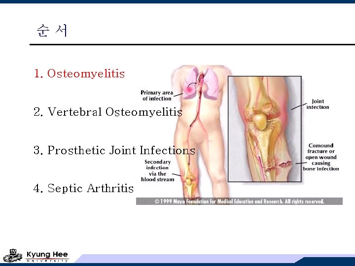
순서 1. Osteomyelitis 2. Vertebral Osteomyelitis 3. Prosthetic Joint Infections 4. Septic Arthritis
Osteomyelitis의 분류 v acute; newly recognized, over days to weeks v chronic; over weeks to months, can persist for years relapse of a previously treated or untreated infection v Waldvogel classification (1970) 1. hematogenous (소아 long bone / 성인 vertebra) 2. secondary to a contiguous focus of infection without vascular insufficiency 3. secondary to a contiguous focus of infection with vascular insufficiency (diabetic foot) 4. chronic osteomyelitis (necrotic bone)
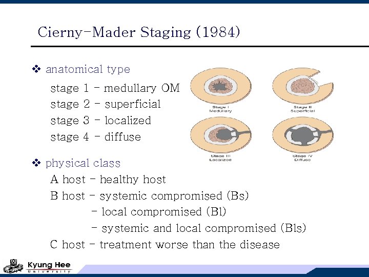
Cierny-Mader Staging (1984) v anatomical type stage 1 2 3 4 - medullary OM superficial localized diffuse v physical class A host - healthy host B host - systemic compromised (Bs) - local compromised (Bl) - systemic and local compromised (Bls) C host - treatment worse than the disease
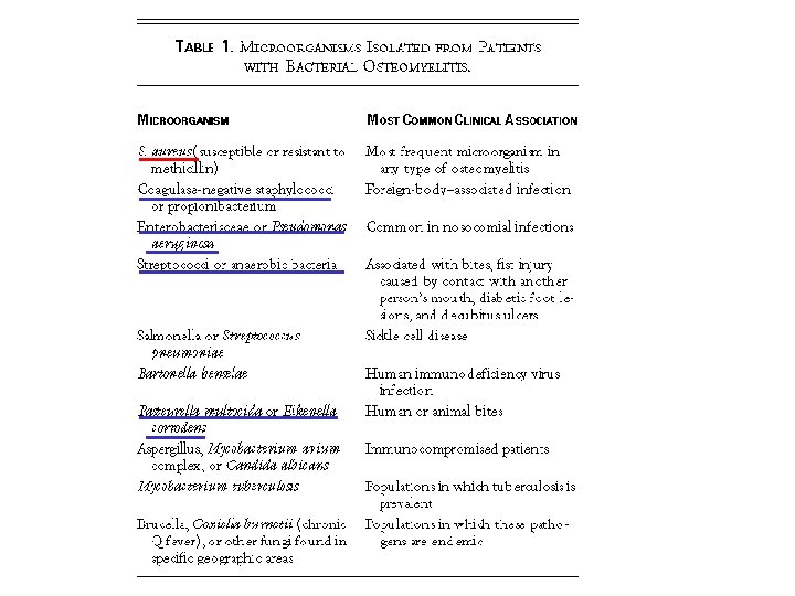
Osteomyelitis의 원인균 v hematogenous (mono-microbial) infant (<1 yr) S. aureus, GBS, E. coli, children (1 -16 yrs) S. aureus, S. pyogenes, H. influenzae adults (>16 yrs) S. aureus, CNS, Salmonella, E. coli, Pseudomonas v contiguous (poly-) diabetic foot (poly-) depends on the primary site S. aureus, strep-/enterococci, GNB, anaerobes
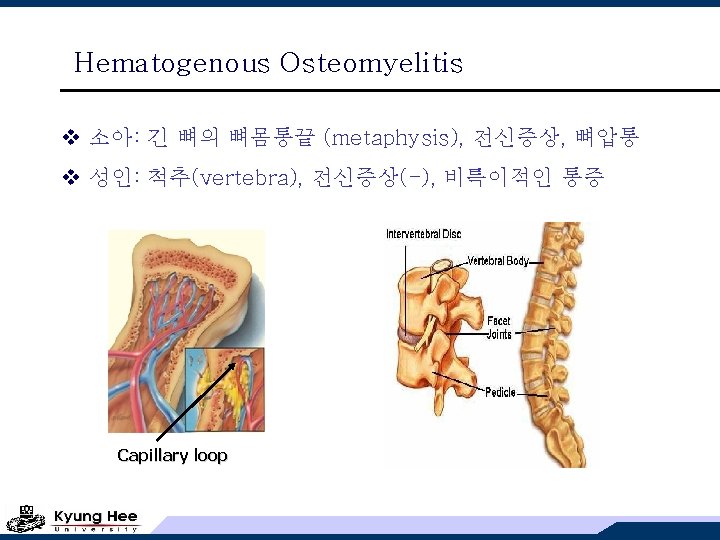
Hematogenous Osteomyelitis v 소아: 긴 뼈의 뼈몸통끝 (metaphysis), 전신증상, 뼈압통 v 성인: 척추(vertebra), 전신증상(-), 비특이적인 통증 Capillary loop
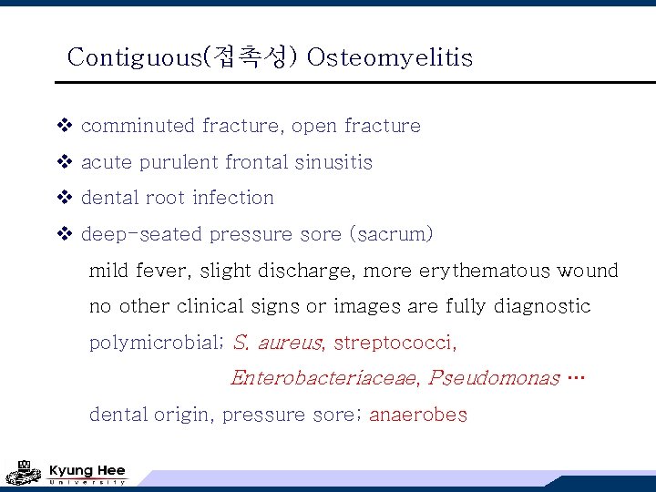
Contiguous(접촉성) Osteomyelitis v comminuted fracture, open fracture v acute purulent frontal sinusitis v dental root infection v deep-seated pressure sore (sacrum) mild fever, slight discharge, more erythematous wound no other clinical signs or images are fully diagnostic polymicrobial; S. aureus, streptococci, Enterobacteriaceae, Pseudomonas … dental origin, pressure sore; anaerobes
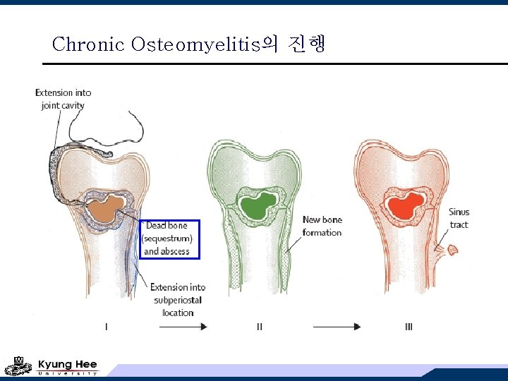
Chronic Osteomyelitis의 진행
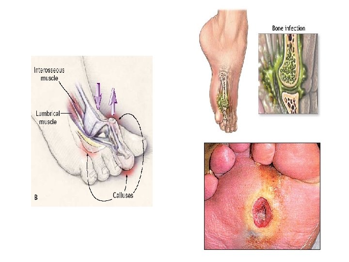
Diabetic Foot Infection v vascular insufficiency v cellulitis may be minimal, progressively burrow to bone v no pain with advanced neuropathy v polymicrobial, mixed; anaerobes or Enterobacteriaceae v S. aureus still predominates v gently advancing a sterile probe with plain film if bone is detected on probing, treatment for OM v occult OM; radiography, repeated in two weeks
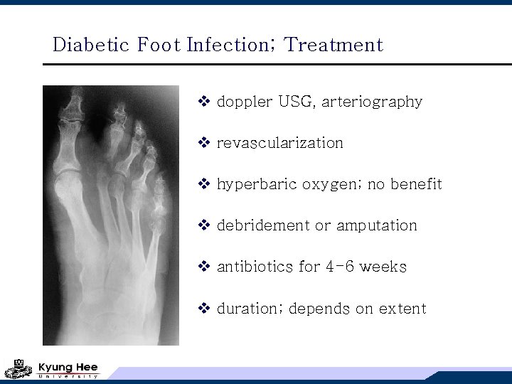
Diabetic Foot Infection; Treatment v doppler USG, arteriography v revascularization v hyperbaric oxygen; no benefit v debridement or amputation v antibiotics for 4 -6 weeks v duration; depends on extent
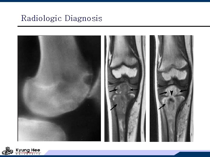
Osteomyelitis의 진단 v long bone; fever/chill, pain + soft tissue swelling (소아) v vertebral; fever (<50%), back pain & tenderness, ESR v plain X-ray; 2 -3주 후 변화 인지 (bone Ca++ 소실 > 50%) sensitivity(60%), specificity(82%) 1. periosteal elevation 2. demineralization (bone margin = "moth eaten") 3. soft tissue swelling 4. late stage (calcification or sclerosis)
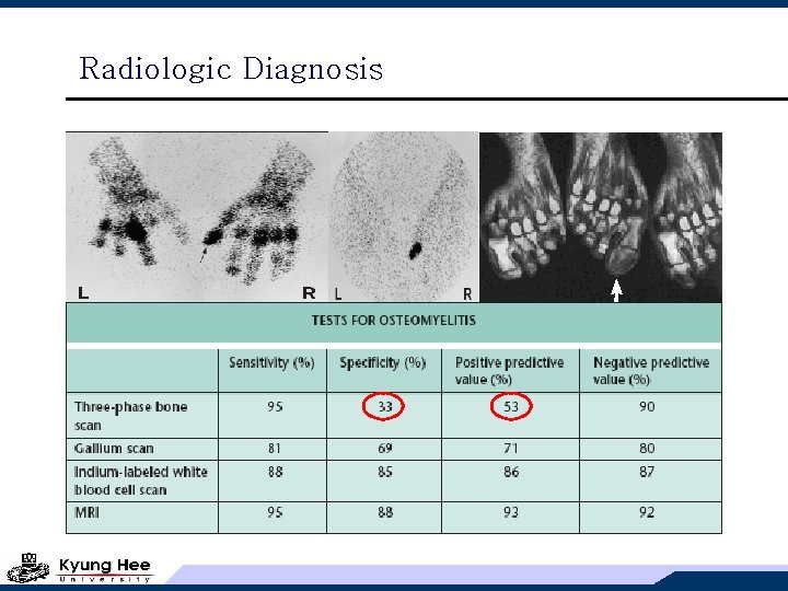
Osteomyelitis의 진단 v CT; more sensitive v MRI; detect early changes 신경성 병변, 통풍, 퇴행성 관절염 수술 후 변화, 골절의 치유 과정 등 v bone scan; detect early disease, false positive Gallium preferred for vertebral OM v tissue sample for culture & pathology, when blood (-) long bone OM in children may be treated empirically long bone OM in adults required operative culture vertebral OM; CT-guided needle biopsy 부적절한 배양 부위; open wound, sinus tract
Radiologic Diagnosis
Radiologic Diagnosis
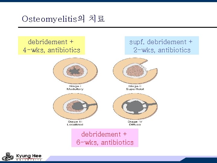
Osteomyelitis의 치료 debridement + 4 -wks, antibiotics supf. debridement + 2 -wks, antibiotics debridement + 6 -wks, antibiotics
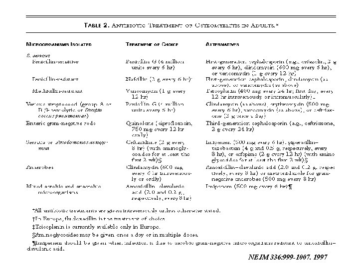
NEJM 336: 999 -1007. 1997
순서 1. Osteomyelitis 2. Vertebral Osteomyelitis 3. Prosthetic Joint Infections 4. Septic Arthritis
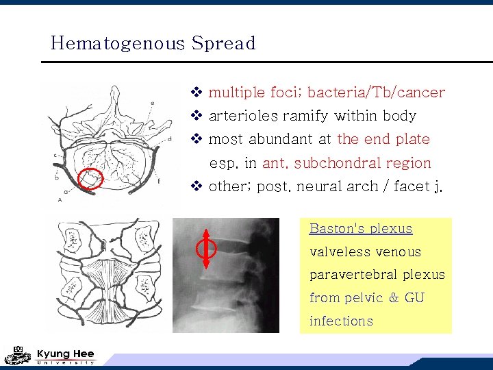
Hematogenous Spread v multiple foci; bacteria/Tb/cancer v arterioles ramify within body v most abundant at the end plate esp. in ant. subchondral region v other; post. neural arch / facet j. Baston's plexus valveless venous paravertebral plexus from pelvic & GU infections
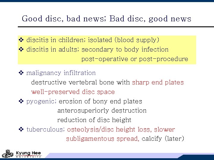
Good disc, bad news; Bad disc, good news v discitis in children; isolated (blood supply) v discitis in adults; secondary to body infection post-operative or post-procedure v malignancy infiltration destructive vertebral bone with sharp end plates well-preserved disc space v pyogenic; erosion of bony end plates anterosuperiorly destruction reduction of disc height v tuberculous; osteolysis/disc height loss, slower subligamentous spread, calcify (later)
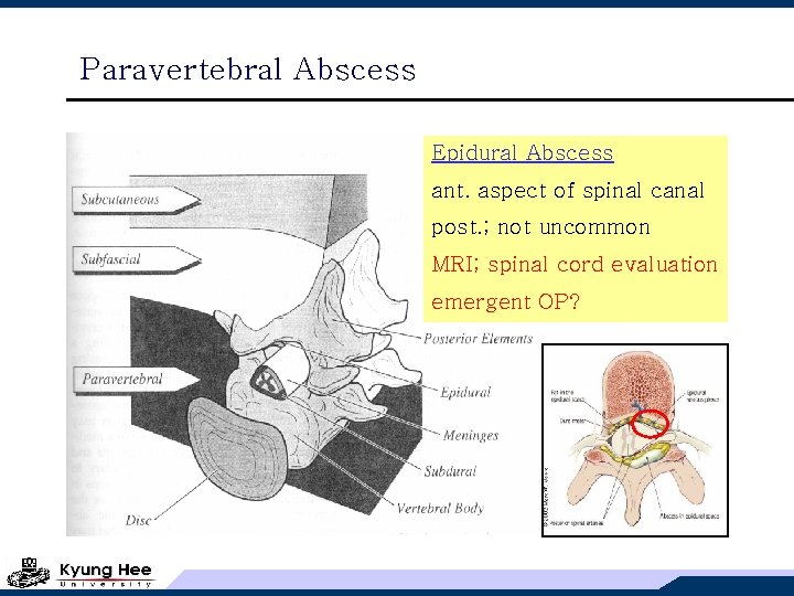
Paravertebral Abscess Epidural Abscess ant. aspect of spinal canal post. ; not uncommon MRI; spinal cord evaluation emergent OP?
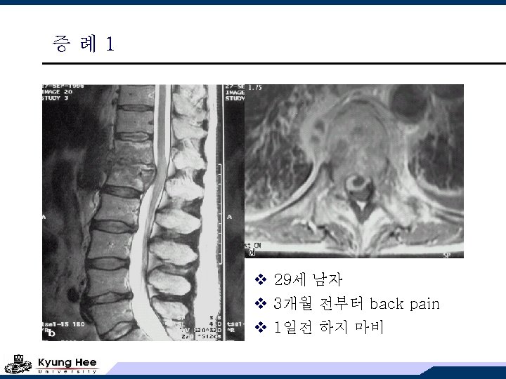
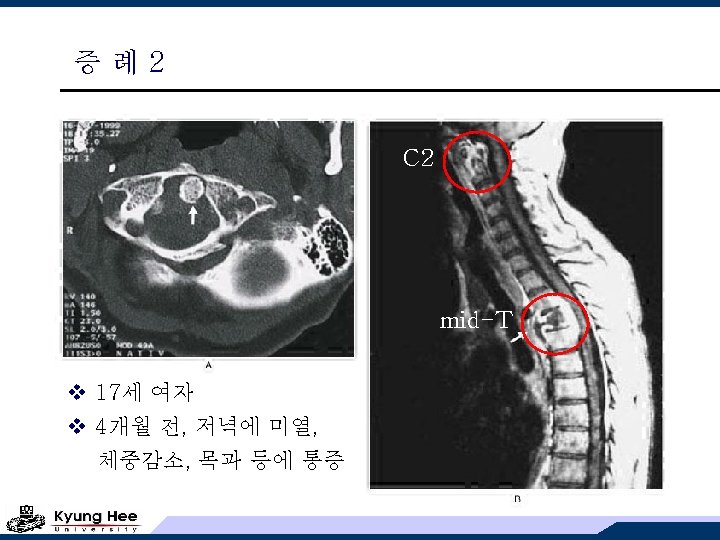
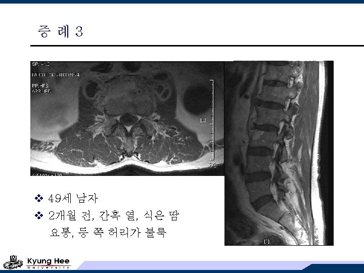
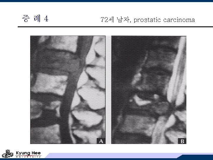
증례4 72세 남자, prostatic carcinoma
Vertebral OM의 진단과 치료 v 임상적, MRI 소견으로는 감별할 수 없다! v CT-guided or operative biopsy & culture v surgical indication 1. open biopsy 2. clinically significant abscess 3. refractory course to adequate medical management 4. significant deformity or destruction leading to deformity
순서 1. Osteomyelitis 2. Vertebral Osteomyelitis 3. Prosthetic Joint Infections 4. Septic Arthritis
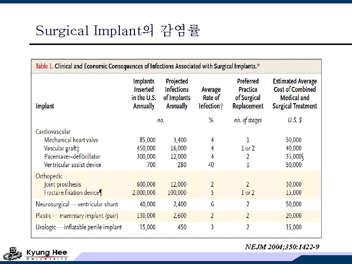
Surgical Implant의 감염률 NEJM 2004; 350: 1422 -9
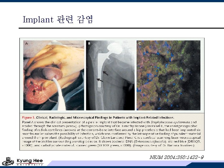
Implant 관련 감염 NEJM 2004; 350: 1422 -9
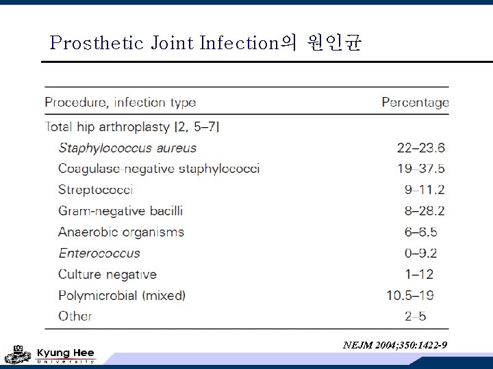
Prosthetic Joint Infection의 원인균 NEJM 2004; 350: 1422 -9
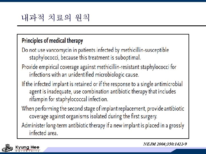
내과적 치료의 원칙 NEJM 2004; 350: 1422 -9
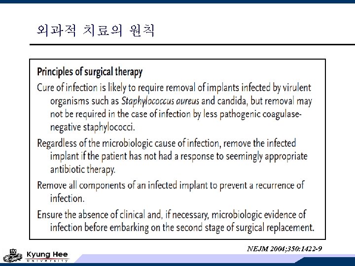
외과적 치료의 원칙 NEJM 2004; 350: 1422 -9
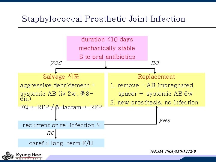
Staphylococcal Prosthetic Joint Infection yes duration <10 days mechanically stable S to oral antibiotics Salvage 시도 aggressive debridement + systemic AB (iv 2 w, 총 36 m) FQ + RFP / -lactam + RFP recurrent or re-infection ? no Replacement 1. remove - AB impregnated spacer + systemic AB 6 w 2. new prosthesis, no infection yes no careful long-term F/U NEJM 2004; 350: 1422 -9
순서 1. Osteomyelitis 2. Vertebral Osteomyelitis 3. Prosthetic Joint Infections 4. Septic Arthritis
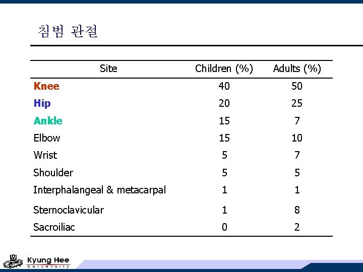
침범 관절 Site Children (%) Adults (%) Knee 40 50 Hip 20 25 Ankle 15 7 Elbow 15 10 Wrist 5 7 Shoulder 5 5 Interphalangeal & metacarpal 1 1 Sternoclavicular 1 8 Sacroiliac 0 2
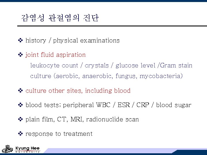
감염성 관절염의 진단 v history / physical examinations v joint fluid aspiration leukocyte count / crystals / glucose level /Gram stain culture (aerobic, anaerobic, fungus, mycobacteria) v culture other sites, including blood v blood tests; peripheral WBC / ESR / CRP / blood sugar v plain film, CT, MRI, radionuclide scan v response to treatment
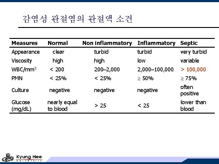
감염성 관절염의 관절액 소견 Measures Normal Non inflammatory Inflammatory Septic Appearance clear turbid very turbid Viscosity high low variable WBC/mm 3 < 200– 2, 000– 100, 000 > 100, 000 PMN < 25% 50% 75% Culture negative often positive > 25 < 25 lower than blood Glucose (mg/d. L) nearly equal to blood
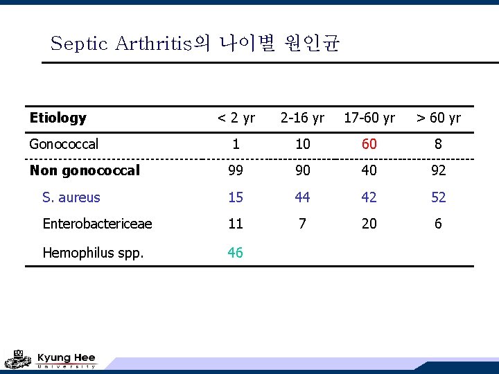
Septic Arthritis의 나이별 원인균 Etiology < 2 yr 2 -16 yr 17 -60 yr > 60 yr Gonococcal 1 10 60 8 Non gonococcal 99 90 40 92 S. aureus 15 44 42 52 Enterobactericeae 11 7 20 6 Hemophilus spp. 46
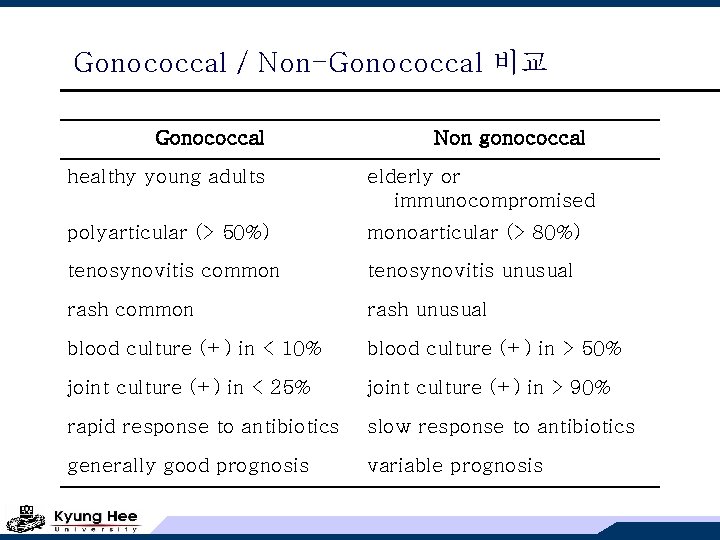
Gonococcal / Non-Gonococcal 비교 Gonococcal Non gonococcal healthy young adults elderly or immunocompromised polyarticular (> 50%) monoarticular (> 80%) tenosynovitis common tenosynovitis unusual rash common rash unusual blood culture (+) in < 10% blood culture (+) in > 50% joint culture (+) in < 25% joint culture (+) in > 90% rapid response to antibiotics slow response to antibiotics generally good prognosis variable prognosis
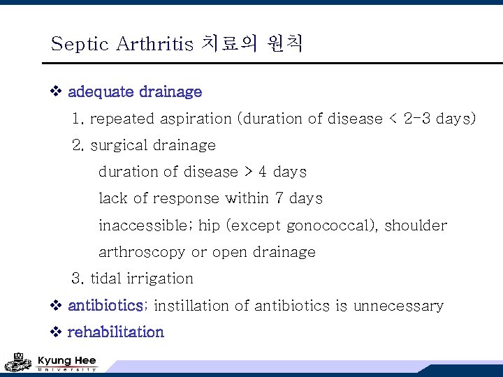
Septic Arthritis 치료의 원칙 v adequate drainage 1. repeated aspiration (duration of disease < 2 -3 days) 2. surgical drainage duration of disease > 4 days lack of response within 7 days inaccessible; hip (except gonococcal), shoulder arthroscopy or open drainage 3. tidal irrigation v antibiotics; instillation of antibiotics is unnecessary v rehabilitation
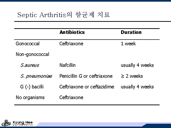
Septic Arthritis의 항균제 치료 Antibiotics Duration Ceftriaxone 1 week S. aureus Nafcillin usually 4 weeks S. pneumoniae Penicillin G or ceftriaxone ≥ 2 weeks G (-) bacilli Ceftriaxone or ceftazidime usually 4 weeks Gonococcal Non-gonococcal No organisms Ceftriaxone
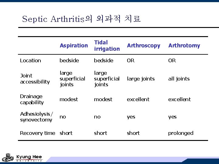
Septic Arthritis의 외과적 치료 Aspiration Tidal irrigation Arthroscopy Arthrotomy Location bedside OR OR Joint accessibility large superficial joints large joints all joints Drainage capability modest excellent Adhesiolysis/ synovectomy no no yes short prolonged Recovery time short
Septic Arthritis의 재활 치료 v weight bearing should be prevented until all signs of inflammation subside v strict immobilization is not required v splinting vs. passive motion is controversial v Recent recommendations: 1. isometric exercise (1 st post-op. day) 2. passive range of motion (during 1 st post-op. wk) 3. active motion (within post-op. 1 -2 wks)
- Slides: 47