FUNGAL INFECTIONS Fungal infections Dermatophyte infections RingwormTinea Candidiasis

 • Candidiasis (Yeasts) • Pityriasis Versicolor (Yeasts)")


: • The most common fungal infection in")


 • in ringworm acquired from")


 appear on")


. • The skin lesions are firm")


.")

- Slides: 36
FUNGAL INFECTIONS
Fungal infections: • Dermatophyte infections (Ringworm/Tinea) • Candidiasis (Yeasts) • Pityriasis Versicolor (Yeasts)
Dermatophyte infections • Also known as ringworm infections. • Three types of fungus: • Trichophyton: skin, hair and nail infections. • Microsporum: skin and hair. • Epidermophyton: skin and nails. • Dermatophytes invade keratin only (keratinophilic), and the inflammation they cause is due to metabolic products of the fungus or to delayed hypersensitivity • • TRANSMISSION transmitted by animals : cats by direct contact By fomites (fallen hair and desquamated skin as in shared hats & brushes • Zoophilic fungi (transmitted to humans by animals) cause a more severe inflammation than anthropophilic fungi (spread from person to person).
Dermatophyte infections • The presentation and course of disease depends on; • The site of infection. • Strain involved. • Sites of infection: • Foot : Tinea pedis (athlete’s foot) • Nails : Onychomycosis • Hands : Tinea of the hands • Trunk and limbs : Tinea corporis • Scalp : Tinea capitis. • Groin : Tinea cruris
Dermatophyte infections • Tinea pedis (athlete’s foot): • The most common fungal infection in humans. • Sharing wash places and swimming pools predisposes infection. • Occlusive foot wear encourages relapses. • Caused by 3 organisms : 1. Trichophyton rubrum (most common and stubborn) 2. Trichophyton mentagrophytes var. interdigitale 3. Epidermophyton floccosum • Clinical presentation • Soggy interdigital scaling; in the fourth and fifth interspace. • A diffuse dry scaling of the soles. • Recurrent episodes of vesication.
Tinea pedis. Scaly area spreading to the sole from the toe webs. Soggy interdigital scaling Powdery scaling, most obvious in the skin creases, caused by a Trichophyton rubrum infection. A diffuse dry scaling of the soles The toe web space contains macerated scale. The fourth web is the most commonly involved web space.
Tinea of the nails • Onychomycosis: chronic painless thickening of the nails and discoloration with some scales • Toenail infection is usually associated with tinea pedis. • The initial changes occur at the free edge of the nail, which becomes yellow and crumbly, subungual hyperkeratosis, separation of the nail from its bed, and thickening may then follow. • Usually only a few nails are infected but rarely all are. • Fingernail lesions are similar, but less common, and are seldom seen without a chronic T. rubrum infection of the skin of the same hand.
Tinea of the nails Chronic tinea of the big toe nail. Starting distally, the thickness and discoloration are spreading proximally.
Tinea of the hands • Asymmetrical. • Associated with tinea pedis and unilateral onychomychosis. Ø T. rubrum may cause : • a barely perceptible erythema of one palm • with a characteristic powdery scale in the creases.
Tinea of the trunk and limbs • Tinea corporis. • characterized by plaques with scaling and erythema. • Mostly at the periphery. • Might be associated with a few small vesicles and pustules. • Lesions expand slowly and heal in the center (ring-like pattern). • In some patients the fungus elicits almost no inflammation, in which case the infection is a marginated patch of rough scaling skin.
Tinea of the trunk and limbs
Tinea of the scalp • Tinea capitis. • Usually a disease of children. • The causative organism varies from country to country. • Fungi coming from human sources (anthropophilic organisms) cause bald and scaly areas, with minimal inflammation and hairs broken off 3– 4 mm from the scalp. • Fungi coming from animal sources (zoophilic fungi) induce a more intense inflammation
v. Kerion: (an abscess caused by fungal infection ) • in ringworm acquired from cattle, presents as boggy swelling with • Inflammation, Pustulation, Lymphadenopathy, Hair loss (may be permanent) • is often so intense that a bacterial infection is suspected. v. Favus: (severe capitis) form of tinea • caused by Trichophyton schoenleini • Foul-smelling yellowish crusts surrounding many scalp hairs, and sometimes leading to scarring alopecia. • Black children are especially prone to infection with Trichophyton tonsurans. • Tinea of the beard area is usually caused by zoophilic species and shows the same features.
Tinea of the scalp Kerion Favus Acquired from cattle boggy swelling with • Inflammation • Pustulation • Lymphadenopathy • Hair loss (may be permanent) Trichophyton schoenleini so intense that a bacterial infection is suspected Foul-smelling yellowish crusts. sometimes leading to scarring Alopecia.
Tinea of the groin • Tinea cruris • Common, affects men more often than women. • The eruption is sometimes unilateral or asymmetrical. • The upper inner thigh is involved and lesions expand slowly to form sharply demarcated plaques with peripheral Scaling. • In contrast to candidiasis of the groin area, the scrotum is usu. 0 ally spared (in contrast to candidiasis which more extensive & Bilateral). • A few vesicles or pustules may be seen within the lesions. • Other organisms may cause it are the same as those causing tinea pedis.
Tinea complications 1. Fierce animal ringworm of the scalp can lead to a permanent scarring alopecia. 2. A florid fungal infection anywhere can induce vesication on the sides of the fingers and palms (a trichophytide). 3. Epidemics of ringworm occur in schools. 4. The usual appearance of a fungal infection can be masked by mistreatment with topical steroids (tinea incognito).
Differential diagnosis
Investigations • Microscopic examination of a skin scraping, nail clipping or plucked hair , Specimens are cleared in potassium hydroxide, Branching hyphae can easily be seen (quicker). • Cultures (take as long as a month). • Wood’s light (ultraviolet light) examination of the scalp; • usually reveals a green fluorescence of the hairs in: • Microsporum audouini. • M. canis infection. • The technique is useful for screening children in institutions where outbreaks of tinea capitis still sometimes occur, but the most common fungi causing tinea capitis (e. g. Trichophyton tonsurans) do not fluoresce.
Treatment q. Local: This is all that is needed for minor skin Infections • Imidazole, allylamines, benzoic acid ointment (Whitfield’s ointment), tolnaftate, Magenta paint (Castellani’s paint). q. Topical nail preparations • Amorolfine, Ciclopirox • Both amorolfine and tioconazole nail Solutions can be used as adjuncts to systemic therapy. q. Systemic: This is needed for widespread infection or chronic infections of the skin that have not responded to local measures. • Terbinafine • Itraconazole • Griseofulvin
Candiasis • Candida albicans is a classic opportunistic pathogen. • predisposing factors : • Obesity • Moisture • Maceration • Immobility • Diabetes • Pregnancy • Broad-spectrum antibiotics and contraceptive pills • Immunosupression
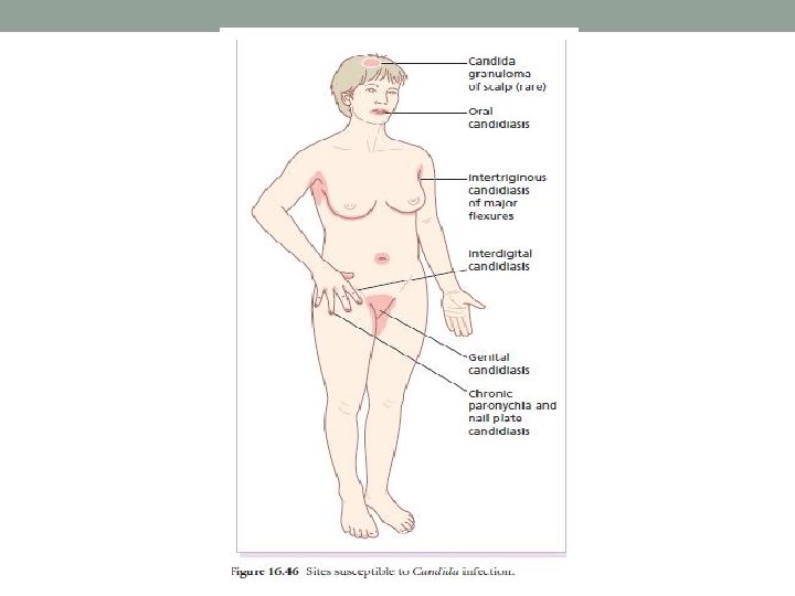
Oral candidiasis • One or more whitish adherent plaques (like bread sauce) appear on the mucous membranes. If wiped off they leave an erythematous base. • Under dentures, candidiasis will produce sore redareas. • Angular stomatitis, in denture wearers may be candidal.
Candidal intertrigo • A moist glazed area of erythema and maceration appears in a body fold. • The edge shows soggy scaling and outlying satellite papulo-pustules. • Sites; • Breast, armpit and groin areas. • In between the fingers of those whose hands are often in water.
Genital candidiasis • Presentation; • Most commonly presents as a sore itchy vulvovaginitis, with white curdy plaques adherent to the inflamed mucous membranes, and a whitish discharge. • Eruption may extend to the groin folds. • In males similar changes occur under the foreskin and in the groin.
Paronychia • Acute paronychia is usually bacterial, but in chronic paronychia Candida may be the sole pathogen. • The proximal and sometimes the lateral nail folds of one or more fingers become bolstered and red. The cuticles are lost and small amounts of pus can be expressed. • The adjacent nail plate becomes ridged & discoloured. • Predisposing factors: • Wet work • Poor peripheral circulation • Vulval candidiasis
Chronic mucocutaneous candidiasis • Persistent candidiasis, affecting most or all of the areas described above, can start in infancy. • Candida granulomas may appear on the scalp. • Sometimes the nail plates as well as the nail folds are involved • Several different forms have been described including those with autosomal recessive and dominant inheritance patterns. • In the Candida endocrinopathy syndrome, chronic candidiasis occurs with one or more endocrine defects, the most common of which are hypoparathyroidism, Addison’s disease and few late- onset cases have underlying thymic tumours.
Systemic candidiasis • severe illness( leucopenia & immunosuppression). • The skin lesions are firm red nodules, which can be shown by biopsy to contain yeasts and pseudohyphae.
Investigations • Swabs from suspected areas should be sent for culture. • Urine can be tested for sugar. • In chronic mucocutaneous candidiasis, a detailed immunological work-up focusing on cell mediated immunity.
Treatment • Predisposing factors should be sought and eliminated (e. g. denture hygiene may be important). • Infected skin folds should be separated and kept dry. • Topical antifungal: Amphotericin, nystatin and imidazole • For the oral candidiasis, lozenges and oral gels are available. • False teeth should be removed at night, washed and steeped in antiseptic or a nystatin solution. • For other areas of candidiasis, creams, ointment and pessaries are available.
• Magenta paint is also a useful but messy remedy for skin flexures. • Imidazole cream or solution (chronic paronychia) • Itraconazole or fluconazole for: • Genital candidiasis • Recurrent oral candidiasis of the immunocompromised • Chronic mucocutaneous candidiasis
Pityriasis versicolor • Tinea versicolor • Caused by commensal yeasts Pityrosporum orbiculare (Malassezia furfur (it is part of the normal flora)) and not by dermatophyte fungi. • Pathogenesis; • Overgrowth of these yeasts, particularly in hot humid conditions and bad hygiene • Carboxylic acids released by the organisms inhibit pigment production by melanocytes that • Occurs normally after exposure to sunlight. • Superficial scaly patches, fawn or pink on non-tanned skin become paler than the surrounding skin after exposure to sunlight. • keeping in mind its lipophilic so doesn’t affect children (because their lipid profile isn’t well developed yet) • The condition should be regarded as non-infectious.
Pityriasis versicolor Presentation and course; • Depigmented areas • Slightly scaling • Fine wrinkling • Symptom-free or only slightly itchy. • Lesions are most common on the upper trunk but can become widespread. • Untreated lesions persist, and depigmented areas, even after adequate treatment, are slow to regain their former color. • Recurrences are common. Investigations • Scrapings, prepared and examined as for a dermatophyte infection show a mixture of short branched hyphae and spores (a ‘spaghetti and meatballs’ appearance). • Culture is not helpful because the organism does not grow on Sabouroud’s medium. (The central area was • Wood’s lamp, appears yellowish fluorescence scraped to demonstrate white, powdery scale).
Pityriasis versicolor
Differential diagnosis üIn vitiligo : the border is clearly defined, scaling is absent, lesions are larger, the limbs and face are often affected, and depigmentation is more complete; however, it may sometimes be hard to distinguish vitiligo from the pale non-scaly areas of treated versicolor. üSeborrheic Eczema of the trunk tends to be more erythematous, and often confined to the presternal or interscapular areas. üPityriasis alba often affects the cheeks. ü Pityriasis rosea, tinea corporis, secondary syphilis, leprosy and erythrasma seldom cause real confusion p. alba
Treatment • Topical • Imidazole group • 2. 5% selenium sulphide mixture (Selsun shampoo). • Ketoconazole shampoo. • Systemic • Itraconazole • Fluconazole • Ketoconazole (Recurrence is common after any treatment).
Thank you