Respiratory infections Respiratory tract infections Serious Bacterial infections

Respiratory infections

Respiratory tract infections Serious Bacterial infections Self-limiting Viral infection
 Lower respiratory tract")
Respiratory tract infections is divided into: Upper respiratory tract infections (URTIs) Lower respiratory tract infections (LRTIs)
 -Colds and flu -Influenza -Sore throat (pharyngitis) -Acute epiglottitis -Otitis media -Acute sinusitis")
(URTIs) -Colds and flu -Influenza -Sore throat (pharyngitis) -Acute epiglottitis -Otitis media -Acute sinusitis (LRTIs) -Acute bronchitis -Bronchiolitis -Pneumonia -Severe acute respiratory syndrome -Cystic fibrosis

For each case you need to know the followings: • Definition • Causative organisms • Clinical features • Diagnosis • Treatment

Upper respiratory tract infections
 Influenza • Causative organisms: True influenza is caused by one of the influenza")
1) Influenza • Causative organisms: True influenza is caused by one of the influenza viruses (influenza A, B or rarely C). • Characterised by: fever (>38 °C), myalgia, headache, sore throat and cough. • Potentially complicated by life-threatening secondary bacterial infections such as staphylococcal pneumonia.
 • Note: Amantadine (as")
Prevention and Treatment • Influenza Vaccine… • Neuraminidase Inhibitors (NAIs) • Note: Amantadine (as antiviral), not recommended.

About influenza vaccine Vaccination is used in patients at higher risk of severe disease and healthcare workers: • • • people older than 65 years people with chronic respiratory disease, people with chronic kidney disease, people with chronic liver disease, people who are immunosuppressed, people with chronic neurological disease, asplenic patients, pregnant women, people with a body mass index greater than 40. Unfortunately, the virus mutates so rapidly that the circulating strains tend to change from season to season, necessitating annual revaccination against the prevailing virus.
 such as zanamivir and oseltamivir, • Oseltamivir is")
Neuraminidase Inhibitors • Neuraminidase inhibitors (NAIs) such as zanamivir and oseltamivir, • Oseltamivir is the first-line agent, given orally. • Zanamivir is administered by dry powder inhalation, IV.
 Sore throat (pharyngitis) • Pharyngitis: is a common condition. In most cases, it")
2) Sore throat (pharyngitis) • Pharyngitis: is a common condition. In most cases, it never comes to medical attention and is treated with simple therapy directed at symptom relief. Many cases are not due to infection at all but are caused by other factors, such as smoking. • Most cases are viral (Epstein–Barr virus (EBV), which causes glandular fever) • The only common bacterial cause of sore throat is Streptococcus pyogenes (βhaemolytic Streptococcus).

Clinical features • For viral: fever and the usual symptoms of the common cold. • In more severe cases----marked inflammation of the pharynx with a whitish exudate on the tonsils, plus enlarged and tender cervical lymph nodes. • For bacterial: Scarlet fever (a toxin-mediated manifestation of streptococcal infection), is associated with a macular rash and sometimes considerable systemic illness.


The Centor score • A clinical scoring system used to identify those at higher risk of bacterial infection. Each feature scores one point. • The criteria are the presence of tonsillar exudate, history of fever, tender anterior surgical lymphadenopathy or adenitis and absence of cough. • Those with a Centor score of 3 or 4 have a 40– 60% risk of group A streptococcal infection. Those with a Centor score of zero or one are unlikely to have group A streptococcal infection

Diagnosis ØThroat Swab for microbiological culture • Distinguish the streptococcal sore throat from viral infections. • The test directed towards detecting β-haemolytic streptococci. ØSerological test • Detect group A streptococcal antigens

Treatment • Most people will recover from sore throat after 7 days. • Analgesics as paracetamol and ibuprofen for reducing pain and fever. • Most patients should not be prescribed an antibiotic

When to prescribe AB • Patients with a Centor score of 3 or 4 are considered for an immediate or delayed antibiotic prescription. • People who are at increased risk of complications and those with Valvular heart disease should be given an antibiotic. • Those who are at risk of immunosuppression. • Those with previous rheumatic fever and those at risk of severe disease.

Streptococcal Sore Throat • Penicillins such as penicillin V are recommended as first-line treatment for group A streptococcal pharyngitis for 5 days. • Erythromycin or clarithromycin is recommended for patients with penicillin allergy.
 Acute epiglottitis • A medical emergency of rapidly progressive cellulitis of the epiglottis")
3) Acute epiglottitis • A medical emergency of rapidly progressive cellulitis of the epiglottis and adjacent structures. • Local swelling has the potential to cause rapid-onset airway obstruction. • Common patient is a child between 2 and 4 years old.
 mainly, • Pneumococci, streptococci and staphylococci")
ØCausative organisms: • Haemophilus influenzae type b (Hib) mainly, • Pneumococci, streptococci and staphylococci (less). ØClinical feature: • Fever • Difficulty speaking and breathing. • Drooling because of impaired swallowing. ØDiagnosis: • By visualization of the epiglottis as ‘cherry-red’. • By Microbiological confirmation by culturing the epiglottis and the blood (but not until the airway is secure).

Treatment • Treatment of choice is a cephalosporin (second-generation cefuroxime and third-generation cefotaxime or ceftriaxone) • High-dose parenteral amoxicillin may be substituted (If a sensitive organism is recovered because amoxicillin resistance among encapsulated H. influenza)
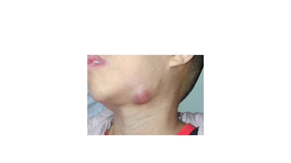
Otitis Media • It is an inflammation of the middle ear, a common condition")
4)Otitis Media • It is an inflammation of the middle ear, a common condition seen most frequently in children under 3 years of age. • Caused by influenza virus and rhinoviruses (mainly) • S. pneumoniae and H. influenza (may be) • Moraxella catarrhalis and S. pyogenes account for a smaller proportion of cases,
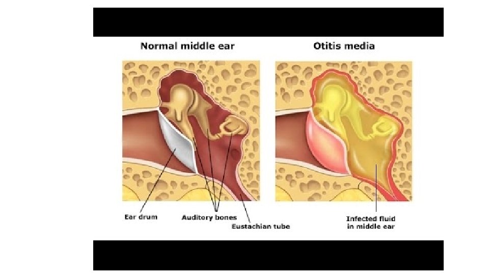

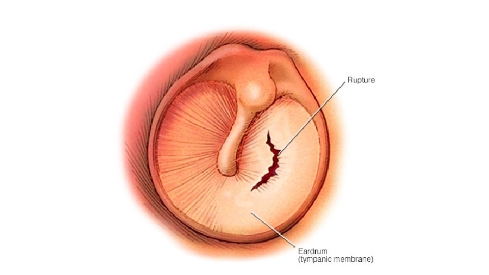
Clinical features • Ear pain, which may be severe. • If the drum perforates, the pain is relieved and a purulent discharge may follow. • There may be a degree of hearing impairment plus non-specific symptoms such as fever or vomiting. • Complications include mastoiditis, meningitis and, particularly, septicaemia and disseminated infection.


Diagnosis • The diagnosis of otitis media is essentially made clinically and laboratory investigations have little role to play. • A swab of the external auditory canal---- if the drum is perforated. • For this reason, a causative organism is rarely isolated and treatment has to be given empirically.

Treatment ØAntibiotics are currently only recommended for Otitis Media in people: • who are systemically unwell, • who are at risk of serious complications, • whose symptoms have lasted more than 4 days and are not improving. • children under 2 with bilateral acute otitis media and for children with ear discharge. Ø If decide to start AB, we need to be effective against the three main bacterial pathogens: S. pneumoniae, H. influenza and S. pyogenes ----- (Amoxicillin or Ampicillin) • Erythromycin or clarithromycin for patients with penicillin allergy
Acute Sinusitis • Normally, the paranasal sinuses are sterile but they can become infected")
5)Acute Sinusitis • Normally, the paranasal sinuses are sterile but they can become infected following damage to the mucous membrane which lines them. • Usually occurs following a viral URTI but is sometimes associated with the presence of dental disease. • Sinusitis is caused by a virus in 98% of cases. • Bacterial acute sinusitis is usually caused by (S. pneumoniae and H. inluenzae), • Other organisms such as Staphylococcus aureus, viridans streptococci and anaerobes may be found.

Clinical features & Diagnosis • Facial pain and tenderness, • Often accompanied by headache and a purulent nasal discharge. • Complications include frontal bone osteomyelitis, meningitis and brain abscess. ØDiagnosis: • No specific test. • Therapeutic sinus washouts may yield specimens for microbiological culture.

Bacterial infection should be suspected when three or more of the following criteria are present: • Discoloured or purulent discharge greater on one side, • Severe local pain greater on one side, • A fever above 38 °C, • Deterioration after an initial milder illness and • A raised erythrocyte sedimentation rate (ESR) or C-reactive protein (CRP)

Treatment ØConsider the symptomatic treatment of cold at the beginning. ØIf the patient is at high risk of complications or bacterial infection is thought likely, then: • First-line agents are Amoxicillin and Doxycycline • Second-line options include co-amoxiclav and azithromycin (If there is no response after 48 hours or if the agent is poorly tolerated)

Lower respiratory tract infections

Acute bronchitis ØAcute bronchitis is acute inflammation of the bronchial tree leading to cough which lasts up to 3 weeks. ØCausative organisms: Most cases are thought to be viral ØClinical features: • Cough (productive of purulent sputum), • Phlegm (yellow or green), the color reflecting the presence of pus cells. • Wheezing and breathlessness ØDiagnosis: • A sputum sample should be sent for bacteriology, to perform antibiotic sensitivity tests on potential pathogens.

Treatment EMPIRIC ANTIBIOTIC TREATMENT: • First-line agents • Amoxicillin • Doxycycline • Second-line agents • Co-amoxiclav • Clarithromycin • Cefixime

Pneumonia ØPneumonia is defined as inflammation of the lung parenchyma, that is, of the alveoli rather than the bronchi or bronchioles, of infective origin and characterized by consolidation. ØA pulmonary consolidation is a region of normally compressible lung tissue that has filled with liquid instead of air.

Clinical classification of Pneumonia: Bronchial pneumonia affects the lungs in patches around the tubes (bronchi or bronchioles). Lobar pneumonia is an infection that only involves a single lobe, or section, of a lung. Interstitial pneumonia involves the areas in between the alveoli.
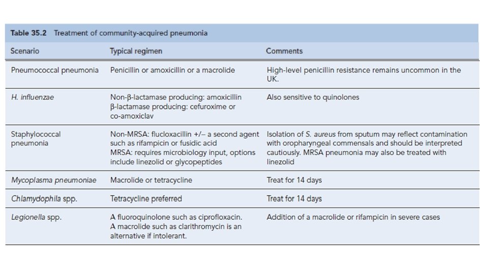
 Clinical studies have defined community-acquired pneumonia (CAP) differently, but fever greater")
Community-acquired pneumonia (CAP) Clinical studies have defined community-acquired pneumonia (CAP) differently, but fever greater than (38 °C), pleural pain, dyspnoea, tachypnea and new signs on examination of the chest seem to be useful for separating CAP from bronchitis in the absence of a chest X-ray. • Typical (classic symptoms of pneumonia) • Atypical (extra-pulmonary symptoms)

Causative organisms ØBacterial: • Typical pneumonia: S. pneumoniae. H. inluenzae and S. aureus, • Atypical pneumonia: Legionella pneumophila, Mycoplasma pneumoniae, Chlamydophila psittaci, Coxiella burnetii. ØViral: • Influenza can cause a primary viral pneumonia as well as be complicated by secondary bacterial (particularly staphylococcal pneumonia), • Chickenpox can be complicated by primary varicella pneumonia particularly in adults, • Cytomegalovirus is capable of causing a variety of infections, including pneumonia.

Clinical features • Cough, with purulent or blood-stained sputum. • Dyspnea and pleuritic chest pain. • Fever. • Fatigue • Confusion or changes in mental awareness (in elderly)
 • Blood")
Diagnosis • Sputum and culture (dependent upon the quality of the specimen) • Blood cultures • Plasma and urine testing for pneumococcal antigen • Bronchoalveolar lavage may be required to tailor treatment (for sever cases).

Empirical treatment
 • Hospital-acquired pneumonia (HAP) is defined as pneumonia developing in a")
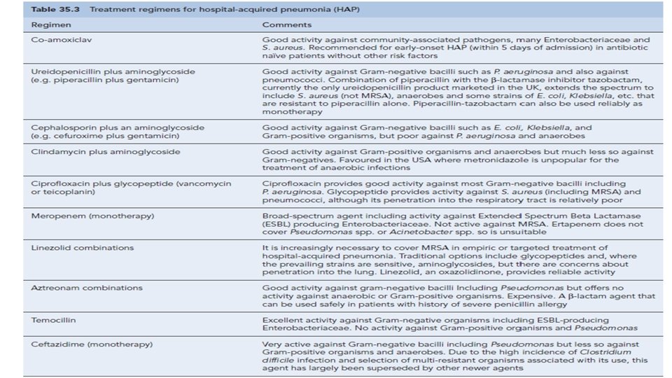
Hospital-acquired pneumonia (HAP) • Hospital-acquired pneumonia (HAP) is defined as pneumonia developing in a hospital at least 48– 72 hours after being admitted. • It is a major cause of morbidity and mortality in hospital patients in the developed world. • Up to 50% of cases are acquired on intensive care units. Predisposing features include stroke, mechanical ventilation, chronic lung disease, recent surgery and previous antibiotic exposure.

Clinical features • present with sepsis and/or respiratory failure. Causative organisms • It is usually caused by a bacterial infection, rather than a virus. • Gram-negative organisms (P. aeruginosa, E. coli, K. pneumonia). • Gram-positive organism (S. aureus, including MRSA) • S. pneumonia and Haemophilus inluenzae.

Diagnosis • Sputum is commonly sent for culture (it may be contaminated by mouth flora. If the patient has received antibiotics, the normal mouth flora is often replaced by resistant organisms such as staphylococci or Gram-negative bacilli, making the interpretation of culture results difficult). • Bronchoalveolar lavage is often more helpful. • Blood cultures.

Empiric therapy • Broad spectrum AB • The choice of antibiotics will be influenced by: -preceding antibiotic therapy, -the duration of hospital admission

Aspiration pneumonia • Initiated by inhalation of stomach contents contaminated by bacteria from the mouth. • Risk factors include alcohol, hypnotic drugs and general anaesthesia (Make a patient vomit while unconscious). • Gastric acid is very destructive to lung tissue and leads to severe tissue necrosis. • Damaged tissue is then prone to secondary infection often with abscess formation. • Treatment with metronidazole plus amoxicillin is usually adequate
 usually refers to the presence of organisms")
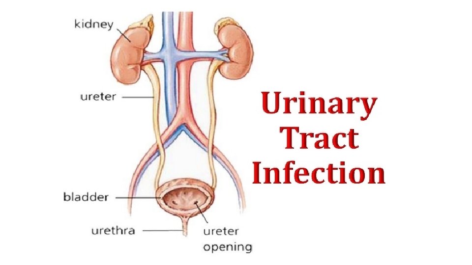
Definition: The term urinary tract infection (UTI) usually refers to the presence of organisms in the urinary tract together with symptoms, and sometimes signs, of inflammation.

It is more precise to use one of the following terms:

Normally: • Small numbers of bacteria are found in the anterior urethra and may be washed out into urine samples. ØUrethral contaminants: Counts of fewer than 1000 bacteria/m. L ØSignificant Bacteriuria: Defined as the presence of at least 100, 000 bacteria/m. L of urine along with symptoms.
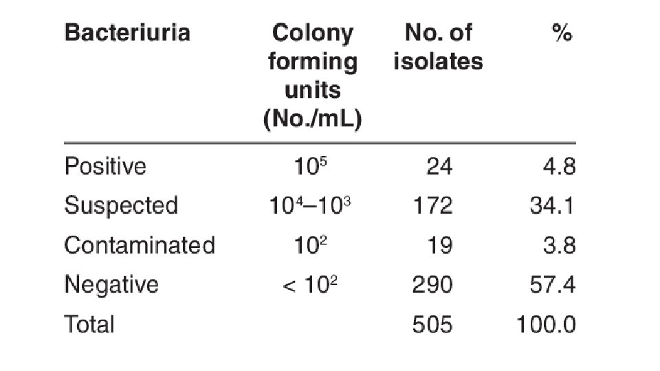

ØAsymptomatic Bacteriuria: • Significant bacteriuria in the absence of symptoms in the patient. ØCystitis: • Syndrome of frequency, dysuria and urgency, • Usually suggests infection restricted to the lower urinary tract (the bladder and urethra). ØUrethral syndrome: • Syndrome of frequency and dysuria in the absence of significant bacteriuria with a conventional pathogen

ØAcute pyelonephritis: • An acute infection of one or both kidneys. • Usually, the lower urinary tract is also involved.

Chronic pyelonephritis: ØIt is a term used in different ways: • Continuous excretion of bacteria from the kidney, • Frequent recurring infection of the renal tissue, • Particular type of pathology of the kidney seen microscopically or by radiographic imaging, which may or may not be due to infection. ØAlthough chronic infections of renal tissue are relatively rare, they do occur in the presence of kidney stones and in tuberculosis.

Relapse and Reinfection • Relapse: is recurrence caused by the same organism that caused the original infection. • Reinfection: is recurrence caused by a different organism, and is therefore a new infection.

Aetiology and risk factors • Age & Gender • Causative Bacterium • Underlying Structural Abnormalities • Hospital-acquired urinary infections

AGE & GENDER • UTI is a problem in all age groups. • In infants up to the age of 6 months… much more common in boys than in girls. • In preschool children and adult … the prevalence is more in girls. • In the elderly, the prevalence of bacteriuria rises dramatically in both sexes.
 Escherichia coli is the most common • (20%) Gram-negative enteric")
Causative Bacterium • (80%) Escherichia coli is the most common • (20%) Gram-negative enteric bacteria such as Klebsiella and Proteus species, and by Gram-positive cocci, particularly enterococci and Staphylococcus saprophyticus. • Rare causes: anaerobic bacteria and fungi • Viruses (in immunocompromised patients, particularly children)

Underlying Structural Abnormalities • Congenital anomalies, • Neurogenic bladder, • Obstructive uropathy, is often caused by more resistant organisms such as • Pseudomonas aeruginosa, • Enterobacter and Serratia species

Acquired • Hospital-acquired urinary infections, • Especially in patients with urinary catheters.

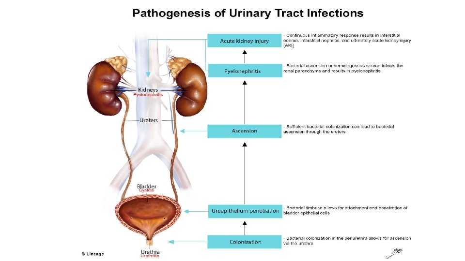
Pathogenesis • There are three possible routes by which organisms might reach the urinary tract: • The Ascending, • Blood-borne, • Lymphatic routes.

Why women more than men? 1. The urethra in women is shorter than in men, 2. The urethral meatus is closer to the anus, 3. Further, sexual intercourse appears to be important in forcing bacteria into the female bladder, 4. The risk is increased by the use of diaphragms and spermicides, which have both been shown to increase E. coli growth.

Natural defence mechanisms üHigh urea concentration and üExtremes of osmolality and p. H inhibit pathologic growth. üThe flushing mechanism of bladder emptying, üThe bladder mucosa, by virtue of a surface glycosaminoglycan, is intrinsically resistant to bacterial adherence. q. If infection occur---WBC are mobilized to the bladder surface to ingest and destroy invading bacteria.

Abnormalities of the urinary tract ØStructural abnormality leading to the obstruction of urinary flow increases the likelihood of infection. • Such abnormalities: • Congenital anomalies of the ureter or urethra, • Renal stones and, • Enlargement of the prostate (in men). ØRenal stones can become infected with bacteria, particularly Proteus and Klebsiella species, and thereby become a source of ‘relapsing’ infection.
 • Is a condition caused by failure of physiological valves at")
Vesicoureteric reflux (VUR) • Is a condition caused by failure of physiological valves at the junction of the ureters and the bladder which allows urine to reflux towards the kidneys when the bladder contracts. • It is probable that VUR plays an important role in childhood UTIs that lead to chronic renal damage (scarring) and persistence of infection.
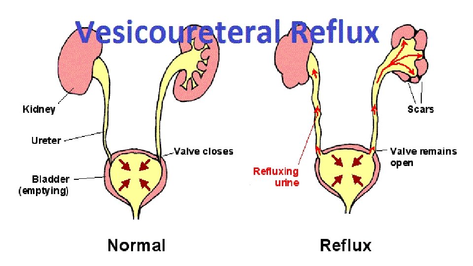

Clinical manifestations in Babies and Infants • Failure to thrive, vomiting, fever, diarrhoea and apathy • Misdiagnosed because the signs may not be referable to the urinary tract. ØPrognosis: • Renal scarring, • Chronic pyelonephritis in adulthood, • Hypertension and • Renal failure.

Clinical manifestations in Children • Classic symptoms such as frequency, dysuria, haematuria. • Acute abdominal pain and vomiting

Clinical manifestations in Adults ØLower UTI, • Frequency, Dysuria, Urgency, Haematuria. ØAcute pyelonephritis (upper UTI) • Fever, loin pain in addition to lower tract symptoms. • Systemic symptoms may vary from insignificant to extreme malaise. ,

Clinical manifestations in Elderly • UTI is one of the most frequent causes of admission to hospital. • Majority of cases are asymptomatic. • Symptoms are not diagnostic because frequency, dysuria, hesitancy and incontinence are common in elderly people without infection. • The infection may be the cause of deterioration in pre-existing conditions such as diabetes mellitus or congestive cardiac failure.

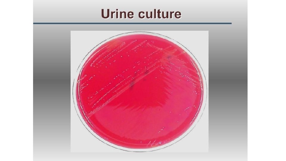
Investigations • The key to successful laboratory diagnosis of UTI lies in obtaining an uncontaminated urine sample for microscopy and culture. • Specimens must reach the laboratory within 1– 2 h or should be refrigerated; otherwise, any bacteria in the specimen will multiply and might give rise to a false-positive result.

Dipsticks

Microscopy

Management

Non-specific treatments • Drink a lot of fluids • Frequent bladder emptying. • Urinary Analgesics such as potassium or sodium citrate, which • Alkalinise the urine, but these should be used as an adjunct to antibiotics (but not Nitrofurantoinwhich is active only at acidic p. H).

Decision of Treatment • Symptomatic UTI---- need AB to eradicate both symptoms and pathogen. • Asymptomatic bacteriuria---- may or may not need treatment (depending upon the circumstances of the individual case). • Bacteriuria in children and in pregnant----- need AB

Antimicrobial agents • Blood levels of antibiotics appear to be unimportant in the treatment of lower UTI; what matters is the concentration in the urine. • However, blood levels probably are important in treating pyelonephritis, which may progress to bacteraemia.

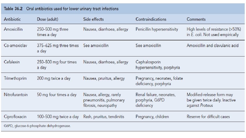
Uncomplicated lower UTI ØTreatment in adult • Trimethoprim, • Oral cephalosporin such as cefalexin, • Co-amoxiclav or • Nitrofurantoin, • Quinolones ØTreatment in adult • β-lactams, • Trimethoprim and • Nitrofurantoin.

Cystitis ØOral treatment include: • Trimethoprim, • β-lactams, particularly amoxicillin, co-amoxiclav and cefalexin, • Fluoroquinolones (ciprofloxacin, norfloxacin and ofloxacin), • Nitrofurantoin. ØIntravenous administration include: • β-lactams such as amoxicillin and cefuroxime, • Quinolones, • Aminoglycosides such as gentamicin.
 • Short-course")
Duration of treatment • Traditionally, a course of 7– 10 days (β-Lactams) • Short-course regimens for 3 -days (trimethoprim and quinolones). • Or even single-dose therapy. • Single-dose therapy, advantages: • Low cost, good adherence and the minimisation of side effects, • Disadvantages: • Less effective than when the same agent is used for longer.

Acute pyelonephritis Ø Severely ill patient ----- • A first-choice agent would be Parenteral antibiotic: • Cefuroxime, • Gentamicin or • Ciprofloxacin. • When the patient is improving, switch to oral therapy, like Quinolone for 10– 14 days. Ø Less severely ill patient… • Oral antibiotic with a shorter course

Catheter-associated infections ØEven with the very best catheter care, most will have infected urine after 10– 14 days of catheterisation, ØThe principles of antibiotic therapy for catheter-associated UTI as follows: • Do not treat asymptomatic infection. • If possible, remove the catheter before treating symptomatic infection.
 of: • Trimethoprim (100")
Prevention and prophylaxis For Adult only: long-term, low dose (Once) of: • Trimethoprim (100 mg) or • Nitrofurantoin (50 mg) at night will suffice.

Thank you
- Slides: 91