
Ultrasonography in Anorectal disorders Dr Suman Sharma Associate





- Slides: 60
Ultrasonography in Ano-rectal disorders Dr. Suman Sharma Associate Professor, Department of Shalya Tantra National Institute of Ayurveda E-mail-sumanhp 2006@gmail. com Phone- 9418159666 rabiezahran@Gawab. com
Ultrasonography Definition • Ultrasonography is study of internal organs, structures or blood vessels using high frequency sound waves, the actual test called ultrasound scan or sonogram.
Definition (contd. ) Ultrasound are sound waves of frequencies greater than audible to human ear i. e. greater than 20, 000 Hz.
Principles • A sound waves travels in a pulse & when it is reflected back it becomes an echo. The pulse-echo principle is used for ultrasound imaging. • A pulse generated by one or more piezo-electric crystals in an ultrasound probe or transducer. • Ultrasound probe crystal is shocked by single extremely short pulse of electricity to vibrate at a frequency determined by its thickness.
Principle • Once echo are converted into electrical signals, these are processed & transformed into a visual display of the measure of the amplitude of the echo this is echo quantification. • The transducer picks up the return echo & record any changes in the pitch or direction of the sound, the image is immediately visible on the screen.
sound waves travel faster in solids than liquids or gases. The major cause of attenuation in soft tissue is absorption,
1. Ultrasound waves – They are waves of very high frequency ranging between 3. 5 – 10 MHz and up to 20 MHz in endo-sonography. – When the frequency penetration. the resolution and
Different Patterns found in USG Echo free Hypo echoic Hyper echoic Heterogeneous
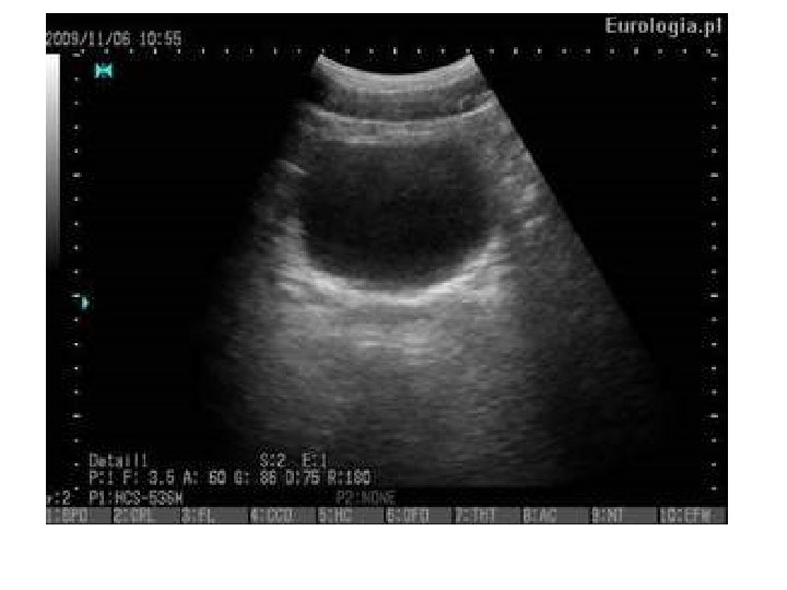
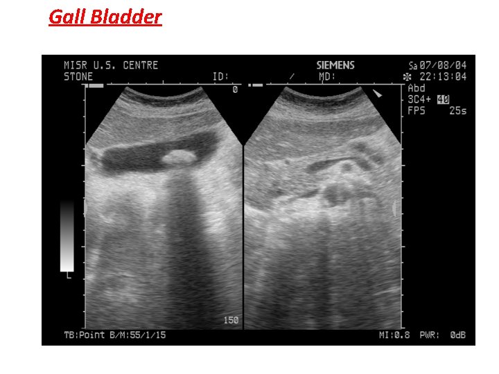
Echo pattern Echofree : When ultrasound waves pass through fluids ( ascites- simple cyst- blood vessels) no reflection occurs and these areas appears as black areas with posterior enhancement.
Echo pattern Echogenic : When ultrasound waves pass through solids (bones – stone) all waves are reflected and appears as white color with posterior shadow.
Gall Bladder
Normal Anatomy of Gall bladder
Liver metastases Heterogeneous echogenicity
Trans Rectal Ultra-sonography (TRUS) or (Endo-rectal Ultra-sonography)
Uses of Endo-Rectal USG This is becoming increasingly popular • Detection of ano-rectal and pelvic disorders. • Useful in detecting prostatic abnormalities. • Getting USG guided Biopsies
Rectal and Pelvic disorders (Indications) • Benign diseases Faecal incontinence Inflammatory conditions
Rectal and Pelvic disorders (Indications) • Detection and staging of CA Rectum (commonest indication) – – Early detection Staging Guiding while chemotherapy or surgery alone is required EUS can assess suitability for transanal excision • Peri-rectal lesions in the pelvis • Retro-rectal masses can be visualized and assessed. • Diagnosis and management of submucosal lesions such as submucosal lipoma, carcinoid tumour and occasionally in gastro intestinal tumours [GIST] as well.
Advantages of TRUS • TRUS provides excellent visualization of the layers of the rectal wall. • Accurate /useful tool for staging primary rectal cancer and determining rectal wall integrity. • It is a fast and minimally invasive technique performed with portable equipment. • It accurately assesses the anal sphincter and provides critical information for planning the appropriate treatment of peri-anal fistulae and fecal incontinence. • TRUS is better than MRI for the evaluation of superficial tumors, whereas MRI provides a better visualization of locally advanced or stenosing cancers. • TRUS is comparable to MRI in the staging of peri-rectal lymph nodes. • Although TRUS is limited in the evaluation of the circumferential resection margin due to its small field of view, 3 D TRUS can improve the accuracy of diagnosis of anorectal diseases and therefore should have an expanded role in the management of patients with anorectal diseases.
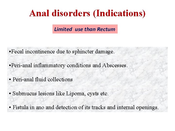
Anal disorders (Indications) Limited use than Rectum • Fecal incontinence due to sphincter damage. • Peri-anal inflammatory conditions and Abscesses. • Peri-anal fluid collections • Submucus lesions like Lipoma, cysts etc. • Fistula in ano and detection of its tracks and internal openings.
Preparation of the patient • The patient must kept nil orally prior to examination. • The patient must be prepared with enemas to remove all air, stool and mucus from the rectum because they can create artifacts during the study.
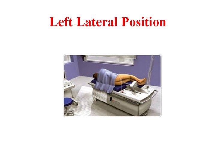
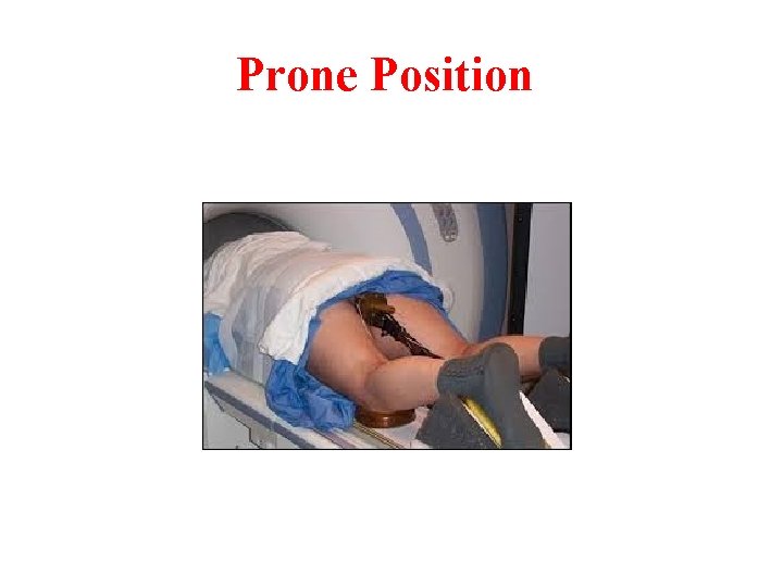
Position • Left lateral decubitus position ( commonly used ). • Knee-chest position. • Prone Position
Left Lateral Position
Prone Position
Technique • Digital rectal exam to evaluate the size, fixation, location and morphology of the rectal lesion. • Following proctoscopy and after estimating the tumor size and distance from the anal verge ERUS is prepared. • A baloon is placed over the crystal of the probe inserted into the rectum, past the proximal border of rectal mass • Then balloon is inflated with water (about 30 -60 ml of water) • Imaging of rectum is initiated as the probe is withdrawn.
Technique • To evaluate the lesions of anal canal the balloon may be replaced with sonolucent plastic cap and Rectum is filled with 150 cc of water.
Technique • Images are usually obtained by using an ultrasound frequency of 5 -15 MHz, depending on which part of rectum or anal canal is being examined.
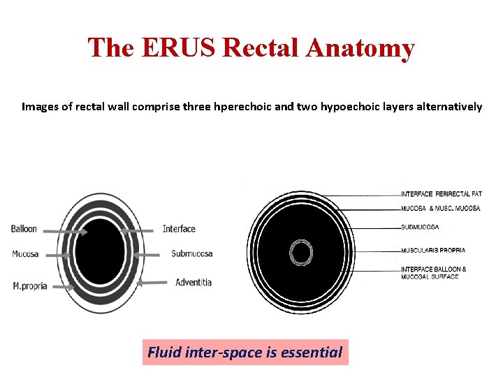
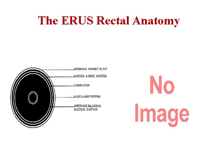
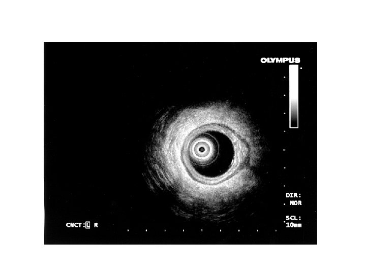
The ERUS Rectal Anatomy Images of rectal wall comprise three hperechoic and two hypoechoic layers alternatively T Fluid inter-space is essential
The ERUS Rectal Anatomy
The ERUS Rectal Anatomy
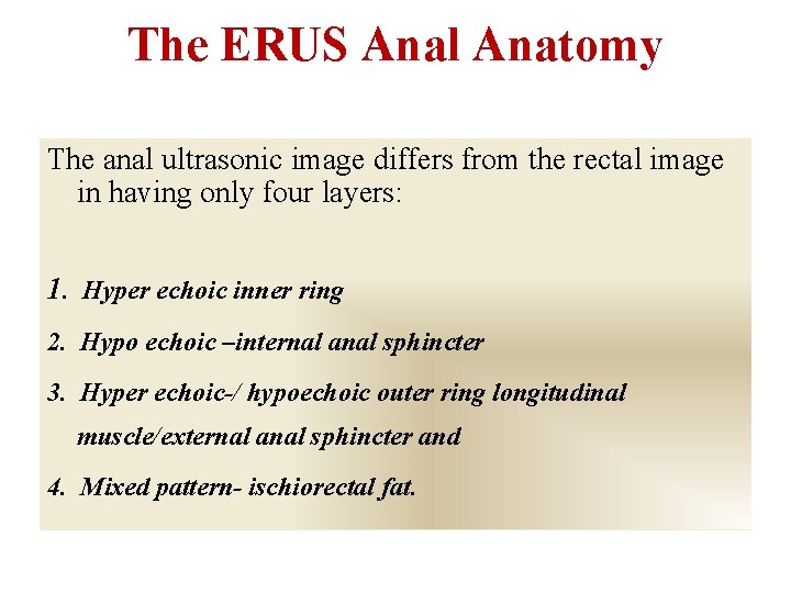
The ERUS Anal Anatomy The anal ultrasonic image differs from the rectal image in having only four layers: 1. Hyper echoic inner ring 2. Hypo echoic –internal anal sphincter 3. Hyper echoic-/ hypoechoic outer ring longitudinal muscle/external anal sphincter and 4. Mixed pattern- ischiorectal fat.
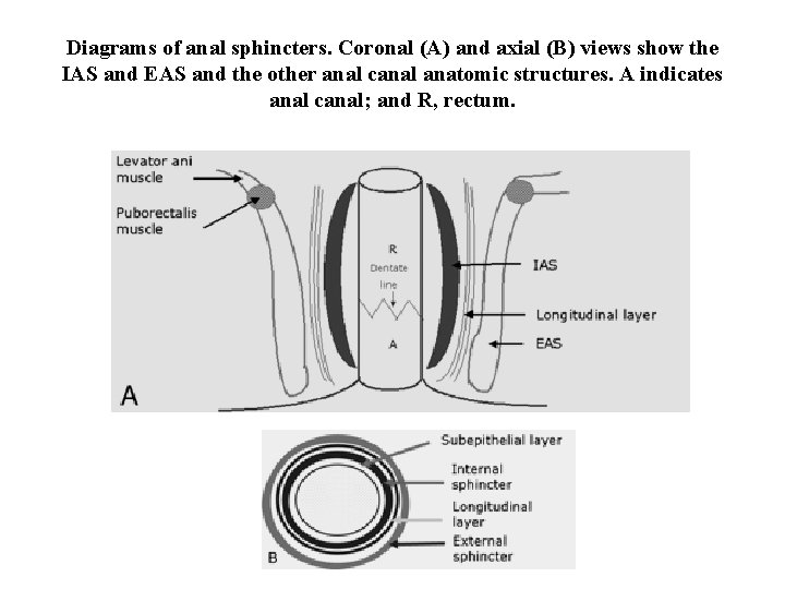
Diagrams of anal sphincters. Coronal (A) and axial (B) views show the IAS and EAS and the other anal canal anatomic structures. A indicates anal canal; and R, rectum.
Upper anal canal level, identified by the horseshoe sling of the puborectalis muscle posteriorly (arrows) and loss of the EAS in the midline anteriorly. The IAS is also shown by arrowheads.
Middle canal level, identified by the completion of the EAS ring anteriorly (black arrows) and maximum IAS thickness (white arrows).
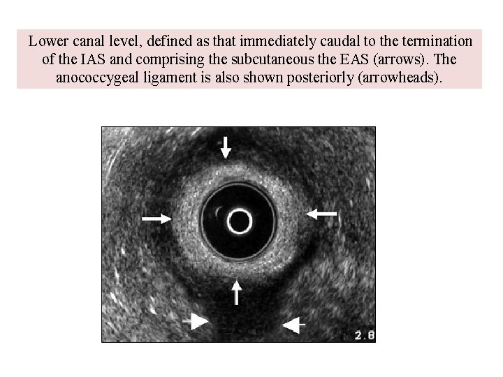
Lower canal level, defined as that immediately caudal to the termination of the IAS and comprising the subcutaneous the EAS (arrows). The anococcygeal ligament is also shown posteriorly (arrowheads).
Common Diseases
Fecal incontinence • To identify sphincter damage, type of sphincter damages, fibrosis and atrophy. • To identify those patients with sphincter damage who may benefitted from surgical repair.
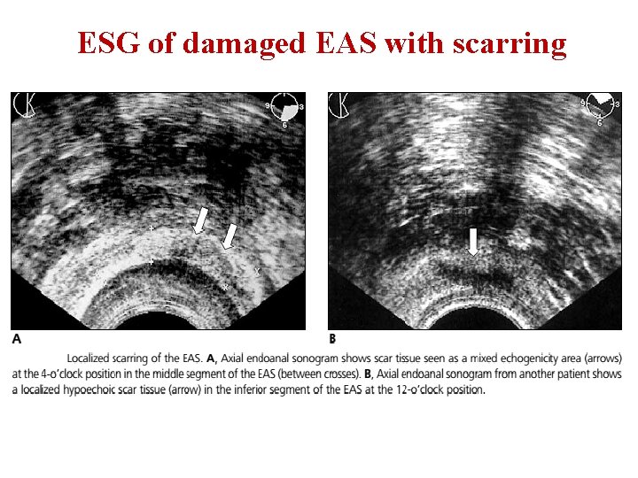
ESG of damaged EAS with scarring
Peri-anal Abscesses and Fistulae Axial (left) and sagittal (right) endoanal sonograms show a transsphincteric fistula with an abscess within ischioanal fossa at the 2 -o’clock position
Peri-anal Abscesses and Fistulae Axial and sagittal magnified endoanal sonograms show the intersphincteric component of the abscess (a) at the 12 o’clock position
Peri-anal Abscesses and Fistulae The IAS is shown between black arrows, and the EAS is shown between white arrows
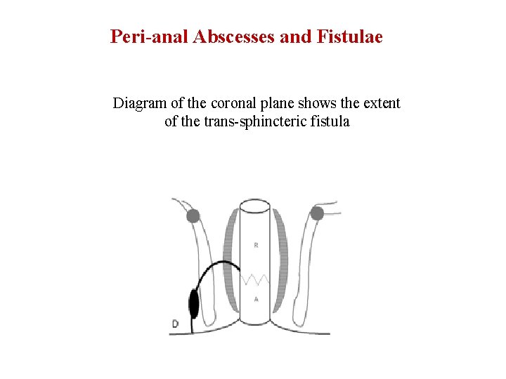
Peri-anal Abscesses and Fistulae Diagram of the coronal plane shows the extent of the trans-sphincteric fistula
Peri-anal Abscesses and Fistulae Endoanal sonogram shows an ischioanal abscess at the 3 - to 6 -o’clock position (black arrows) and its transsphincteric extension through both the markedly thinned EAS and IAS (white arrows) at the level of the middle anal canal
Peri-anal Abscesses and Fistulae After cannulation of the fistula and injection of peroxide, on a subsequent endoanal sonogram through the high anal canal level, prompt visualization of the peroxide was noted in the abscess (black arrows) and fistula extending through both the markedly thinned IAS and puborectalis muscle (PRM), which extends into a small superficial submucosal abscess cavity (white arrows).
Peri-anal Abscesses and Fistulae Endoanal sonogram through the supralevatoric level obtained before administration of peroxide reveals a horseshoe supralevatoric abscess cavity (black arrows) and a deficiency area in the rectal wall at the 6 o’clock position (white arrow).
Peri-anal Abscesses and Fistulae After peroxide injection, an endoanal sonogram clearly depicts the suprasphincteric abscess cavity (black arrows) communicating with the anal lumen (white arrow). The internal opening is shown as a subepithelial breach connecting with the intersphincteric tract through an internal sphincteric defect.
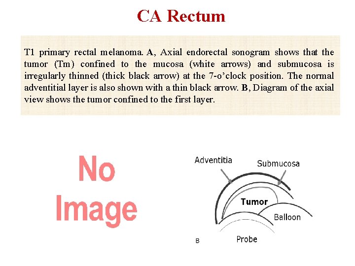
CA Rectum T 1 primary rectal melanoma. A, Axial endorectal sonogram shows that the tumor (Tm) confined to the mucosa (white arrows) and submucosa is irregularly thinned (thick black arrow) at the 7 -o’clock position. The normal adventitial layer is also shown with a thin black arrow. B, Diagram of the axial view shows the tumor confined to the first layer.
CA Rectum T 1 overstaging adenocarcinoma. Axial endorectal sonogram after endoscopic biopsy shows a mass confined with an adventitial layer (arrows) at the 6 o’clock position. Overstaging of the T 1 tumor was caused by presence of hemorrhage in the biopsy area.
CA Rectum T 2 adenocarcinoma. Axial endorectal sonogram shows a mass confined with the adventitial layer (arrows) at the 12 -o’clock position. There is also a lymph node (4 mm in diameter; arrowhead) adjacent to the tumor (Tm).
CA Rectum T 2 overstaging adenocarcinoma. A, Axial endorectal sonogram shows extension of the tumor (large arrows) through the muscularis propria (small arrow) into the perirectal tissue at the 3 -o’clock position. B, Continuity of the muscularis propria layer (arrows) with probe rotation; the T 2 tumor can be seen.
CA Rectum T 3 adenocarcinoma. Axial endorectal sonogram shows that the thin and echogenic fifth (adventitial) layer is focally disrupted at the 10 -o’clock position (arrows).
CA Rectum T 4 adenocarcinoma. A, Axial endorectal sonogram shows a fatty tissue plane between the tumor (Tm) and the right seminal vesicula (S). There is no invasion to the seminal vesicula. B, Axial endorectal sonogram from the same patient at another plane. There is considerable invasion of the left seminal vesicula.
Villous adenoma. A and B, pedunculated polypoid tumor (Tm; arrows) -3 to 6 -o’clock position. The rectal submucosa (small arrowhead) and adventitia (large arrowhead) are shown in B. C and D, Magnified endorectal sonogram and diagram show the polyp stalk (C, white arrows) and its relationship to the rectal submucosal (C, long black arrow) and adventitial (C, short black arrow) layers. SV indicates seminal vesicula.
CA Rectum Pathologic lymph node in rectal non-Hodgkin malignant lymphoma. Axial endorectal sonogram shows a pathologic lymph node (arrow) with a diameter of 10 mm, round and hypoechoic adjacent to the tumor (Tm).
Treatment Options in Ayurveda • • Improvement of status of Agni Rasayan Sevan Snehana Basti Chikitsa Aushadh Prayoga Vyayama Ahara Vyavastha Avoidance of vata prakopaka Ahara Vihara