ACUTE GINGIVAL INFECTIONS Contents n Introduction n Classification


: q Terminologies: Trench mouth , Vincent's stomatitis, Vincent’s infection,")
is an inflammatory destructive disease of the gingiva , which presents")
.")


-classified into 3 forms Acute Subacute Chronic (Now considered")
 proposed following stages: Stage 1: Only tip of the")
 extended the staging of these oral necrotizing diseases as follows: Ø")
described four zones namely: 1. Zone I—Bacterial zone: It is")


 Alleviation of the acute inflammation plus treatment")


 n It is a viral infection of the oral mucous")


- Slides: 52
ACUTE GINGIVAL INFECTIONS
Contents: n Introduction n Classification n Acute necrotizing ulcerative gingivitis. Terminology Definition Clinical features Oral symptoms Classification Histopathology Etiology Diagnosis Differential diagnosis Treatment.
n Acute Herpetic Gingivostomatitis Clinical features Oral symptoms History Histopathology Diagnosis Differential diagnosis n Pericoronitis Definition Types Clinical features complications Treatment
Introduction
Classification Of Various Acute Gingival Lesions: A. Traumatic lesions of gingiva: • Physical injury • Chemical injury B. Viral infections: • Acute herpetic gingivostomatitis • Herpangina • Hand, foot and mouth diseases • Measles • Herpes varicella/zoster virus infection • Glandular fever
C. Bacterial infections: • Acute necrotizing ulcerative gingivitis Tuberculosis • Syphilis D. Fungal diseases: • Candidiasis. E. Gingival abscess F. Apthous ulceration G. Erythema multiforme. H. Drug allergy and contact hypersensitivity n
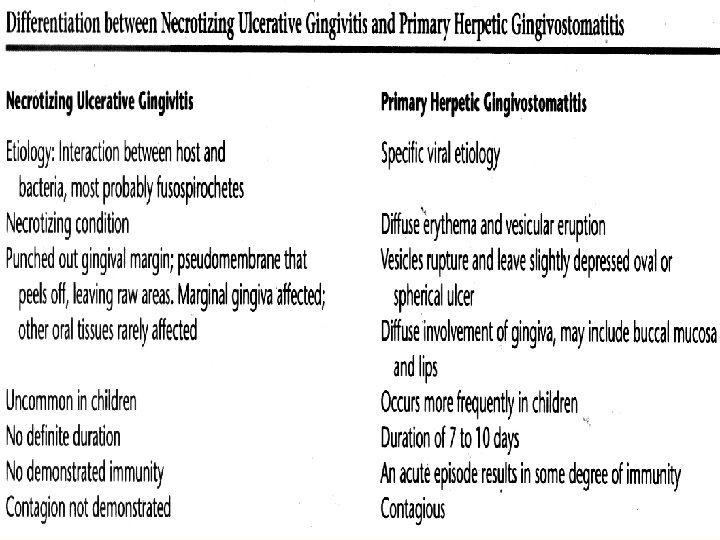
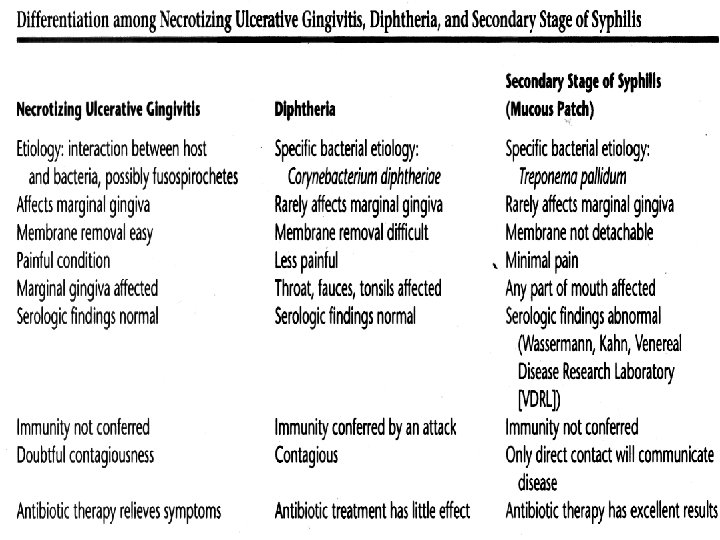
Acute Necrotizing Ulcerative gingivitis (ANUG): q Terminologies: Trench mouth , Vincent's stomatitis, Vincent’s infection, Plaut-Vincents stomatitis, Stomatitis Ulcerosa. o NUG (Necrotizing Ulcerative Gingivitis) can cause destruction of the supporting structures- bone loss NUP (Necrotizing ulcerative periodontitis) q Maybe localized or generalized.
Definition: Necrotizing Ulcerative gingivitis(NUG)is an inflammatory destructive disease of the gingiva , which presents characteristic signs and symptoms. -
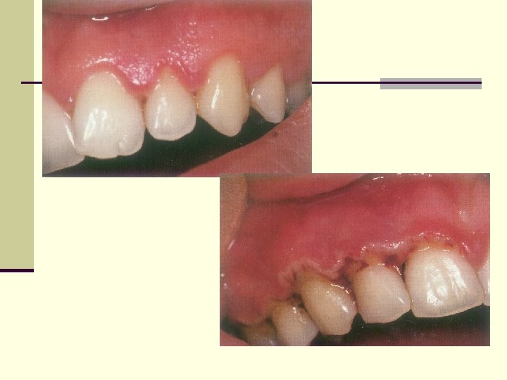
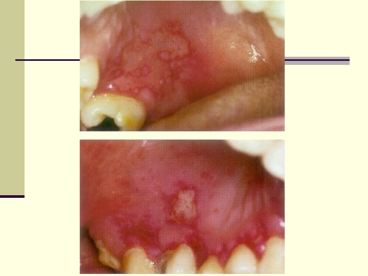
Clinical features: n Painful ulceration and engorgement of the interdental papillae(punched out inverted papillae). These craters are covered by greyish pseudo membranous slough, which is demarcated from the remaining of the mucosa by a pronounced linear erythema. n Pseudo membrane- easily removed exposing raw bleeding tissue. Ø .
Ø Pronounced tendency to gingival bleeding. Ø Halitosis. Ø Sialorrhoea. Ø Fever, Malaise and lymphadenopathy. Ø Other sites of involvement: Occasional spread to contiguous or adjacent tissues.
Oral Symptoms 1. The lesions are extremely sensitive to touch. 2. Complains of a constant radiating, gnawing pain that is intensified by eating spicy or hot foods and chewing. 3. There is a metallic foul taste, and the patient is conscious of an excessive amount of “pasty saliva”.
Extraoral Signs and Symptoms n Mild to Moderate stages- local lymphadenopathy, elevated temperature. n Severe -Marked systemic complications such as high fever, increased pulse rate, leukocytosis, loss of appetite and general lassitude are common. n Systemic reactions -more severe in children. Insomnia, constipation, gastrointestinal disorders, headache, and mental depression also accompany.
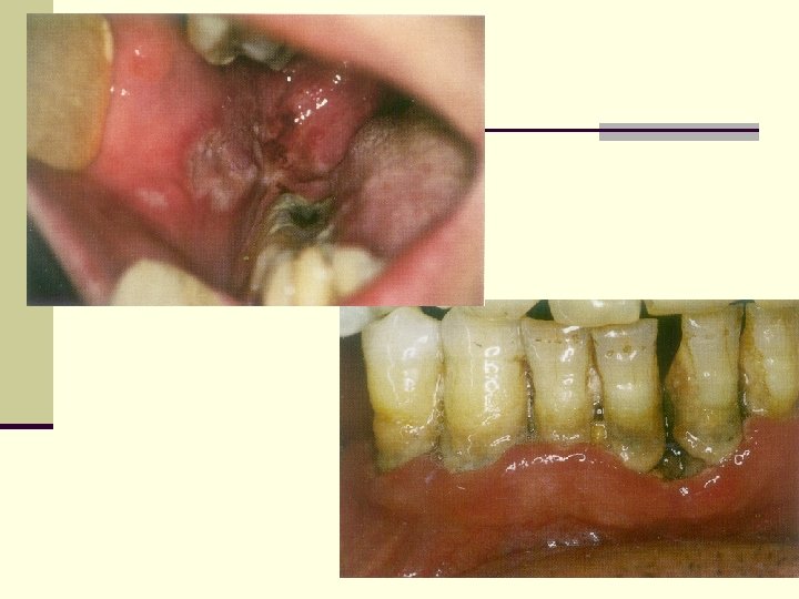
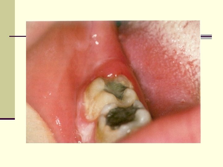
Ø ANUG in malnourished, debilitated and immunocompromised patients may extend onto the oral mucosa and skin resulting in cancrum oris (noma; gangrenous stomatitis). .
Spreading necrosis Gangrene penetrates buccal mucosa Oro-cutaneous fistula and scarring. n.
q most significant criteria in diagnosis of ANUG are : v Interproximal necrosis and ulceration. v History of soreness and bleeding in the area.
Classification: n According to Pindborg J(1951)-classified into 3 forms Acute Subacute Chronic (Now considered a misidentification of recurrence of the disease).
STAGES OF ANUG Pindborg (1967) proposed following stages: Stage 1: Only tip of the papilla was affected. Stage 2: Marginal gingiva involved with punched out papillae. Stage 3: Attached gingiva was also involved. Stage 4: Bone was exposed.
Horning and Cohen(1995) extended the staging of these oral necrotizing diseases as follows: Ø Stage 1: Necrosis of the tip of the interdental papilla Ø Ø Ø (93%) Stage 2: Necrosis of the entire papilla (19%) Stage 3: Necrosis extending to the gingival margin (21%). Stage 4: Necrosis extending also to the attached gingiva(1%). Stage 5: Necrosis extending into buccal or labial mucosa (6%). Stage 6: Necrosis exposing alveolar bone (1%). Stage 7: Necrosis perforating skin of cheek (0%).
Histopathology: Listgarten and colleagues (1965)described four zones namely: 1. Zone I—Bacterial zone: It is the most superficial zone, consists of varied bacteria, including a few spirochetes of the small, medium -sized and large types. 2. Zone II—Neutrophil-rich zone: Contains numerous leukocytes predominantly neutrophils with bacteria including spirochetes of various types.
3. Zone III—Necrotic zone: Consists of a dead tissue cells, remnants of connective tissue fragments, and numerous spirochetes. 4. Zone IV—Zone of spirochetal infiltration: Consists of a well preserved tissue infiltrated with spirochetes of intermediate and large-sized without other organisms.
Etiology: n Role of Bacteria n Plaut and Vincent introduced the concept that acute n necrotizing ulcerative gingivitis is caused by a specific bacteria namely, a fusiform bacillus and a spirochetal organisms( Treponema Microdentium. ) n More recently Loesche and colleagues described a constant and a variable flora associated with ANUG. n Constant flora is composed of Fusospirochetal organisms and also Prevotella Intermedia. The variable flora consists of a heterogeneous array of bacterial types. n Role of Host response
Predisposing factors 1. Local predisposing factors n Preexisting gingivitis n Injury to gingiva n Smoking, tobacco 2. Systemic predisposing factors n Nutritional deficiency(vit B 2, vit C) n Debilitating diseases (AIDS, Blood Dyscrasias etc) n Psychosomatic factors: stress
Diagnosis n Diagnosis is based on clinical findings. n A bacterial smear may be used to corroborate the clinical diagnosis, but it is not necessary nor definitive because the bacterial picture is not appreciably different from the other conditions (Gingivitis, periodontal pockets etc).
Differential diagnosis include: a. Gonococcal stomatitis. b. Agranulocytosis. c. Vincent’s angina. d. Desquamative gingivitis. e. Acute necrotizing ulcerative gingivitis in leukaemia. f. Acute necrotizing ulcerative gingivitis in AIDS. g. Streptococcal gingivostomatitis.
The treatment of NUG consists of (1) Alleviation of the acute inflammation plus treatment of chronic disease either underlying the acute involvement or else where in the oral cavity, (2) Alleviation of generalized toxic symptoms such as fever and malaise, and (3) Correction of systemic conditions that contribute to the initiation or progress of the gingival changes.
Treatment 1. Non-ambulatory patient: With symptoms of generalized systemic complications. 2. Ambulatory patient: With no serious systemic complications.
Treatment for Non-ambulatory Patients Day 1: n Local treatment limited to gently removing the necrotic pseudomembrane with a pellet of cotton saturated with hydrogen peroxide (H 2 O 2) n Advised bed rest and rinse the mouth every 2 hours with a diluted 3 percent hydrogen peroxide (H 2 o 2) n Systemic antibiotics like penicillin or metronidazole can be prescribed. Day 2: n If condition is improved, proceed to the treatment described for ambulatory patients. If there is no improvement at the end of the 24 hours, a bedside visit should be made. The treatment again includes gently swab the area with hydrogen peroxide, instructions of the previous day are repeated. Day 3: n Most cases, the condition will be improved, start the treatment same as for ambulatory patients.
Treatment for Ambulatory Patients n First visit: Cotton pellet to remove pseudomembrane and non-attached surface debris. n Superficial calculus is removed with ultrasonic scalars. n Patients with moderate or severe necrotizing ulcerative gingivitis and local lymphadenopathy, Antibiotic regime - Penicillin 500 mg thrice daily, Penicillin-sensitive patients erythromycin or metronidazole 200 mg or 400 mg twice daily for seven days. n Sub-gingival scaling and curettage are contraindicated.
Instructions to the patient: Avoid smoking and alcohol. n 2. Rinse with 3 percent hydrogen peroxide and warm water for every two hours. n 3. Confine toothbrushing to the removal of surface debris with a bland dentifrice, use of interdental aids and chlorhexidine mouth rinse are recommended. n 1. Second visit: n Scalers and curettes are added to the instrumentarium, shrinkage of the gingiva may expose previously covered calculus which is gently removed. Same instructions are reinforced. Third visit: n Scaling and root planing are repeated, plaque control instructions are given. Hydrogen peroxide rinses are discontinued.
Fourth visit: n Oral hygiene instructions are reinforced and thorough scaling and root planing are performed. Fifth visit: n Appointments are fixed for treatment of chronic gingivitis, periodontal pockets and pericoronal flaps, and for the elimination of all local irritants. Patient is placed on maintenance programme.
Further Treatment Considerations 1. Gingivoplasty. 2. Systemic antibiotics—only in patients with toxic systemic complications. 3. Supportive systemic treatment—copious fluid consumption and administration of analgesics and adequate bed rest. 4. Nutritional supplements—vitamin B/C supplements.
ACUTE HERPETIC GINGIVOSTOMATITIS (AHG) n It is a viral infection of the oral mucous membrane caused by HSV (Herpes simplex virus). It occurs most frequently in infants and children younger than 6 years of age but is also seen in adults. Clinical Features: Oral Signs 1. Diffuse, shiny erythematous, involvement of the gingiva with edema and gingival bleeding. 2. Initial stage-appears as discrete, spherical, Gray vesicles dispersed in different areas, e. g. labial and buccal mucosa, soft palate, pharynx and tongue. After 24 hours the vesicles rupture and form painful small ulcers with a red, elevated, halo-like margins and a depressed yellow and greyish white central portion.
3. Diffuse, edematous, erythematous enlargement of the gingiva with a tendency towards bleeding is seen (primary herpes occuring without ulceration). 4. The course of the disease is 7 to 10 days. It may appear in a localized form following operative procedures in the oral cavity and by placement of cotton rolls or by vigorous application of digital pressure.
Oral Symptoms 1. Generalized soreness of the oral cavity 2. The ruptured vesicles are sensitive to touch, thermal changes and foods. Extraoral and Systemic Signs and Symptoms n Involvement of the lips, face (Herpes labialis, “cold sore”) with vesicles and surface scale formation. n Cervical adenitis, fever -101 to 105°F and generalized malaise.
History n The condition frequently occurs after an episode of febrile diseases such as pneumonia, meningitis, influenza and typhoid. It also tends to occur during periods of anxiety, strain or exhaustion. n The location of virus is in the gasserian ganglion. The virus may descend to the lip through the trigeminal nerve.
Diagnosis n Patients history and clinical findings. n Confirmatory testsü ü Direct smear Inoculation of the virus - tissue culture. .
Histopathology n Virus targets epithelial cells n Ballooning degeneration n Acantholysis n Nuclearing n Nuclear enlargement n Infected cells form multinucleated cells n Intraepithelial vesicle formation.
Differential Diagnosis 1. Acute necrotizing ulcerative gingivitis 2. Erythema multiforme 3. Steven’s-Johnson syndrome 4. Lichen planus 5. Desquamative gingivitis 6. Apthous stomatitis (canker sores).
Management n Acyclovir-15 mg 5 times daily for 7 days. n NSAIDS n Antibiotics to prevent opportunistic infections.
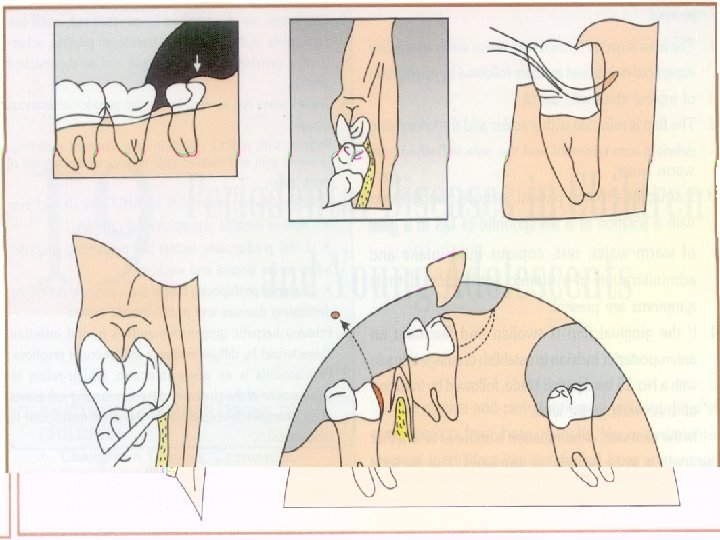
PERICORONITIS Definition n It is an acute infection which refers to inflammation of gingiva and surrounding soft tissues of an incompletely erupted tooth. n It occurs most frequently in the mandibular third molar area. n Types Acute, sub-acute or chronic.
Clinical Features Signs and Symptoms: n Markedly red, edematous suppurating lesion , extremely tender with radiating pain to the ear, throat and floor of the mouth. n Foul taste n Inability to close the jaws. n Swelling of the cheek in the region of the angle of the jaw is seen.
Acute Pericoronitis n Varying degrees of involvement of pericoronal flap as well as with systemic complications. n An influx of inflammatory fluid and cellular exudates results in an increase in bulk of the flap which interferes with complete closure of the jaws. The flap is traumatized by contact with the opposing jaw and inflammatory involvement is aggravated. n Lymphadenitis, toxic systemic complications- fever, leukocytosis and malaise.
Complications • Involvement may become localized-pericoronal abscess. • Occurrence in a partly-erupted vital tooth- cyst formation. • Spread posteriorly -oropharyngeal area and medially into the base of the tongue- difficulty in swallowing. • Involvement of the submaxillary, cervical, deep cervical and retro pharyngeal lymph node. • Peritonsillar abscess formation, cellulitis and Ludwig’s angina occur infrequently.
Treatment is based on • Severity of the inflammation. • The systemic complications and, § The advisability of, retaining the involved tooth.