Plasma Cell Dyscrasias Miten R Patel MD Cancer


- Slides: 41

Plasma Cell Dyscrasias Miten R. Patel, MD Cancer Specialists of North Florida

Disclosures • None

Objectives • • • Review types of Plasma cell dyscrasias Epidemiology and definitions Presenting signs and symptoms Myeloma staging Couple of cases Treatment overview

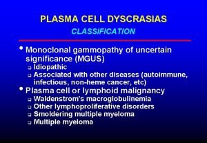
Plasma Cell Dyscrasias • Monoclonal Gammopathy of Unknown Significance (MGUS) • Smoldering Myeloma (SMM) • Multiple Myeloma (MM) • Waldenstroms Macroglobulinemia (WM) • Amyloidosis • Solitary plasmacytoma • POEMS syndrome

MGUS • Mean age at diagnosis – 70 yo • M>F, incidence increases with age, found in 1 -2% of adults • African Americans 2 -3 x > Caucasians • Present in 3% of persons >50 and 5% of persons over the age of 70 • Most cases sporadic but relatives of patients with MGUS/myeloma have an increased risk (RR 2 -3)

MGUS • Definition – Serum monoclonal protein <3 g/d. L, bone marrow plasma cells <10%, and absence of end organ damage • • Hypercalcemia Renal Failure Anemia/Thrombocytopenia Lytic bone lesions

MGUS • Non-Ig. M MGUS – Progresses to myeloma – Rarely, to AL amyloid, light chain disease or other lymphoproliferative disorder • Ig. M MGUS – Progresses to Waldenstroms macroglobulinemia – Rarely, to myeloma, AL amyloidosis or lymphoma

MGUS • Part of the spectrum of smoldering myeloma and multiple myeloma • Characterized by a monoclonal protein level of <30 g/L (m spike of 3. 0 g/d. L), <10% plasma cells in the bone marrow • Progression to MM or related disorder is about 1% per year. • Clinically asymptomatic premalignant condition

MGUS • No symptoms? – Pts. identified when undergoing testing for other conditions such as neuropathy, vasculitis, hemolytic anemia, rashes, hypercalcemia, elevated ESR, elevated total protein – Likely had existed for years before diagnosis – Finding on labs - usually elevated total protein, immunoglobulin, abnormal SPEP/UPEP, positive IFE, or abnormal light chain ratio

MGUS – Workup • Labs - CBC + diff, BMP (Cr, Ca), SPEP + IFE, Quant Igs, 24 urine IFE, B 2 microglobulin, albumin, serum free light chains • Imaging – Skeletal survey, Certain cases - MRI & PET scan • Bone Marrow biopsy & aspirate – Cytogenetics & FISH studies

MGUS • Once confirmed (Monoclonal protein <3 g/d. L, <10% plasma cells, no CRAB symptoms) – no treatment • Serial followup, every 3 -6 months, exam, assessment and labs, less often after 2 years. • Monitor for developing symptoms (bone pain, hypercalcemia, kidney dysfunction, anemia, etc) • SMM – every 3 months until progression • No treatment unless symptomatic MM

Smoldering Myeloma • Smoldering Myeloma (aka asymptomatic multiple myeloma) – Same as MM but without symptoms and • Hgb > 10. 5, • Monoclonal Ig Peak (usually > 3 g/d. L and/or >10 but <60% clonal plasma cells) • Normal serum Ca and Cr levels • No lytic bone lesions • REQUIRES NO TREATMENT • >60% plasma cells or FLC ratio >100 even without end organ damage, best classified as multiple myeloma

Multiple Myeloma • Presentation – Weakness, Fatigue, Pallor (32%) – Weight Loss (24%) – Radiculopathy, cord compression – Bone pain (58%), fractures – Recurrent infections (dysfunctional immunoglobulin) – Labs – Anemia (73%), hypercalcemia (28%), elevated total protein, kidney dysfunction (48%)

Multiple Myeloma Rouleaux Formation Elevated Sed Rate Elevated C Reactive Protein Normocytic, normochromic anemia Renal Disease, Cr > 2 mg/d. L in 19% Renal insufficiency caused by cast nephropathy (myeloma kidney) or hypercalcemia • Altered mental status caused by hypercalcemia and hyperviscosity • • •

Multiple Myeloma • Monoclonal Ig – – Ig. G 60% Ig. A 20% Light chain only 18% (K>L) Ig. M, Ig. D, Ig. E, non-secretory < 1% • For nonsecretory myeloma (no M protein) or light chain only, diagnosis is often made with bony lesions and plasmacytosis by bone marrow

Myeloma Bone Marrow Aspirate

Myeloma Blood Smear

Myeloma Staging Durie Salmon • Stage 1 – Ca <12, Hgb >10, Normal skeletal survey or solitary plasmacytoma, low M protein with Ig. G <5 g/d. L or Ig. A <3 g/d. L, Bence Jones protein <4 g/24 h • Stage 2 – Neither stage 1 or 3 • Stage 3 – One of the following: Hgb <8. 5, Ca >12, multiple lytic lesions, high M component (Ig. G >7, Ig. A >5 or Bence Jones >12 gm)

• Stage 1 Myeloma Staging ISS – B 2 M ≤ 3. 5 and albumin ≥ 3. 5 • Stage 2 – B 2 M > 3. 5 but <5. 5 or albumin >3. 5 • Stage 3 – B 2 M ≥ 5. 5

Case Presentation • 41 yo WF with no symptoms goes to annual physical at office based clinic. • No symptoms other than mild fatigue • Routine annual labs drawn

Lab draw #1

Lab draw #2

Lab draw #3

Lab draw #4

Bone Marrow Biopsy

Myeloma Treatment • Determine eligibility for HDT and SCT – Age, overall health, psychological evaluation – Avoid alkylating agents (marrow toxic) – Chemotherapy to induce remission – Refer for stem cell collection and transplant – Maintenance therapy – Regular monitoring – ASCT improves median OS by >12 months

Myeloma Treatment • Transplant ineligible – Immunomodulators (Thalidomide, Revlimid, Pomalyst) • DVT risk – Proteosome inhibitors (Velcade, Kyprolis) – Chemotherapy (MP, VAD, Doxil, Hyper. CVAD) – Supportive care • Bisphonates

Myeloma Treatment

Solitary Plasmacytoma • Approx 3% of myeloma patients • No or very low myeloma protein in serum or urine • MR must be done to evaluate patient as it may detect other bone lesions (upstage the patient to myeloma) • Presence of monoclonal protein >1 yr after irradiation denotes progression to myeloma • Treat with XRT (at least 45 Gy). • MM manifests overtime, only 20% remain disease free at 10 yrs • Median time to progression is 2 yrs

Waldenstrom’s • • • Lymphoplasmacytic bone marrow infiltration Malignant B lymphocytes producing monoclonal Ig. M Median age 65; Whites > Blacks; M>F 1500 cases in the US per year Hyperviscosity syndromes – neurologic complaints including vision change, headache, vertigo, nystagmus, dizziness, deafness, diplopia and ataxia • 10 -20% of pts. Have a cryoglobulin (Type I) which precipitates at high T >22 C on cold exposure may cause symptoms such as Raynauds phenomenon, urticaria, purpura and acral cyanosis. • Diarrhea and Steatorrhea (Ig. M infiltration into GI tract)

Waldenstrom’s

Waldenstrom’s Treatment • Goal to reduce hyperviscosity (Ig. M is a pentamer!) and lymphoproliferation – Pheresis if symptoms from the hyperviscosity causing severe neurologic deficits • Chemotherapy – Chlorambucil, other CCU, Cladribine and Fludarabine, Rituxan, IFN, Thalidomide, Ibrutinib, Velcade

Case #2 • 73 yo WF presenting with neuropathy, dizziness, 40 lb weight loss, Ig. M 5454, immediately hospitalized due to neurologic symptoms, started plasma exchange

Case #2 • Bone Marrow – Bone marrow, flow cytometric immunophenotypic analysis: Monoclonal plasma cells and lymphocytes identified, consistent with Waldenstrom's macroglobulinemia.

Case #2 • Dramatic decline in Ig. M (>5000 to <2000) after plasma exchange • Symptoms have improved • Treated with Rituxan and Velcade, Ig. M started rising again. • Changed to Ibrutinib, symptoms remain improved, Ig. M stabilized below 1500 • Ig. M stable for > 2 years now

Amyloidosis • Occurs in 10% of patients with MM • Infiltrative process resulting from amyloid fibril deposition in organs • Fibril is made from the terminal amino acid residue (NH 2) of the variable portion of the light chain Ig molecule • Produced by clonal plasma cells

Amyloidosis • Renal disease - asymptomatic proteinuria to nephrotic syndrome • Cardiomyopathy • Hepatomegaly • Neuropathy • Pseudohypertrophy of muscles • Bleeding problems (Factor X def, Liver dz) • Lung and Skin infiltration

Amyloidosis

Amyloidosis • AL should be suspected in patients with plasma cell dyscrasia history • Biopsy results as shown • SPEP/UPEP - confirms monoclonal protein (plasma cell population) • Treatment - treat underlying plasma cell dyscrasia

Summary • MGUS, SMM and MM are a spectrum of the same disease process • MGUS and SMM patients need close observation • WM patients present with hyperviscosity symptoms and very high Ig. M, total protein • Amyloidosis (AL) is characterized by fibril deposition in organs with damage (most commonly heart/liver/kidney) • Treatment is dependant on the disease process

Questions Miten R. Patel, MD Office 904 -516 -3737 Cell phone 904 -451 -9820 miten. patel@csnf. us
 Waldenstrom macroglobulinemia mnemonic
 Plasma cell neoplasm
 Plasma cell mastitis treatment
 Lipids cell membrane function
 Smoldering multiple myeloma
 Työturvallisuus tilaaja ja toimittaja
 Järviekosysteemi
 Miten dekolonisaatio vaikutti kylmään sotaan?
 Logopedian kandiohjelma
 Miten lattanat lisääntyvät
 Mihin hauskuutta tarvitaan
 Miten referoidaan
 Nestelista tulostettava
 Luetelma yo
 Arvoitusdekkari
 Ympyrälieriön tilavuus
 Mitä kappaleeseen jatkuvasti vaikuttava voima saa aikaan
 Voittoprosentin laskeminen
 Hinnoittelualue
 Lukualueet
 Itseisarvo matematiikka
 Lemcba
 Tunnetuimmat tulivuoret
 Työ ja teho
 Miten magneettinen vuorovaikutus ilmenee
 Laktoosi operoni
 Lentokoneen aerodynamiikka
 Vaihto-opiskelu tanska
 Laadullisen tutkimuksen luotettavuuden arviointi
 Hygieniaosaamistodistus
 Pyramidien rakentaminen
 Op mobiiliavain käyttöönotto
 Millainen englanniksi
 Dr vikas patel
 Dr vikas patel
 Kanika kitchlu-connolly
 Dr niraj patel
 Is it really
 Jaydeep patel youtube income
 Dr niraj patel
 Dr megha patel
 Ushma patel uci
