MUSCLES OF FACIAL EXPRESSION The muscles of facial


, depressor labii inferioris (quadratus) and the mentalis muscles")


")


 of the trigeminal nerve forms")

 is the largest division of")


 emerges from the same point at the")


 of the neck lie beneath the platysma and")


- Slides: 68
MUSCLES OF FACIAL EXPRESSION �The muscles of facial expression arise from the second branchial arch, �are innervated by the seventh cranial nerve (i. e. the facial nerve, cranial nerve VII), �Fluidity of facial movements is orchestrated by their interaction with the SMAS
�The epicranius or occipitofrontalis muscle of the scalp has an anterior and posterior region connected by the galea aponeurotica. Contraction of these muscles allows the skin to slide over the scalp. �The frontalis muscle is a member of the epicranius complex that begins at the anterior hairline and inserts into the forehead and eyebrow skin.
� Midline vertical forehead skin tension lines occur due to variation in distance between the left and right frontalis bellies. � Horizontal skin tension lines occur perpendicular to the frontalis contractile orientation. � Loss of frontalis function results in flattening of forehead skin tension lines and a drooping eyebrow. This occurs when the temporal branch of the facial nerve is disrupted. � Patients with compromised frontalis function may be unable to open their eyes widely, due to the synergistic effect this muscle has with the orbicularis oculi muscle.
�The small periauricular muscles or the temporoparietalis group arise from the superficial temporalis SMAS and the lateral galea. �They help draw back the temporal skin and are innervated by the posterior ramus of the temporal branch of the facial nerve.
� The orbicularis oculi muscle complex is the major group that acts on the eyelid and periorbital skin. It inserts into the medial and lateral canthal tendons and encircles the eye region. � Its palpebral portion has a preseptal component overlying the orbital septum and a pretarsal portion overlying the tarsal plate of the eyelid. The palpebral orbicularis oculi muscle aids in tear excretion. • Upper pretarsal and preseptal muscles depress the upper lid. The orbital component of this muscle group allows voluntary tight closure of the eye. The palpebral portion allows gentle eye closure and blinking.
� The corrugator supercilii muscle is located over the medial upper orbital rim. � It contributes to a 'scowling' facial expression by drawing the eyebrows medially and downward. � It interdigitates with and is covered by the frontalis and orbicularis oculi muscles. � The vertical and oblique skin tension lines of the glabella are caused by contraction of this muscle, which is innervated by the temporal branch of the facial nerve
� The procerus muscle overlies the nasal bone and attaches to the nasal root skin. � It causes foreshortening of the nose and 'rabbit lines‘ (i. e. skin tension lines exaggerated by wrinkling up the nose).
� The nasalis muscle courses across the nasal dorsum and facilitates alar 'flaring‘ and compression. These muscles are innervated by the zygomatic and buccal branches of the facial nerve. � The depressor septi nasi muscle lies deep to the orbicularis oris and can form a transverse skin tension line across the philtrum. It plays a minor role in facial expression by pulling the columella down toward the lip
� The orbicularis oris muscle allows pursing and puckering of the lips, apposition of the corners of the mouth, and pulling of the lips up against the teeth and gingivae. � It has no bony or cartilaginous attachment and is innervated by the buccal or marginal mandibular branches of the facial nerve. � This circumferential muscle is necessary for correct speech and allows enunciation of the letters M, V, F, P and O. � The facial arteries and veins are covered and protected from damage by the lip elevator muscles.
� The quadratus labii superioris muscle group is comprised of several lip elevators. � The levator anguli oris and risorius muscles are mouth angle retractors and elevators. � The zygomaticus major muscle travels from the zygoma downward and diagonally to the upper corner of the mouth, where it contributes to the nasolabial fold. � Zygomaticus major and minor muscles are the main contributors to smile formation. � The risorius muscle also contributes to a smiling facial expression by drawing back the corners of the mouth. � The modiolus platform is formed by the convergence of fibers from the orbicularis oris and lip elevators and depressors. � It is located 1 cm lateral to the mouth angle and accounts for cheek 'dimples' in some patients. It works in synergy with the perioral muscles to facilitate speech enunciation.
�The buccinator muscle constitutes a large area of the cheek as it courses from the posterior maxillary area to the upper medial surface of the mandible, where it interdigitates with the orbicularis oris. The buccinator is innervated by the buccal branch of the facial nerve and contracts synergistically with the orbicularis oris muscle. Together, these muscles allow whistling of the lips. The buccinator also keeps the cheek flat against the teeth, which prevents food accumulation during chewing. It also prevents overextension of the cheek when high intraoral pressures are generated.
� The depressor anguli oris (triangularis), depressor labii inferioris (quadratus) and the mentalis muscles are lip depressors and retractors that antagonize the superioral muscle groups. � They are innervated by the marginal mandibular branch of the facial nerve. � The deep mentalis muscle permits chin elevation and depression and protrusion of the lower lip. The bellies of the mentalis muscles have variable proximities to each other. � A patient with a chin 'dimple' or 'cleft chin‘ has a larger distance between mentalis muscles. This is a normal anatomic variant.
�The platysma muscle runs from the superficial fascia of the chest across the anterior and lateral neck over the mandible to intercalate with the lower lip depressors and retractors. �It is innervated by the cervical branch of the facial nerve.
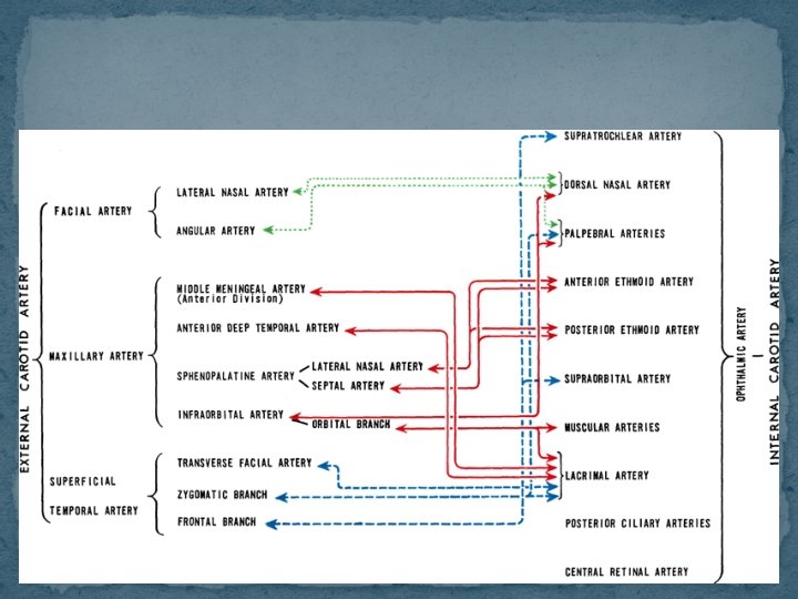
VASCULAR ANATOMY �The internal carotid artery supplies the eyelids, upper nose and nasal dorsum, forehead and scalp via subbranches of its ophthalmic branch. �The ophthalmic artery arises behind the eye and branches into the orbital and ocular group.
� The orbital group includes the supraorbital, dorsal nasal and the anterior ethmoidal artery. � The supraorbital artery exits the orbit through the supraorbital foramen alongside the supraorbital nerve and perforates the frontalis muscle to ultimately course in the subcutaneous tissue of the forehead and scalp. � The supratrochlear artery exits the medial orbit and courses medial to the supraorbital artery to supply the nasal root and the low midline forehead. This artery serves as the axial blood supply for the midline forehead flap often used to repair nasal defects. The success of this flap depends upon isolation and preservation of the supratrochlear artery. � The anterior ethmoidal artery exits the nasal passage at the interface of the nasal bone and nasal cartilage to provide arterial supply to the nasal dorsum. � The dorsal nasal artery crosses the midline over the nasal root and anastomoses with the angular artery, which originates from the external carotid artery.
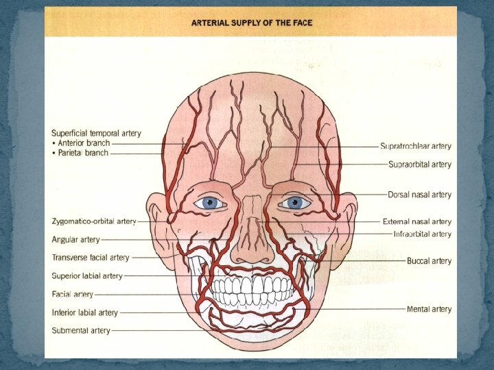
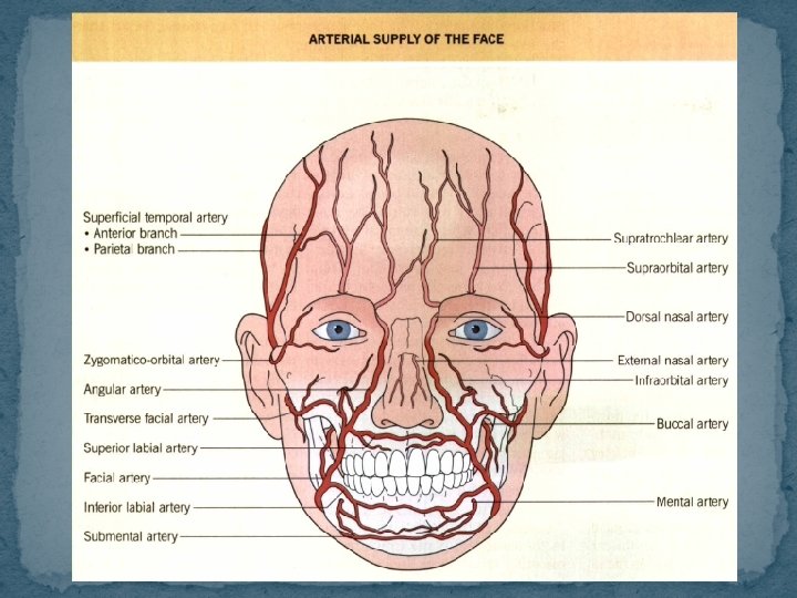
The facial artery � The facial artery branches off from the external carotid artery and courses deep to the mandible up through or behind the submandibular salivary gland. � It passes over the mandibular ridge onto the face anterior to the masseter muscle, where it can be palpated. Its tortuous course maintains a diagonal and superior direction passing alongside the nose and terminating at the medial canthus. � The platysma and risorius muscles protect the facial artery near the mandible. As the vessel traverses up the face, it becomes covered by the zygomaticus muscles of the midface and the orbicularis oculi as it nears the medial canthus.
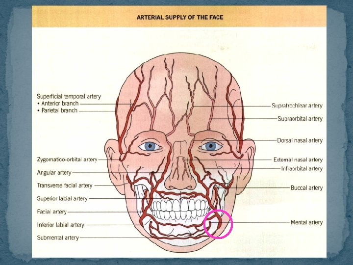
� The facial artery gives rise to the inferior and superior labial arteries( found deep to the lip mucosa), but can course more superficially into the submucosa of the lip in elderly patients. � After forming the superior labial branch, it becomes known as the angular artery. � At its endpoint near the medial canthus tendon, the angular (or facial) artery anastomoses with the dorsal nasal branch of the ophthalmic artery. � Since the ophthalmic artery is a branch of the internal carotid system, this anastomosis joins the internal and external carotid arterial systems. � During most of their course, the facial artery and vein are covered by the superficial muscles of facial expression. � The facial artery also anastomoses with branches of the internal maxillary (the infraorbital branch) and superficial temporal (transverse facial) arterial tree. � This allows excellent blood supply to the facial skin even if distal arterial branches are cut or tied during surgery. The high density of arterial supply to the head and neck accounts for its excellent healingpotential and viability of local flaps and grafts.
Superficial temporal artery � The lateral face, scalp and forehead are primarily supplied by the superficial temporal artery and its branches. � This artery arises in the superficial lobe of the parotid gland as the terminal branch of the external carotid artery. It courses superficially to the main facial nerve trunks, � then gives off the transverse facial artery before exiting the parotid gland superficially. The latter transverse artery runs parallel to and 2 cm below the zygomatic arch. � The superficial temporal artery exits the parotid and enters the subcutaneous fat in the preauricular crease, where it assumes an ascending vertical course over the zygomatic arch. � It can be easily palpated just medial to the upper tragus of the ear. It runs alongside and superficial to the auriculotemporal nerve within, then above the SMAS layer of the temple and lateral forehead.
� most superficial portion of the superficial temporal artery is often visible in aged patients within the subdermal fat above the galea aponeurotica as it courses cephalad above and anterior to the ear. � Here it forms the parietal and frontal (anterior) arterial branches that originate just above the uppermost attached portion of the ear. The forehead, eyebrows and lateral scalp receive their arterial supply from these branches of the superficial temporal artery. � There arc many anastomoses on the scalp between the bilateral superficial temporal arteries. Because of this rich supply chain, the entire scalp tissue remains viable even if one of these arteries is occluded. � This rich anastomotic network also explains why scalp surgery can be a very bloody process. � Surgical dissection at the level of the galea aponeurotica may serve to avoid transection of the copious subdermal vascular supply.
Facial vein � Most of the arteries of the face run anterior to and parallel with their corresponding veins. � The veins lack valves : permit two-way flow of venous blood. � The facial vein connects to the deep facial vein as it drains the cheek: parallels the internal maxillary artery and anastomoses with the pterygoid venous plexus medial to the upper mandibular ramus. � The facial vein crosses over the submandibular glands, while its corresponding artery passes beneath them. � It then drains into the internal jugular vein, which connects with the external jugular vein via the retromandibular vein. � The facial vein can communicate with the cavernous sinus of the brain via the ophthalmic vein or the pterygoid plexus. The paranasal area and upper lip are the regions drained by this network. This interface may permit skin or wound infections to gain access to the cavernous sinus of the brain from the draining facial or ophthalmic veins, with potentially devastating consequences.
� Arterial blood supply to the face is delivered by a rich subdermal plexus that is fed by larger perforating arteries. � Wound healing and flap success depend on maximal blood supply to the area. � Axial flaps, such as the midline forehead flap, incorporate a known subcutaneous artery (e. g. the supratrochlear artery) into their design. � Random pattern flaps are maintained by the subdermal arterial plexus and do not rely on a single feeder artery to maintain blood flow. � The anastomotic vascular network permits facial arteries to be clamped or tied off during surgery without compromising tissue viability. � Nearby ipsilateral or contralateral anastomotic arterial branches can often compensate for any loss in local blood supply.
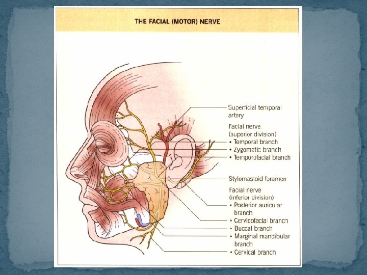
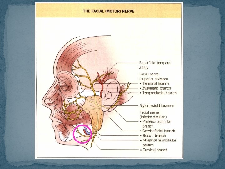
THE FACIAL NERVE � The facial nerve courses between the SMAS and the deep fascia before its branches penetrate the lateral underside of the facial muscles. � Cranial nerve VII has two major roots, the smaller of which provides sensory innervation and taste sensation to the anterior two-thirds of the tongue via the chorda tympani branch. � Sensory innervation to a portion of the external auditory meatus, soft palate, and pharynx is also derived from this small facial nerve root. � The submaxillary, submandibular and lacrimal glands contain parasympathetic fibers of the facial nerve that have secretory effects. In addition to the muscles of facial expression , the buccinator, stapedius, posterior belly of the digastric, stylohyoid and platysma muscles are all innervated by branches of the facial nerve.
� Upon exiting the skull at the stylomastoid foramen near the level of the earlobe, the facial nerve immediately gives off the posterior auricular branch, which provides motor innervation to the occipitalis and posterior auricular muscles. � The remainder of the nerve trunk enters the parotid gland bifurcates into the horizontally oriented temporofacial branch and lower cervicofacial branch. � If a line is drawn from the superior border of the tragus to the angle of the mandible, the entrance site of the facial nerve trunk into the parotid gland lies at the midpoint. � The temporal, zygomatic, buccal, marginal mandibular, and cervical branches arise from the two major rami of the facial nerve. � It courses in between the glandular parotid tissue and becomes more superficial as it follows an upward curvilinear pathway on its way to the ventral surfaces of the muscles of facial expression.
�While the facial nerve trunk is well protected in adults at its exit from the skull by the mastoid process, the surgeon must beware when performing procedures in this anatomic region in children. Because the mastoid process is not hilly developed until age five, the main facial nerve trunk lies in a superficial subcutaneous plane behind the earlobe and may be damaged in superficial cutaneous procedures. �The major facial nerve branches on the cheek are protected only by a small amount of parotid tissue, parotid fascia and subcutaneous fat. If a surgical procedure requires violation of the parotid fascia, meticulous dissection is necessary to avoid major facial nerve damage and subsequent functional disability. The motor nerves of the face course deeper than the sensory nerves or axial vasculature.
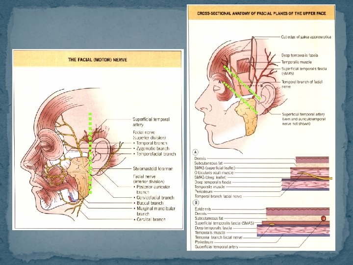
The temporal branch � The temporal branch of the facial nerve provides motor innervation to the frontalis, upper orbicularis oculi, and corrugator supercilii muscles � It usually has four rami that originate over the middle third of the zygomatic arch for a combined approximately 2. 5 cm 'danger' zone in this area. � The most posterior ramus of the temporal branch can be topographically located on the temple 1 cm anterior to a vertical line drawn from the anterior insertion of the ear to the scalp. This posterior ramus runs anterior to the superficial temporal artery and vein. � The most significant 'danger zone' for the temporal branch of the facial nerve lies between a line drawn from the earlobe to the lateral edge of the eyebrow and a line drawn from the trab. TUs to just above and lateral to the highest forehead crease. Within this zone, the temporal branch is at highest risk as it crosses the midzygomatic arch, where it lies most superficially over this bony prominence.
� The temporal branch of the facial nerve courses between the superficial and deep temporalis fascia, penetrating the underside of the frontalis muscle from its lateral edges. � The superficial temporal artery and vein as well as the auriculotemporal sensory nerve run posterior to but more superficial than the temporal nerve branch. � This neurovascular bundle lies in the subcutaneous fat overlying the SMAS of the temple and lateral forehead region. � Remember that once the temporal nerve reaches the lateral underbelly of its ipsilateral target muscles, muscles it is most protected. In order to avoid damaging this facial nerve branch, the surgeon should either remain superficial to the SMAS (i. e. the superficial temporalis fascia) or dissect in the subgaleal plane from the medial to lateral forehead.
The temporal branch consists of long, usually singular and often superficially coursing rami that have few arborizations or cross-innervations. These characteristics make nerve damage and permanent sequelae more likely when cutaneous procedures are performed in the forehead and temporal regions. Even though the upper orbicularis oculi and corrugator supercilii muscles are innervated by the temporal branch, minimal functional or cosmetic compromise occurs, due to cross-innervation by other motor nerves. � However, only 15% of patients will have any crossinnervation to the frontalis muscle by the more inferior zygomatic branch of the facial nerve. Such arborization permits retention of some functional mobility of the frontalis should the temporal branch be sacrificed.
� For the other 85% of patients, violation of the temporal nerve results in motor denervation and the inability to raise a now lowered or 'droopy' eyebrow. � Flattening of the forehead with diminished visibility of wrinkles and skin tension lines on the ipsilateral side is easily noted. � The functional loss of the frontalis muscle significantly hampers a patient's ability to communicate non-verbally via facial expression and may have devastating psychosocial consequences. � Over time, the inability to raise one's eyebrow can lead to eyebrow and eyelid ptosis and upper visual field compromise as muscular disuse atrophy progresses. � Brow lifts and blepharoplasty may be necessary if the temporal nerve branch is permanently damaged.
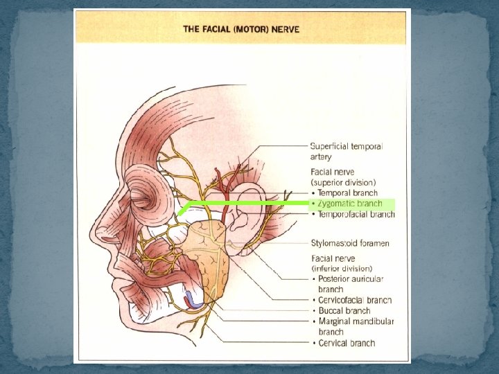
The zygomatic branch � The zygomatic branch of the facial nerve provides motor innervation to the lower orbicularis oculi, procerus, mouth elevators and nasal muscles. � Its fibers overlie the parotid (Stensen's) duct and course horizontally and upwards after emerging from the parotid gland as the second division of the facial nerve. There is marked variability in the innervation it provides to these muscles; thereby, damage to this nerve branch can have unpredictable outcomes � Generally, injury to the zygomatic branch results in decreased orbicularis oculi function and a diminished ability to close the ipsilateral eyelid tightly. � The orbicularis oculi is also innervated in its supraorbital aspect by the temporal branch of the facial nerve. Therefore, complete loss of circumferential periocular motor function is highly unlikely. � Other effects may include dysfunction of nasal muscles and lip elevators
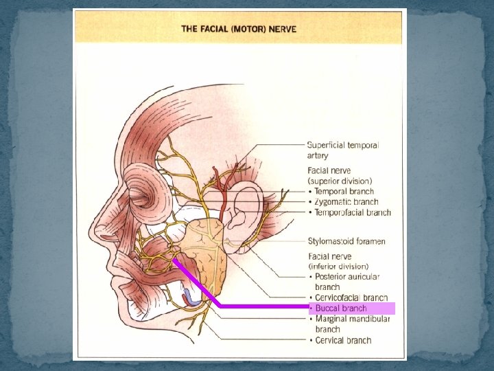
The buccal branch � The buccal branch is the third division of the facial nerve, and it courses inferiorly to the zygomatic branch in a downward direction on the cheek. � It innervates the orbicularis oris muscle, the zygomaticus muscles, the lip elevators, the buccinator muscle and nasal muscles to a variable extent. � Damage to this nerve causes buccinator dysfunction that results in accumulation of food between the teeth and the buccal mucosa with chewing. � Approximately 80% of patients have anastomoses between the fibers of the zygomatic and buccal branches. Each branch has two points of arborization, with the � first occurring 2 cm anterior to the anterior edge of the parotid gland � the second usually occurring under the modiolus 1 cm lateral to the oral commissure at the anterioraspect of the buccal fat pad
� Here the nerves may be damaged due to their superficial anatomy, protected only by the thin fascia of the SMAS, an often underdeveloped risorius muscle, and the subcutaneous fat. � At the first branching site near the parotid, the zygomatic and buccal nerves lie between the masseter muscle and the posterior side of the buccal fat pad. � Although damage to the zygomatic or buccal branches of the facial nerve may occur during surgical procedures, subsequent motor dysfunction is often temporary and far less debilitating than a similar injury to the temporal nerve. The high degree of anastomoses between the zygomatic and buccal branches minimizes functional damage and promotes nerve recovery after trauma.
� Partial paralysis of the perioral muscles may occur, causing variable symptomatic defects in facial expression, including a diminished ability or unilateral defect in forming a smile or pucker, lip pursing and lip seal formation. � Drooling, food accumulation between the cheeks and gingivae, and muffled speech may occur secondary to buccal or zygomatic nerve damage. � Orbicularis oculi defects have already been discussed and include lower eyelid droop, which can lead to chronic conjunctivitis, sicca symptoms, and ectropion. � Difficulty wrinkling up the nose and inability to flare the nostrils may also occur with zygomatic or buccal branch trauma. Fortunately, most of these symptoms resolve within 6 months due to the extensive ramification and cross-innervation of these branches of the facial nerve.
The marginal mandibular branch � The orbicularis oris, mentalis, and lip depressor muscles are innervated by the marginal mandibular branch of the facial nerve. � The nerve courses along the angle of the mandible below the parotid gland continues up over the mandibular body anterior to the facial artery, which can be palpated easily as it courses over the medial mandible. � The nerve is very susceptible to damage, damage due to its superficial location over the bony edge of the jaw (i. e. just inferior and lateral to the lateral oral commissure), where it is covered only by fascia and an often unpredictably thin or poorly developed platysma muscle. � This facial nerve branch is often composed of only one ramus. The marginal mandibular nerve 'communicates' with the buccal branch of the facial nerve in only 10% of patients; patients therefore, damage to the former branch can lead to permanent disfiguring and functional defects in facial expression.
�Normal symmetric facial expression and hmction of the mouth depends upon the equal and opposite effects exerted by the lip depressors and elevators in conjunction with the orbicularis oris muscle. �Characteristically, a patient with marginal mandibular nerve damage cannot form a symmetric smile. There is inability to pull the ipsilateral lower lip downward and laterally or evert the corresponding vermilion border. The end result is a 'crooked' smile. The defect is appreciated upon smiling, but is not as apparent when the patient is at rest.
The cervical branch � The cervical branch of the facial nerve innervates the platysma muscle. � This muscle receives nerve fibers from the marginal mandibular nerve as well. � Damage incurred to the cervical branch rarely causes functional or cosmetic defects.
SENSORY INNERVATION OF THE HEAD AND NECK �The trigeminal nerve, nerve or cranial nerve V, provides the primary sensory innervation to the face, while the upper cervical nerves (C 2, C 3) provide sensory supply to the neck, part of the ear, and posterior scalp � The facial, glossopharyngeal and vagus nerves provide a small portion of sensory innervation to the ear. � The trigeminal nerve is the largest cranial nerve. It has motor (to the muscles of mastication), sensory and parasympathetic functions, supplying secretory fibers (originating from the facial and glossopharyngeal nerves) to the lacrimal and parotid glands.
�The sensory branches of the trigeminal nerve course more superficially than the trunk of the facial nerve, and are thereby readily subject to damage during surgical procedures. �Fortunately, most resulting sensory dysfunction is not debilitating or permanent. �Transmedian re-innervation after unilateral trigeminal root transection has been demonstrated. This is due to collateral sprouting of sensory nerves from the contralateral trigeminal nerve root � The sensory nerves exist in the superficial plane between the subcutaneous fat and the SMAS and often run together with arteries and veins in neurovascular bundles.
�The trigeminal nerve is divided into three main branches, called the �ophthalmic (V 1) �maxillary (V 2) �mandibular (V 3)
The ophthalmic division � The smallest, uppermost sensory branch is the ophthalmic division, which gives off three branches (nasociliary, frontal and lacrimal nerves) before exiting the orbit. � Sensory fibers to the sinuses and upper nasal septal mucosa, as well as secretory parasympathetic fibers (that originate from the facial nerve) to the lacrimal gland are also provided by the ophthalmic branch of the trigeminal nerve. � The nasociliary branch gives rise to the infra trochlear nerve and the external branch of the anterior ethmoidal nerve. Sensory innervation to the root of the nose and part of the medial canthus is supplied by the infra trochlear nerve. � The nasal dorsum, tip, supra tip and columella derive cutaneous innervation from the external nasal branch of the anterior ethmoidal nerve, which emerges between the upper nasal cartilage and nasal bones.
� The nasociliary branch also supplies the corneal surface via the ciliary nerve. If an episode of zoster (varicella zoster virus) involves the nasal tip, then close ophthalmologic followup is warranted due to presumed corneal involvement.
�The frontal nerve forms the � supratrochlear �supraorbital nerves The exit route (called the supratrochlear ridge) of the supratrochlear nerve lies 1 cm lateral to the midline on the supraorbital ridge. This branch of the frontal nerve provides sensory innervation to the medial upper eyelid, medial forehead, and frontal scalp. The supraorbital foramen (through which emerges the supraorbital neurovascular bundle) lies 2. 5 cm lateral to the midline on the supraorbital ridge
�After penetrating the frontalis muscle to emerge above the frontalis SMAS, the supraorbital nerve provides cutaneous sensation to the forehead, scalp and upper eyelid. A small lacrimal nerve branch of the ophthalmic division of cranial nerve V innervates the lateral eyelid skin and lies near the upper lateral orbital rim. Frontal nerve blocks, and specifically the supratrochlear and supraorbital block, offer quick and effective anesthesia for surgical procedures of the forehead.
The maxillary branch � The maxillary branch (V 2) of the trigeminal nerve forms the infraorbital, zygomaticofacial and zygomaticotemporal cutaneous sensory branches. � The infraorbital foramen lies 2. 5 cm lateral to midline and 1 cm inferior to the infraorbital rim, in the same vertical line as the supraorbital and mental foramina. � The infraorbital neurovascular bundle emerges here to provide significant sensory innervation to the medial cheek, upper lip, nasal sidewall and ala, and the lower eyelid.
� Infraorbital nerve blocks offer simple and effective anesthesia for much of the cheek, lower eyelid and nose. � Lateral to the infraorbital foramen, the zygomaticofacial nerve emerges to innervate the skin of the malar eminence. • Cutaneous innervation of the temple and supratemporal scalp region is provided by a third branch of the maxillary division, the zygomaticotemporal nerve. It emerges from the lateral orbital margin at the zygomatic bone. • The superior alveolar and palatine nerves are deeper branches of V 2 that provide sensory innervation to the upper teeth, palate, nasal mucosa, and gingiva
The mandibular branch � The mandibular branch (V 3) is the largest division of the trigeminal nerve and the only one to carry both cutaneous sensory and motor fibers. � The auriculotemporal, buccal and inferior alveolar nerves represent the three main cutaneous branches of V 3. � The auriculotemporal nerve emerges from behind the neck of the mandible to course just deep to the superficial temporal artery from the superior margin of the parotid gland in the preauricular sulcus up towards the lateral scalp. � It provides sensory innervation to the external ear and auditory canal, temple, temporoparietal scalp, temporomandibular joint and tympanic membrane. It also carries parasympathetic secretory fibers to the parotid gland.
� The buccal nerve supplies sensory innervation to the cheek, buccal mucosa, and gingiva. It runs deep to the parotid over the pterygoid muscle to the upper surface of the buccinator, which it pierces to reach the overlyingthe skin. • Because terminal branches of the buccal nerve are small and numerous, regional buccal nerve block techniques are not feasible anesthetic options. • The inferior alveolar branch of V 3 innervates the mandibular teeth as it courses through the mandibular sulcus.
Its terminal branch forms the mental nerve, nerve which emerges from the mental foramen in the same topographic line as the supraorbital and infraorbital neurovascular bundles (i. e. 2. 5 cm lateral to the midline). �The mental vein, artery and nerve emerge from the mental foramen below the lower second premolar. �The lingual nerve supplies sensory innervation to the anterior two-thirds of the tongue, the floor of the mouth, and the lower gingivae. It arises from V 3 and courses parallel and superior to the inferior alveolar nerve.
The cervical plexus � The cervical plexus is a network of arborizing and anastomosingnerve branches of the ventral rami of the four most superior cervical nerves. � It emerges from the mid-posterior margin of the sternocleidomastoid (SCM) muscle at Erb's point to give off three branches, designated as C 2, C 3 and C 4. C 2 and C 3 compose the greater auricular nerve, nerve which runs from the posterior edge of the SCM towards the earlobe in the same plane and path as the external jugular vein. � It innervates the skin of the lateral neck, angle of the jaw, and part of the auricular and postauricular skin.
� The lesser occipital nerve (C 2) emerges from the same point at the SCM and assumes a course parallel to the SCM upward to innervate the skin of the neck and postauricular scalp. � The transverse cervical nerve (C 2 and C 3) likewise emerges from behind the SCM and arcs anteriorly around across the SCM in a transverse direction. Its many terminal branches supply the skin of the anterior neck. � The supraclavicular nerve (C 3 and C 4) emerges from the same point at the SCM posterior margin, then courses inferiorly until it reaches the supraclavicular region, where it terminates, and provides sensory innervation to the anterior chest and shoulder skin
� Perineural invasion of cutaneous tumors such as basal cell carcinomas, neuropathic melanomas and squamous cell carcinomas may be encountered by the clinician. � Patients are often asymptomatic from dermal nerve twig infiltration by tumor cells, with the diagnosis made only by histopathologic tissue examination. � Some patients may experience sensory abnormalities and, rarely, motor dysfunction. � Most of these scenarios occur in the setting of squamous malignancies (incidence rates range from 3% to 14%), with less than 1% of basal cellcarcinomas showing any histologic evidence of perineural spread. � Knowledge of the neuroanatomy of the head and neck assists the surgeon in planning adjuvant therapy, such as radiation, for patients with nerve involvement.
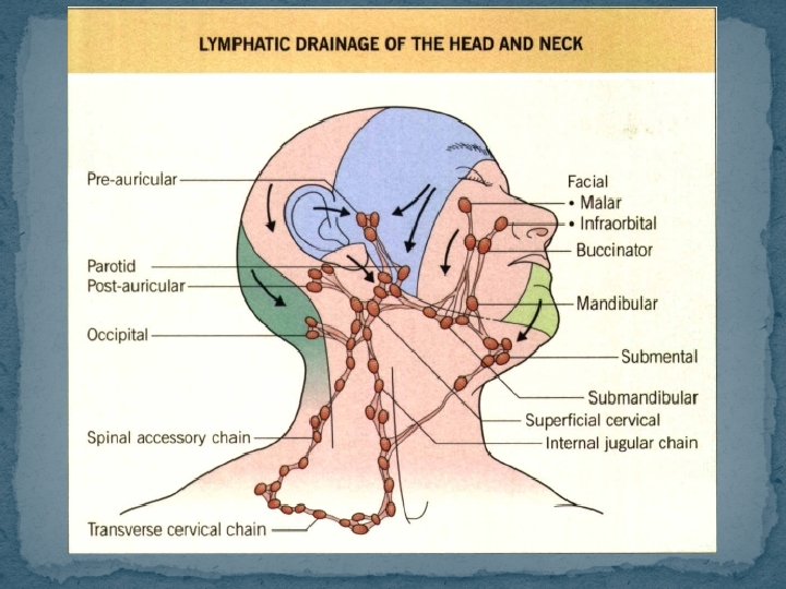
LYMPHATIC DRAINAGE OF THE HEAD AND NECK
�. � Before any surgical endeavor is undertaken for a malignancy in this region, palpation of regional lymph nodes and basins should be performed. � Head and neck cancers usually spread to adjacent lymph nodes in a diagonal direction from cephalad to caudad. � There is a large degree of variability in drainage pathways, but the anatomic location of individual lymph nodes is more consistent from patient to patient. � Cutaneous neoplasms that breach the papillary dermis may spread from small lymph capillaries to progressively larger and deeper lymphatic trunks in the area. � Lymph channels often course along the same directional pathway as the head and neck veins. � They are more numerous and often more superficial than the corresponding veins and lie predominantly between the superficial and deep fascial layers.
� Important primary lymphatic drainage patterns of the head and neck region include the following: � parotid nodes often collect from the forehead and eyelids (upper lateral face); � submandibular nodes from the lower and medial face or from the submental nodes; � submental nodes from the central lower lip and chin. � Lateral cervical nodes are the common subsequent lymph collection site from these areas. � Parotid nodes may be extraglandular or intraglandular. � The extraglandular channels are invested within the parotid sheath. � Two-thirds of all people will also have one to three pretragal and infra-auricular lymph nodes that are considered part of the parotid node basin
�These pre-and infra-auricular nodes drain the ear, the lateral lower cheek, the frontolateral scalp and the forehead, as well as the nasal root. �Drainage of the parotid unit may then follow the external or internal jugular vein in the jugular lymph node chain; therefore, palpation for nodes from the site of the cutaneous lesion to the supraclavicular and even axillary nodal basins is recommended. �Submandibular nodes should be examined as the patient relaxes the neck muscles and tilts the chin down. � This nodal group drains the gingival and mucous membranes, lower eyelids, anterior two-thirds of the tongue, lips, nose and medial cheeks.
�The submental nodes (up to eight) of the neck lie beneath the platysma and drain the anterior third of the tongue and floor of the mouth, in addition to the lower middle lip, chin and medial lower cheeks. � They are best examined by elevating the chin and asking the patient to engage the platysma. �Submental nodes frequently drain bilaterally or contralaterally and empty into the submandibular basin or directly into the internal jugular lymphatic chain. � Note that up to one-quarter of healthy people have small (less than 1 cm) non-fixed palpable submental nodes.
� The superficial lateral cervical nodes are adjacent to the infraauricular parotid nodes and lie near the high external jugular vein. Use the sternocleidomastoid muscle as a landmark to palpate these nodes (up to four) over its cephalad portion. � Deeper lateral cervical nodes include the spinal accessory, internal jugular and transverse cervical chains, which form a triangle on the neck. The internal jugular chain is the main lymphatic collection trunk of the head and neck and may contain up to 25 lymph nodes in each patient. The internal jugular chain on the right often drains into the subclavian vein, whereas the left-sided lymphatic chain empties into the thoracic duct. � These nodes can be palpated by rolling two fingers over the area of the carotid triangle.
� Acute or chronic lymphedema : after transection of larger lymphatic channels or nodes, or smaller channels in areas (e. g. infraorbital) with limited or vulnerable lymphatic drainage. � Adequate drainage can be achieved by : orienting flaps in the same direction as lymphatic patterns. � The surgeon must be mindful of the variability in drainage patterns and sites and the fact that malignancies do not respect the midline � Since cross-communication between lymphatics may result in contralateral drainage, bilateral examination for lymphadenopathy should be undertaken before a neoplasm is excised.
Once a year in Jindo in Korea, the seas mysteriously part and visitors can walk through the sea from the mainland to a nearby island. This phenomenon is caused due to the difference in high tides and low tides, which creates a 2. 8 -kilometer-long road measuring 40 to 60 meters in width.