Muscular System Muscles of the facial expression Occipitofrontalis


")


- Slides: 45
Muscular System
Muscles of the facial expression Occipitofrontalis Orbicularis oculi Auricularis Zygomaticus
Muscles of the facial expression Occipito frontalis: The occipitofrontalis is a muscle of the human skull and consists of two parts: The occipital belly, near the occipital bone, and the frontal belly, near the frontal bone. Function : Draws the scalp back which raises the eyebrows and wrinkles the forehead.
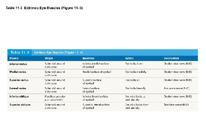
Orbicularis Oculi: Origin: Medial side of eye Insertion: Medial side of eye (returns to same attachment encircling the eye) Action: Closes & squints the eye. Also assists in tear transport and drainage N: CN VII (Facial nerve)
Zygomatic Origin: Zygomatic bone Insertion: Angle of Mouth A: Elevation of Angle of Mouth as in smiling. Also draws laterally at the angle of the mouth Nerve: CN VII (Facial nerve)
Auricularis: Origin: Cartilage of auricle Insertion: Galeal aponeurosis Function: Adjusts position of ear
Auricularis:
Buccinator: Origin: Maxilla and Mandible Insertion: Lips (buccinator muscle is located in the tissue of the cheek) Action: Compression of Cheeks (against teeth) Nerve: CN VII (Facial nerve)
Masseter: Origin: Zygomatic arch Insertion: Angle of mandible Function: Elevates and protracts mandible to close the jaw
Sternocleido mastoid Origin: sternum; clavicle Insertion: temporal bone Action: flexes neck; rotates head side to side; assists you to look down
Trapezius Origin: Vertebral column Insertion: Scapula Action: Elevation, adduction, depression and outward rotation of scapula
Scalenes Scalene origin Origin: Transverse processes of the cervical vertebrae Scalene insertion Insertion: First and second ribs Scalene action Action: Bilaterally - Assists in neck flexion. Unilaterally - Neck lateral bending
Deltoid Origin: Lateral third of clavicle, acromion, and spine of scapula Insertion: Deltoid tuberosity of humerus Action: Anterior part: flexes and medially rotates arm; Middle part: abducts arm; Posterior part: extends and laterally rotates arm
Latissmus dorsi
Pectoralis Major
Biceps Brachii
Brachio radialis
Brachialis
Triceps Originates on scapula Inserts on olecranon Origin: Originates on scapula Insertion - Olecranon Function: Extends elbow
Flexors of the wrist
Extensors of the wrist
Flexors of the Wrist Palmaris longus-Superficial, flexes wrist Flexor carpi ulnaris-Superficial, flexes wrist, adducts wrist Flexor carpi radialis-Superficial, flexes wrist, abducts wrist
Extensors of the Wrist Extensor carpi radialis- Superficial, extends wrist, abducts wrist Extensor carpi ulnaris- Superficial, extends wrist, adducts wrist
Muscles of Respiration
Muscles of respiration: Quiet breathing Inhalation: diaphragm, external intercostals. Exhalation: none. Forced breathing Inhalation: diaphragm, external intercostals, scalene, pectoralis minor, sternocleidomastiod, serratus anterior. Exhalation: internal intercostals, abdominals
Muscles acting on the abdominal wall
Muscles acting on the abdominal wall There are five muscles in the abdominal wall. They can be divided into two groups: The Flat Muscles There are three flat muscles; the external oblique, internal oblique and transversus abdominis. They are located laterally in the abdominal wall, stacked upon one another. These muscles act to flex, laterally flex and rotate the trunk. Vertical Muscles Rectus Abdominis: This is a long, paired muscle, found either side of the midline in the abdominal wall. It is split into two by the linea alba. Pyramidalis:
Muscles acting on the hip and femur Anterior group Posterior group Adductors Abductors
Anterior group It contains the following five muscles: Sartorius: the longest muscle in the human body Quadriceps: Rectus femoris, Vastus lateralis, Vastus intermedius, vastus medialis Function: Knee Extensors and hip flexors
Anterior group of muscles Iliopsoas group: Which consists of the psoas major and iliacus muscles. Quadriceps femoris group: Which consists of the rectus femoris, vastus intermedius, vastus lateralis, and vastus medialis.
Gluteal Muscles Gluteus maximus: Largest, most posterior gluteal muscle Produces extension and lateral rotation at hip Tensor fasciae latae: Works with gluteus maximus Stabilizes iliotibial tract Gluteus medius and gluteus minimus: Originate anterior to gluteus maximus Insert on trochanter
Abductors of thigh Gluteus minimus: Abduction and medial rotation of thigh Gluteus medius: Abduction and medial rotation of thigh Gluteus maximus: External rotation and extension of hip joint Chief anti gravity muscle
Posterior group Flexors of the Knee Hamstrings Biceps femoris Semimembranosus Semitendinosus
Posterior thigh
Adductors Located on medial side of the hip Adductor magnus: Produces adduction, extension, and flexion Adductor brevis: Hip flexion and adduction Adductor longus: Hip flexion and adduction Pectineus: Hip flexion and adduction Gracilis: Hip flexion and adduction
Adductors
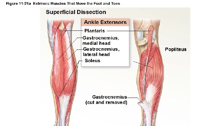
Muscles on the posterior compartment of leg Four Muscles That Produce Extension (Plantar Flexion) at the Ankle Gastrocnemius Soleus Fibularis (group) Tibialis posterior
Gastronemius and Soleus Posterior compartment of the leg. Origin: femur Insertion: Achilles tendon and inserts onto the posterior surface of the calcaneus or heel bone. Function Standing, walking, running and jumping. Plantar flexing the foot at the ankle joint and flexing the leg at the knee joint.
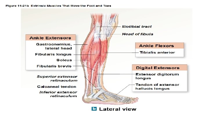
Muscles of lateral compartment of leg Fibularis longus Fibularis brevis Function: Plantar flexion and Eversion of the foot
Muscles in anterior compartment of leg Tibialis anterior: Origin: body of tibia Insertion: medial cuneiform and first meta tarsal of foot Function: Dorsi flexion and inversion of foot
Muscles acting on the feet Muscles That Produce Extension at the Toes Extensor digitorum longus Extensor hallucis longus Extensor retinacula fibrous sheaths Muscles That Produce Flexion at the Toes Flexor digitorum longus Flexor hallucis longus