SCALP Definition Soft tissue covering the cranial vault


")


 § Thin sheet of fibrous tissue, § Attachment to occipito-frontalis")


- Slides: 30
SCALP
Definition Soft tissue covering the cranial vault of the skull Extent Anteriorly posteriorly Each side Thickness : 5 – 6 mm
Supraorbital margin
Superior temporal lines
Superior nuchal line External occipital protuberance
Layers of the scalp S – Skin C – Cutaneous tissue ( Dense ) A – Epicranial aponeurosis or galea aponeurotica L – Loose areolar tissue P - Pericranium
Layers of scalp
Skin § Thick § Hair bearing § Sebaceous and sweat glands § Closely adherent to epicranial aponeurosis through the dense superficial fascia
Sebaceous Cyst
Superficial fascia § Fibro-fatty tissue § Medium for blood vessels and nerves of skin § Walls of the vessels are adherent to fibrous network ü Minor injury causes profuse bleeding ü Infections tend to remain localized and are painful
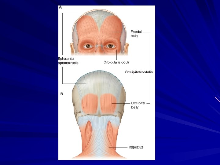
Epicranial aponeurosis (Galea aponeurotica) § Thin sheet of fibrous tissue, § Attachment to occipito-frontalis muscle § Anteriorly – frontal belly § Posteriorly – occipital belly, external occipital protuberance & highest nuchal lines § On each side – superior temporal line, sends thin expansion to zygomatic arch
Occipitofrontalis muscle § Occipital belly – Superior nuchal lines § Frontal belly – skin of the forehead § Insertion – epicranial aponeurosis § Nerve supply – Posterior auricular branch Temporal branch
Gaping of wounds when cut transversely
Loose areolar tissue § Subaponeurotic space § Extent § Emissary veins Valveless, connect superficial veins of scalp with intracranial venous sinuses § Dangerous area of scalp § Accumulation of blood leads to generalized swelling, black eye…
Black eye
Pericranium § Outer periosteum of skull § Continuous with endocranium through the sutures Cephalohematoma – collection of fluid beneath the pericranium produces localised swelling - assumes the shape of related bones
Cephalohematoma
Innervation of scalp
In front of the ear – Supratrochlear N – Supraorbital N – Zygomaticotemporal N – Auriculotemporal N – Temporal branch of the facial nerve Behind the ear – – – Great auricular Lesser occipital Great occipital Third occipital Posterior auricular branch of facial nerve
Blood supply of scalp
In front of the ear – Supratrochlear artery – Supraorbital artery – Superficial temporal artery Behind the ear – Posterior auricular artery – Occipital artery
Venous drainage § The veins of the scalp accompany the arteries and have similar names § The supratrochlear and supraorbital veins unite to form the angular vein and continue further as the facial vein
The superficial temporal vein form the retromandibular vein The posterior auricular vein joins the posterior division of the retromandibular vein to form the external jugular vein The occipital vein terminates in the suboccipital venous plexus
Cont…. § Emissary veins Parietal emissary vein Mastopid emissary vein § Diploic veins Frontal, anterior temporal, posteior temporal and occipital
Lymphatic drainage Anterior part of scalpparotid lymphnodes Posterior part- mastoid & occipital lymph nodes
Applied anatomy
Caput succedaneum Passage through the birth canal Compression of the skull and scalp An interference of venous return Temporary edematous condition of scalp
Black eye
Thank you