Face scalp Muscles of Facial Expression The face


; ü A-aponeurotic layer; ü L-loose connective tissue; ü")
- Slides: 22
Face & scalp
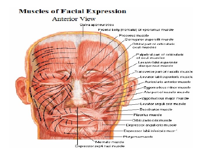
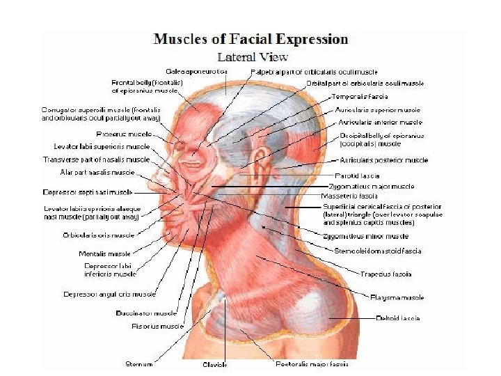
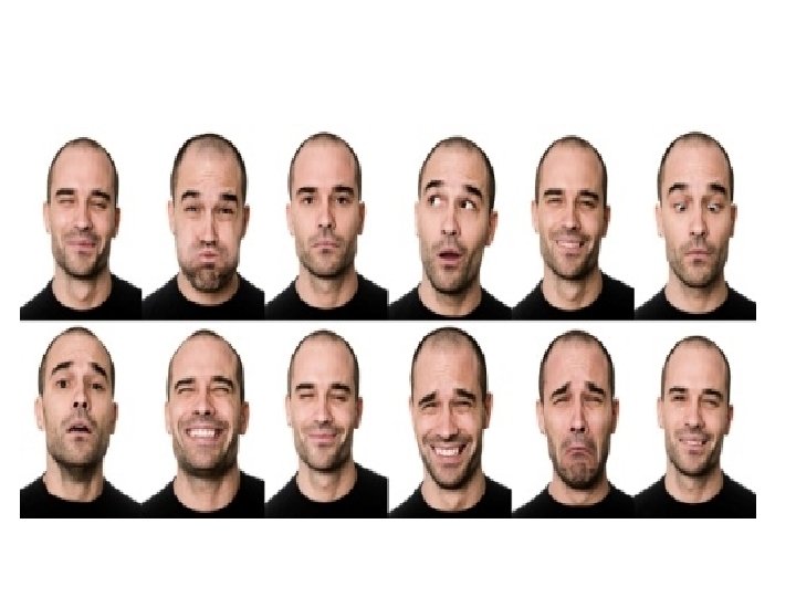
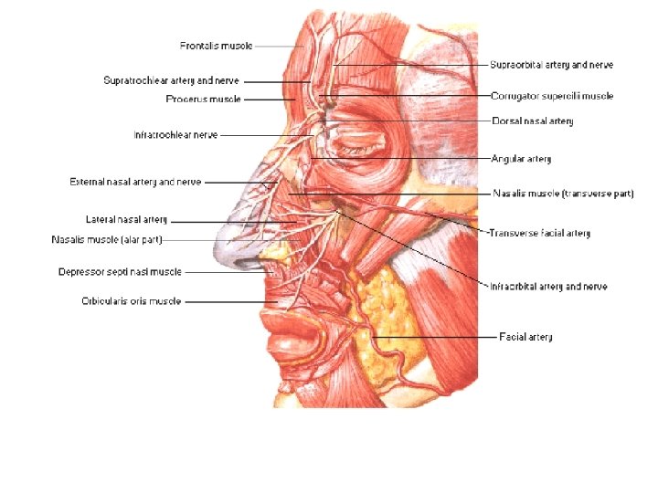
Muscles of Facial Expression • The face is the region of the front of the head that extends from the tragus of one ear to the other one, and from the base of the mandible below to the NORMAL limit of the hair line above. • The muscles of facial expression are a unique group in that they are found in the superficial fascia of the face, they are not bound down by deep fascia, and that they usually have one bony attachment and the other attachment goes to the skin. • These muscles are arranged around the openings of the face: the palpebral fissure ( the opening between the two eyelids), the nostrils, and the mouth. They act as two main groups: a dilator and a constrictor of their opening.
• The fontal belly of occipitofrontalis. This is the muscle of the scalp and is evident from its name that it has two bellies. • The occipital belly lies at the back of the scalp and attaches to the supreme nuchal line. The muscle is attached to the skin of the eyebrow and helps to widen the palpebral fissure. • The orbicularis oculi forms concentric circles around the lids. It closes the palpebral fissure.
• The orbicularis oris forms the sphincter around the opening of the mouth. • the zygomaticus major and minor pull up the upper lip from the side e. g. in smiling. • the buccinator is the muscle of the cheek and helps compress the contents of the oral cavity e. g. during eating or blowing air. • levator labii superioris lifts the upper lip. • depressor labii inferioris pulls down the lower lip.
• The depressor anguli oris and the levator anguli oris act on the angle of the mouth to depress it or lift it respectively. • The ala of the nose has the nasalis with two components: one dilates the nostril and the other compresses it • The levator labii superioris alaeque nasii pulls the upper lip and the nostril at the same time. • the platysma
Scalp • The scalp is the covering of the vault of the skull and is the hair bearing part of the skin of the head. • It extends from the upper margin of the orbital cavities anteriorly to the highest nuchal lines posteriorly and from one ear and zygomatic arch on side to the other side.
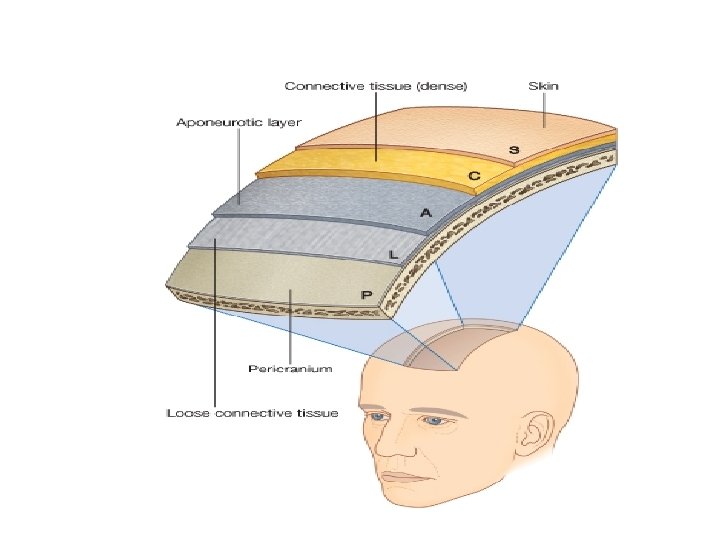
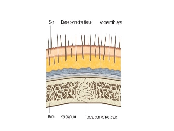
• It is made up of five layers designated by the letters of the word SCALP as follows: • The first is the Skin which has a dense population of hair follicles. • Then comes the Subcutaneous layer of Connective tissue which is mainly fatty tissue that is permeated by a lot of fibrous strands connecting the skin to the third layer. In this layer course the various vessels and nerves that supply the scalp.
• The third layer is the pericranial Aponeurosis or the galea aponeurotica. It is the aponeurosis between the frontal and occipital bellies of the occipitofrontalis muscle. • The aponeurosis is attached anteriorly to the posterior margin of the frontal bellies of the muscle that are then attached to the skin of the eyebrows; posteriorly it is attached to the anterior margin of the occipital bellies of the muscle that are in turn attached to the supreme nuchal lines. At the sides it is attached to the superior temporal lines with the temporal fascia.
• The aponeurosis is tightly bound to the skin by the numerous fibrous strands that travel between them and course in the second layer of the scalp making these three layers- skin, subcutaneous tissue and aponeurisismove as one layer. This is appreciated if one contracts his occipitifrontalis muscle and observes the way it moves the scalp
• The fourth layer is a potential space deep to the galea filled with Loose connective tissue. • This subgaleal space can be filled with blood in cases of blunt trauma to the head. The blood can migrate freely within the limits of the space and can track down into the skin of the eye lids to form a black eye or what is known as the rackoon or panda sign.
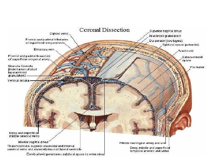
• The last layer is the Periosteum or Pericranium of the skull. It is firmly attached to the underlying bones and becomes continuous with its counterpart on the inside of the skull through the sutures. • The blood supply of the scalp comes from the supraorbital and supratrochlear anteriorly, the superficial temporal at the side, the posterior auricular and the occipital arteries posteriorly.
ü S-skin; ü C-connective tissue (dense); ü A-aponeurotic layer; ü L-loose connective tissue; ü P-pericranium