DISEASE OF THE UVEA EYE HOSPITAL OF THE


 retinal artery occlusion is not commonly seen in clinic, but with very serious")


 § 视神经纤维在各种因素的影响下,发生退行性变, 使视乳头褪色并有视功能损害者, 称为视神经萎缩不是一个独立的病变, 而是某些神经 系统或视神经本身病变的结果. § Optic atrophy is a final")


- Slides: 60
葡萄膜病 DISEASE OF THE UVEA 哈尔滨医科大学第一临床医学院眼科医院 EYE HOSPITAL OF THE 1 ST CLINICAL MEDICAL COLLEGE, HMU
§ Besides the factors of injury, operation, infection, the cause of almost all iridocyclitis belongs to endogenous. By carefully taking history, some correlated systemic disorders may be found out, for example, rheumatic disease (ankylosing spondylitis, juvenile rheumatoid arthritis), ulcerative colitis, tuberculosis, sarcoidosis, urethrits as well as venereal disease and so on. In recent years, it is discovered that the occurrence rate of HLA-27 in acute iridocyclitis may get as high as 60%.
Some specific types of uveitis § Sympathetic ophthalmia It indicates that the eye which got perforating injury or intraocular operation (called exciting eye) undergoes a period of granulomatous panuveitis, then the panuveitis with the same character takes place in another eye (called sympathizing eye). The interval from ocular injury or operation to appearance of inflammation in healthy eye is from tow weeks to two years (the earliest is in 10 days, the longest is after 50 years). But most have attack within 2 months, the probability of onset over 2 years decreases with the time going.
(1) retinal artery occlusion is not commonly seen in clinic, but with very serious prognosis. If there is not proper management in time, the results is the loss of vision finally. The cause leading to retinal central artery or its branch occlusion often is the debris exfoliated from atherosclerotic plague of the carotid. A few may be embolism by exfoliated thrombus on the vegetaiton of cardiac valves.
Or it is due to narrow blood vessel and spasm, vascular inflammation, pulseless disease, oral administration of contraceptive or caused by fat embolus at long bone fracture. Sometimes operation of retinal detachment or intraorbital operation may cause it. Recently occasionally, the cases are seen due to injection of prednisolone and other medicines into inferior nasal concha or behind the globe that induce retinal artery occlusion.
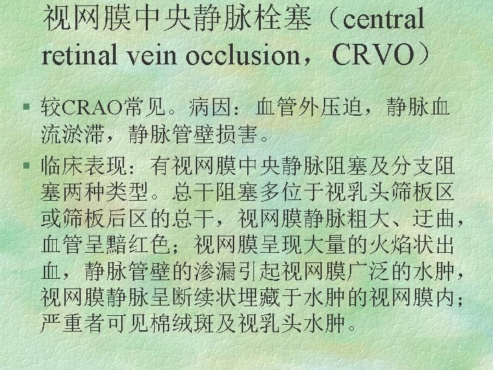
§ retinal vein occlusion Retinal vein occlusion is a common fundus disease; its causes are extravascular compression, stagnation of venous blood stream as well as impairment of venous vascular inner wall. Extravascular compression is most caused by sclerosis of the central retinal artery or its branches within the optic nerve or at the arteriovenous crossing to compress its neighbor veins. So, it is commonly seen in the elder with hypertension and arteriosclerosis. Stagnation of venous blood stream is seen in the cases with insufficient perfusion pressure or increased intraocular pressure or high blood viscosity.
§ So it is often complicated by insufficient blood supply of the carotid, a large quantity of blood loss, lower intraocular pressure, glaucoma, erythrocytosis, diabetes, sickle cell anemia and abnormal albumen in the blood and other diseases. Impairment of vascular inner wall is often caused by trtinal vasculitis, so it is commonly seen in the young and diabetic patients.
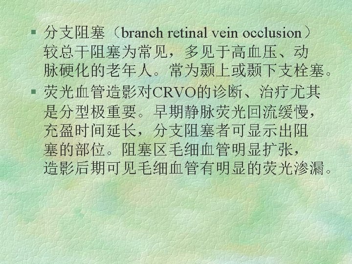
§ Clinicallly, according to the different sites of occlusion, it is divided into central retinal vein occlusion (CRVO) and branch retinal vein occlusion (BRVO).
The sorts of retinal degeneration are varied. Clinically the most common one is retinitis pigmentosa. The etiology is unclear, as a kind of genetic disorders; autosomal recessive inheritance, dominant inheritance as well as sex-linked recessive inheritance may all be seen. Among the cases of three kinds of genetic defects, sex-linked recessive type is the least, but the most severe. The damage of autosomal dominant inheritance is less severe, whereas the autosomal recessive one is
between both types above. Clinically there are not less sporadic cases without genetic evidence. At early stage of ill course, it mainly damages the rods and pigment epithelial cells. Whereas at advanced stage, all the retinal cells and choroidal capillary layer will be impaired.
The disease of the optic nerve and pathway § Optic neuropathy optic neuritis papillitis etiology clinical findings: ocular examination fundus examination of visual field treatment
视神经萎缩(optic atrophy) § 视神经纤维在各种因素的影响下,发生退行性变, 使视乳头褪色并有视功能损害者, 称为视神经萎缩不是一个独立的病变, 而是某些神经 系统或视神经本身病变的结果. § Optic atrophy is a final end of various severe disorders of the retina and optic nerve. Due to extensive damage of the retinal photoreceptor, gangliocyte as well as its axon, and loss of nervous fiber, gliosis, severe disturbance of visual function have been induced.
§ Main sysmptom of the optic atrophy is disturbance of visual function, manifesting as decrease of vision, contraction of visual field. Total loss of vision at ill eye in severe case. Moreover, the pupil may have the corresponding changes, for example, disappearance or retardation of light reflex. Clinically according to fundus manifestationg, it is divided into primary, secondary and ascending atrophy, three kinds.