
Ocular Manifestations of Systemic diseases Dr Ajai Agrawal













- Slides: 51
Ocular Manifestations of Systemic diseases Dr Ajai Agrawal Additional Professor, Department of Ophthalmology, AIIMS, Rishikesh. 1
Acknowledgement • Kanski’s Clinical Ophthalmology, 8 th Ed. (Elsevier) • Endocrinology : Adult and Paediatric. Burch, Henry B. ; Bahn, Rebecca S. . Published January 1, 2016. © 2016. • Some of the images used were taken from eyetext. net 2
Learning Objectives • At the end of this class the students shall be able to : • Describe ocular signs and symptoms associated with selected systemic diseases. • Understand the importance of early detection of ocular features of systemic diseases. 3
Introduction • "The eyes are the window to the soul. " (English proverb) • Occasionally, eye findings may be the first indication of underlying systemic disease leading to early diagnosis and management. 4
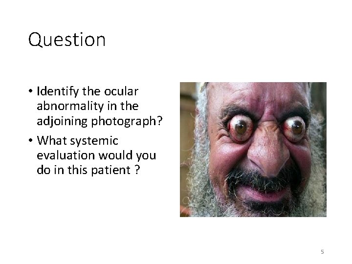
Question • Identify the ocular abnormality in the adjoining photograph? • What systemic evaluation would you do in this patient ? 5
Why do eye problems manifest in systemic forms The eye is linked with rest of the body by • Development • Blood supply • Meninges and nerve fibres of brain 6
Common Systemic Diseases affecting the Eye • Autoimmune disorders • Haematological abnormalities • Infectious diseases • Endocrine disorders • Muscular disorders • Inherited disorders 7
Autoimmune disorders 8
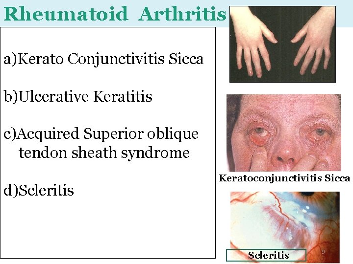
Rheumatoid Arthritis a)Kerato Conjunctivitis Sicca b)Ulcerative Keratitis c)Acquired Superior oblique tendon sheath syndrome d)Scleritis Keratoconjunctivitis Sicca Scleritis 9
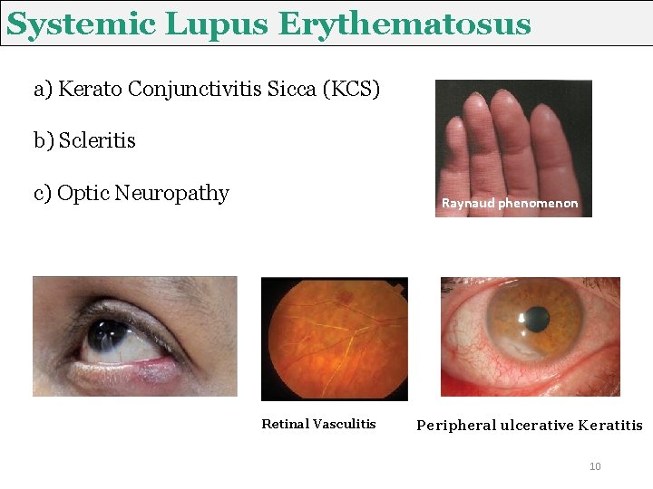
Systemic Lupus Erythematosus a) Kerato Conjunctivitis Sicca (KCS) b) Scleritis c) Optic Neuropathy Raynaud phenomenon Retinal Vasculitis Peripheral ulcerative Keratitis 10
Sarcoidosis • 30% ocular signs Phlebitis Iris nodule Mutton fat kps 11
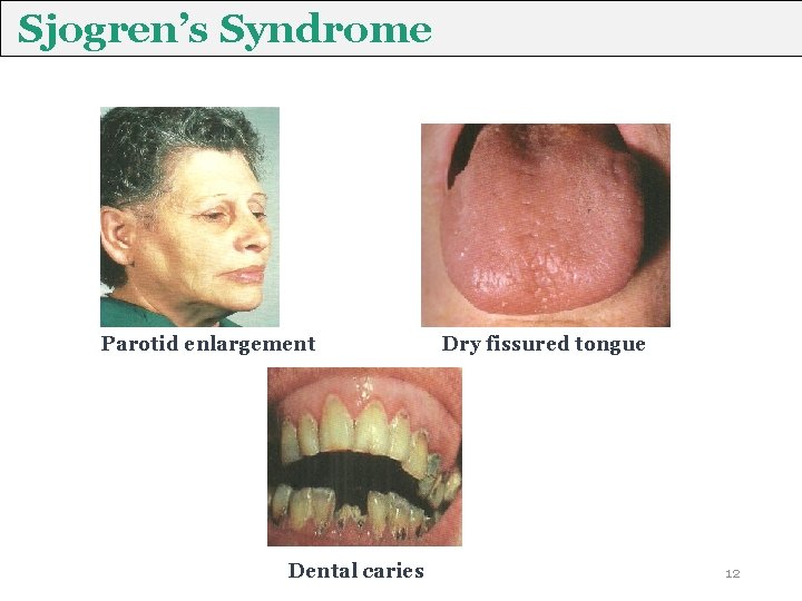
Sjogren’s Syndrome Parotid enlargement Dental caries Dry fissured tongue 12
Ankylosing Spondylitis • Acute anterior uveitis • Scleritis Symptoms: • Photophobia • Redness • Decreased vision • Bilateral sclerosis • Erosion of sacro-iliac joint • Bony fusion of spine 13
Reiter’s Syndrome Triad Arthritis Urethritis Conjunctivitis Eye Signs • Conjunctivitis • • • Acute anterior uveitis Numular Keratitis Episcleritis Scleritis Papillitis Retinal vasculitis Urethritis 14
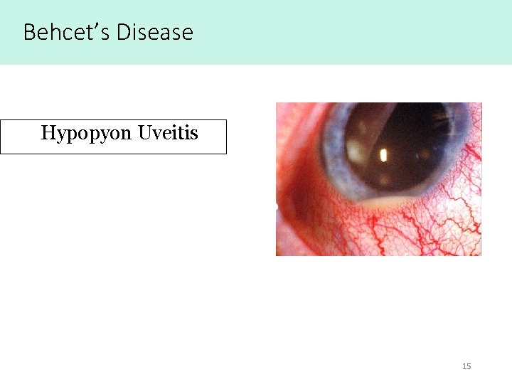
Behcet’s Disease Hypopyon Uveitis 15
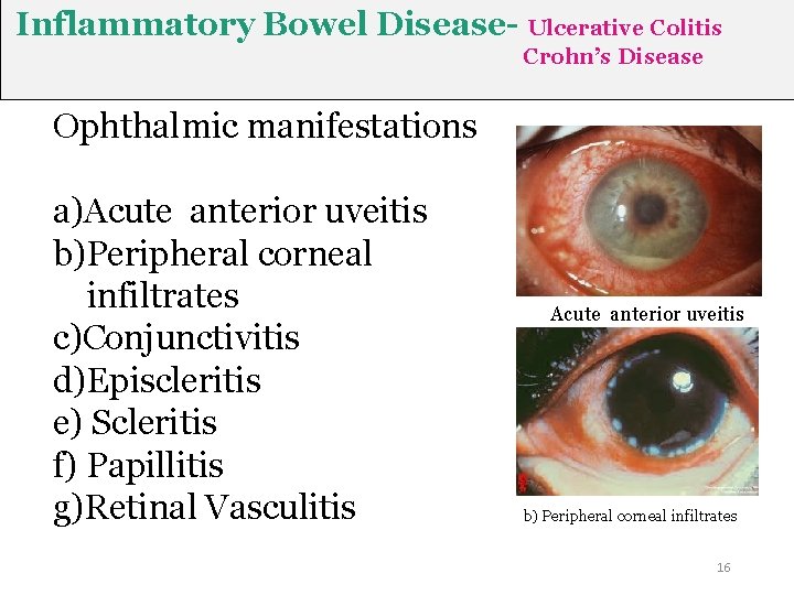
Inflammatory Bowel Disease- Ulcerative Colitis Crohn’s Disease Ophthalmic manifestations a)Acute anterior uveitis b)Peripheral corneal infiltrates c)Conjunctivitis d)Episcleritis e) Scleritis f) Papillitis g)Retinal Vasculitis Acute anterior uveitis b) Peripheral corneal infiltrates 16
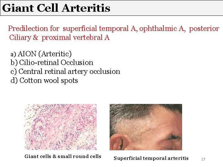
Giant Cell Arteritis Predilection for superficial temporal A, ophthalmic A, posterior Ciliary & proximal vertebral A a) AION (Arteritic) b) Cilio-retinal Occlusion c) Central retinal artery occlusion d) Cotton wool spots Giant cells & small round cells Superficial temporal arteritis 17
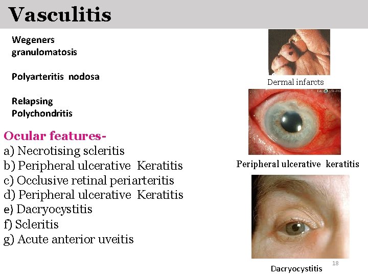
Vasculitis Wegeners granulomatosis Polyarteritis nodosa Dermal infarcts Relapsing Polychondritis Ocular featuresa) Necrotising scleritis b) Peripheral ulcerative Keratitis c) Occlusive retinal periarteritis d) Peripheral ulcerative Keratitis e) Dacryocystitis f) Scleritis g) Acute anterior uveitis Peripheral ulcerative keratitis Dacryocystitis 18
Haematological diseases 19
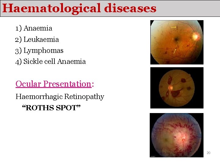
Haematological diseases 1) Anaemia 2) Leukaemia 3) Lymphomas 4) Sickle cell Anaemia Ocular Presentation: Haemorrhagic Retinopathy “ROTHS SPOT” 20
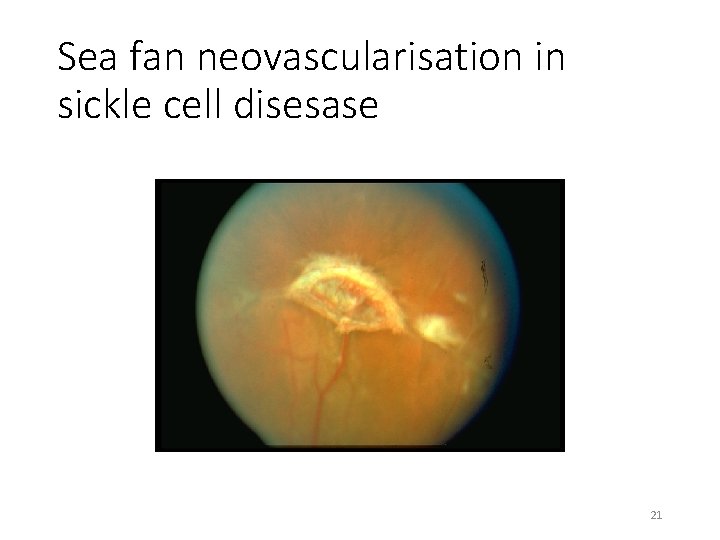
Sea fan neovascularisation in sickle cell disesase 21
Infectious diseases 22
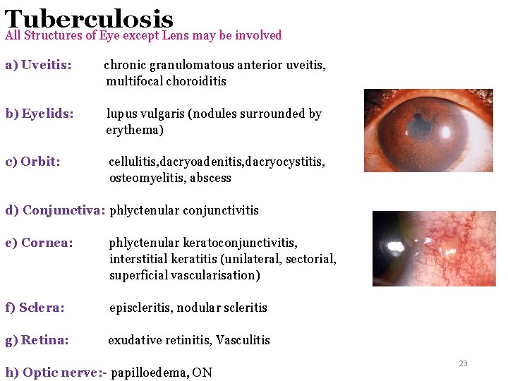
Tuberculosis All Structures of Eye except Lens may be involved a) Uveitis: chronic granulomatous anterior uveitis, multifocal choroiditis b) Eyelids: lupus vulgaris (nodules surrounded by erythema) c) Orbit: cellulitis, dacryoadenitis, dacryocystitis, osteomyelitis, abscess d) Conjunctiva: phlyctenular conjunctivitis e) Cornea: phlyctenular keratoconjunctivitis, interstitial keratitis (unilateral, sectorial, superficial vascularisation) f) Sclera: episcleritis, nodular scleritis g) Retina: exudative retinitis, Vasculitis h) Optic nerve: - papilloedema, ON 23
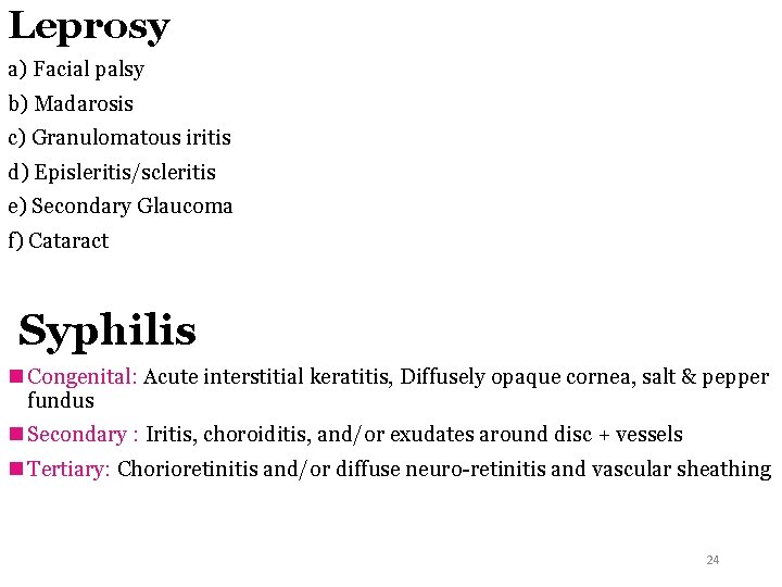
Leprosy a) Facial palsy b) Madarosis c) Granulomatous iritis d) Episleritis/scleritis e) Secondary Glaucoma f) Cataract Syphilis n Congenital: Acute interstitial keratitis, Diffusely opaque cornea, salt & pepper fundus n Secondary : Iritis, choroiditis, and/or exudates around disc + vessels n Tertiary: Chorioretinitis and/or diffuse neuro-retinitis and vascular sheathing 24
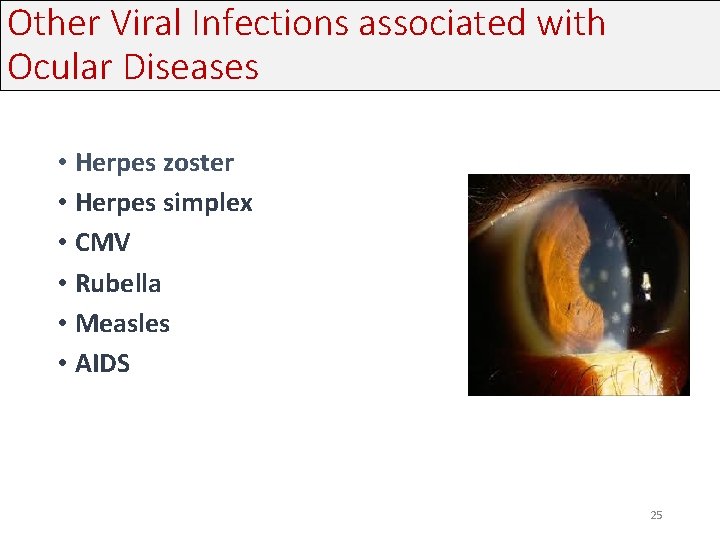
Other Viral Infections associated with Ocular Diseases • Herpes zoster • Herpes simplex • CMV • Rubella • Measles • AIDS 25
AIDS Kaposi’s sarcomamost common tumour Anterior Uveitis AIDS retinopathy- 50 to 70% CMV retinitis- most common opportunistic infection
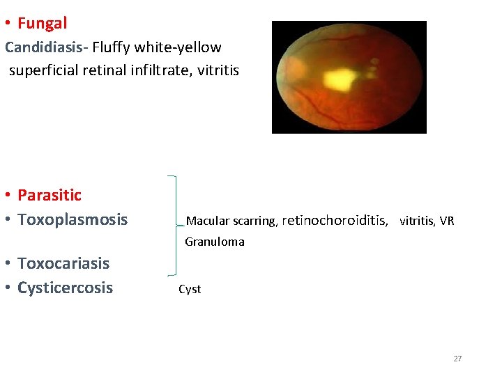
• Fungal Candidiasis- Fluffy white-yellow superficial retinal infiltrate, vitritis • Parasitic • Toxoplasmosis Macular scarring, retinochoroiditis, vitritis, VR Granuloma • Toxocariasis • Cysticercosis Cyst 27
Endocrine & Metabolic disorders 28
Endocrine & Metabolic disorders • Diabetes Mellitus • Muco-polysaccharidoses • Wilson’s disease • Homocystinuria • Hyperthyroidism • Hypothyroidism 29
Diabetes mellitus • Refractive error • Infections • Corneal ulcers • Snowflake cataract • Retinopathy • Maculopathy • Optic nerve papillopathy 30
THYROID EYE DISEASE Periorbital and lid swelling Chemosis Conjunctival hyperaemia 31 Superior limbic keratoconjunctivitis
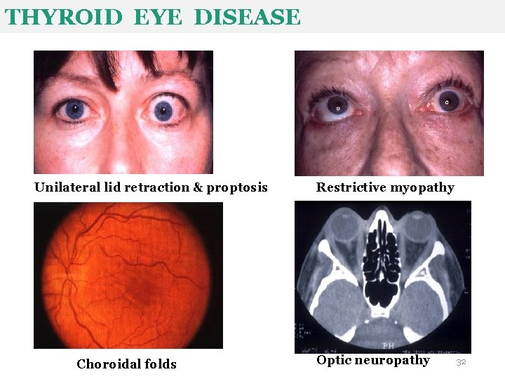
THYROID EYE DISEASE Unilateral lid retraction & proptosis Choroidal folds Restrictive myopathy Optic neuropathy 32
Muscular disorders 33
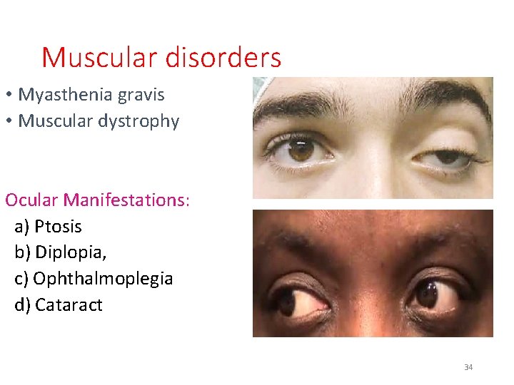
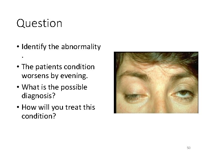
Muscular disorders • Myasthenia gravis • Muscular dystrophy Ocular Manifestations: a) Ptosis b) Diplopia, c) Ophthalmoplegia d) Cataract 34
Inherited disorders 35
Marfan’s Syndrome a) Hypoplasia of dilator pupillae b) Myopia c) Retinal detachment d) Microspherophakia e) Keratoconus f) Cornea Plana Arachnodactyly High-arched palate Ectopia lentis Angle anomaly 36
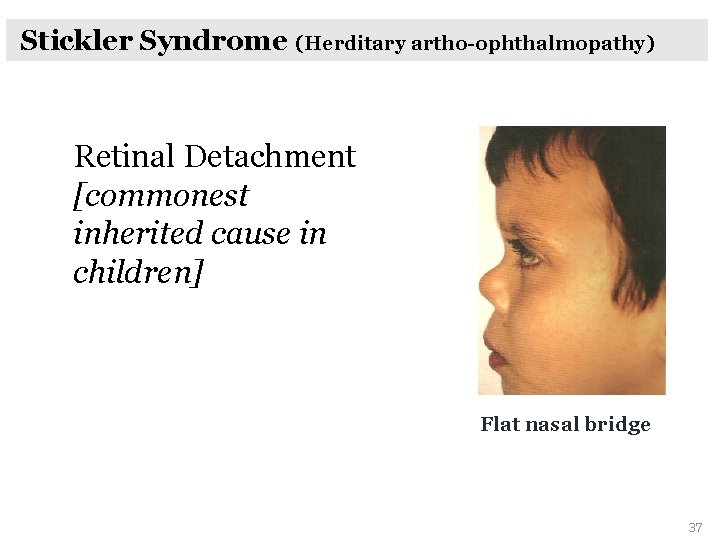
Stickler Syndrome (Herditary artho-ophthalmopathy) Retinal Detachment [commonest inherited cause in children] Flat nasal bridge 37
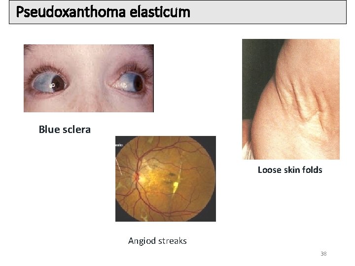
Pseudoxanthoma elasticum Blue sclera Loose skin folds Angiod streaks 38
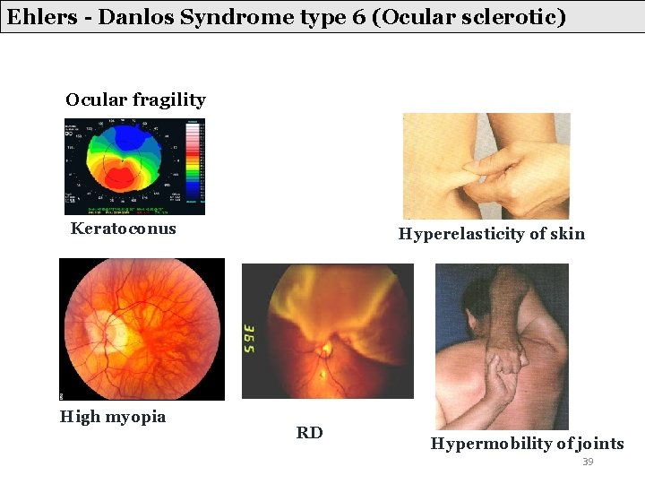
Ehlers - Danlos Syndrome type 6 (Ocular sclerotic) Ocular fragility Keratoconus High myopia Hyperelasticity of skin RD Hypermobility of joints 39
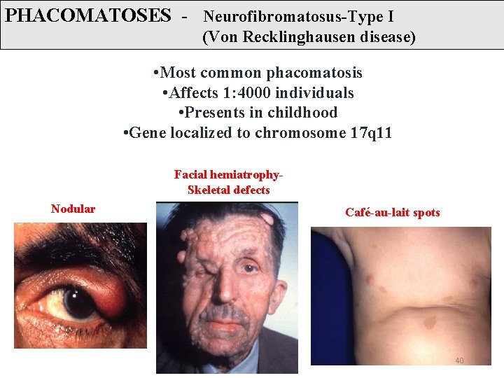
PHACOMATOSES - Neurofibromatosus-Type I (Von Recklinghausen disease) • Most common phacomatosis • Affects 1: 4000 individuals • Presents in childhood • Gene localized to chromosome 17 q 11 Facial hemiatrophy. Skeletal defects Nodular Café-au-lait spots 40
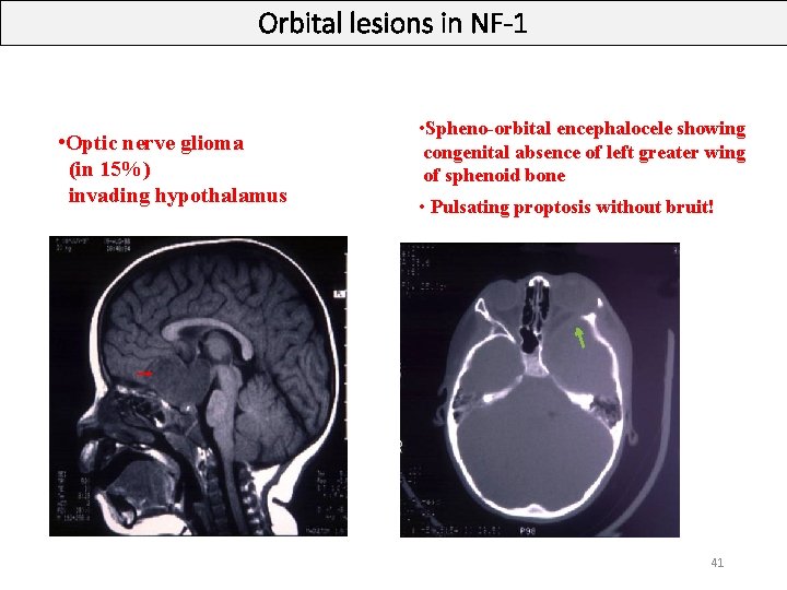
Orbital lesions in NF-1 • Optic nerve glioma (in 15%) invading hypothalamus • Spheno-orbital encephalocele showing congenital absence of left greater wing of sphenoid bone • Pulsating proptosis without bruit! 41
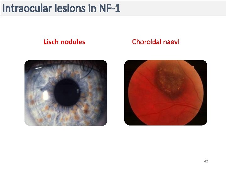
Intraocular lesions in NF-1 Lisch nodules Choroidal naevi 42
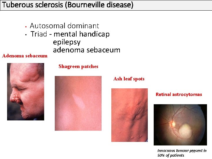
Tuberous sclerosis (Bourneville disease) Autosomal dominant • Triad - mental handicap epilepsy adenoma sebaceum • Adenoma sebaceum Shagreen patches Ash leaf spots Retinal astrocytomas Innocuous tumour present in 43 50% of patients
Von-Hippel-Lindau syndrome • Tumours– renal carcinoma and phaeochromocytoma • Cysts- kidneys, liver, pancreas, epididymis, ovary and lungs • Polycythaemia • Retinal capillary haemangioma Tiny lesion between arteriole and venuole Round orange-red mass Associated dilatation and tortuosity of feeder vessels 44
Malignancy 45
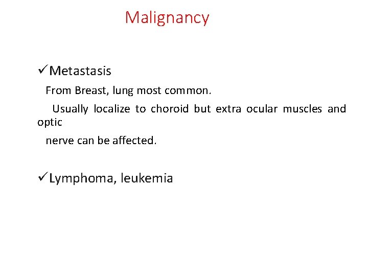
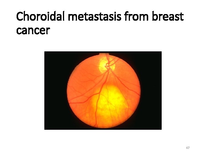
Malignancy üMetastasis From Breast, lung most common. Usually localize to choroid but extra ocular muscles and optic nerve can be affected. üLymphoma, leukemia
Choroidal metastasis from breast cancer 47
Summary • Ocular examination plays a significant role in several systemic diseases/disorders. • Ophthalmologists detect/suspect systemic illness for the first time in a patient when they examine the patient for Eye related complaints. • These patients then need Physician/Paediatrician/Neurologist/ Neurosurgeon/ Orthopaedician/Obstetrician opinion for further management. 48
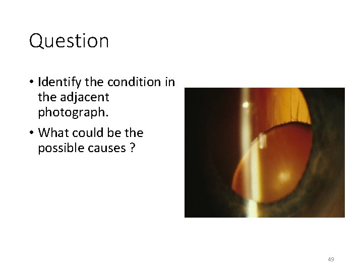
Question • Identify the condition in the adjacent photograph. • What could be the possible causes ? 49
Question • Identify the abnormality . • The patients condition worsens by evening. • What is the possible diagnosis? • How will you treat this condition? 50
THANK YOU 51