MAREKS DISEASE PENYAKIT MIKROBIAL DAN PARASITER II Tim

MAREK’S DISEASE PENYAKIT MIKROBIAL DAN PARASITER II Tim Mikrobiologi & Imunologi FKH-UBP

Marek’s Disease A highly contagious infection in chickens caused by a specific herpes virus that results in a rapidly fatal polyclonal lymphoma. Marek's disease is a neoplastic disease in chickens Chickens are the t natural host for Marek's disease virus Turkeys are also commonly infected with turkey herpesvirus (HVT), an avirulent strain related to Marek's disease virus that is commonly used as a Marek's disease vaccine in chickens. Other birds and mammals appear to be refractory to the disease or infection

Transmission& Epidemiology Marek's disease is one of the most ubiquitous avian infections; it is identified in chicken flocks worldwide. Suggested every flock, except for those maintained under strict pathogen-free conditions, is presumed to be infected. Although clinical disease is not always apparent in infected flocks, a subclinical decrease in growth rate and egg production may be economically important

The disease is highly contagious and readily transmitted among chickens Virus mature epithelium follicle feather environment survive for months in poultry house litter, dust Dust or dander from infected chickens transmission Once the virus is introduced into a chicken flock, infection spreads quickly Infected chickens be carriers for long periods and act as sources of infectious virus.

Birds infected with Ga. HV-2 can be carriers and shedders of the virus for life. Newborn chicks are protected by maternal antibodies for a few weeks. After infection, microscopic lesions are present after one to two weeks, and gross lesions are present after three to four weeks. The virus is spread in dander from feather follicles and transmitted by inhalation

Attenuated Marek's disease virus strains vary greatly in their transmissibility among chickens Marek's disease virus is not vertically transmitted.
 or Gallid herpesvirus")
Etiology Caused by an alphaherpesvirus known as 'Marek's disease virus' (MDV) or Gallid herpesvirus 2 (Ga. HV-2). May an oncogenic virus Pathogenicity: • Classical form (lesion in peripheral nerves) • Tumoral form (lymphoid tumors in different organs or in skin) • Transient paralysis
")
Marek's disease Virus classification Group: Order: Family: Subfamily: Genus: Species: Group I (ds. DNA) Herpesvirales Herpesviridae Alphaherpesvirinae Mardivirus Gallid herpesvirus 2 (Ga. HV-2 Gallid herpesvirus 2 (MDV-1) represents all virulent Marek's disease virus strains. Divided into pathotypes: mild (m), virulent (v), very virulent (vv), and very virulent plus (vv+).
")
Pathogenesis. Four phases of infection by virulent Marek's disease virus strains in vivo 1) Early productive-restrictive virus infection causing primarily degenerative changes Productive infection transiently in B lymphocytes within a few days after infection characterized by antigen production cell death. Because few if any virions are produced, this has also been termed a restrictive-productive infection. Productive infection also occurs in the feather follicle epithelium, in which enveloped virions are produced.
 Latent infection of activated T cells is responsible for the longterm carrier state.")
2) Latent infection of activated T cells is responsible for the longterm carrier state. No antigens are expressed, but virus can be recovered from such lymphocytes by co-cultivation with susceptible cells in tissue cultures.
 A second phase of cytolytic, productive-restrictive infection coincident with permanent immunosuppression Some T")
3) A second phase of cytolytic, productive-restrictive infection coincident with permanent immunosuppression Some T cells, latently infected with oncogenic Marek's disease virus strains, undergo neoplastic transformation. These transformed cells, provided they escape the immune system of the host, may multiply to form characteristic lymphoid neoplasms.
 A proliferative phase involving nonproductively infected lymphoid cells that may or may not")
4) A proliferative phase involving nonproductively infected lymphoid cells that may or may not progress to the point of lymphoma formation. Cell-mediated and humoral immune responses are both directed against viral antigens, with cell-mediated immunity probably being the most important.

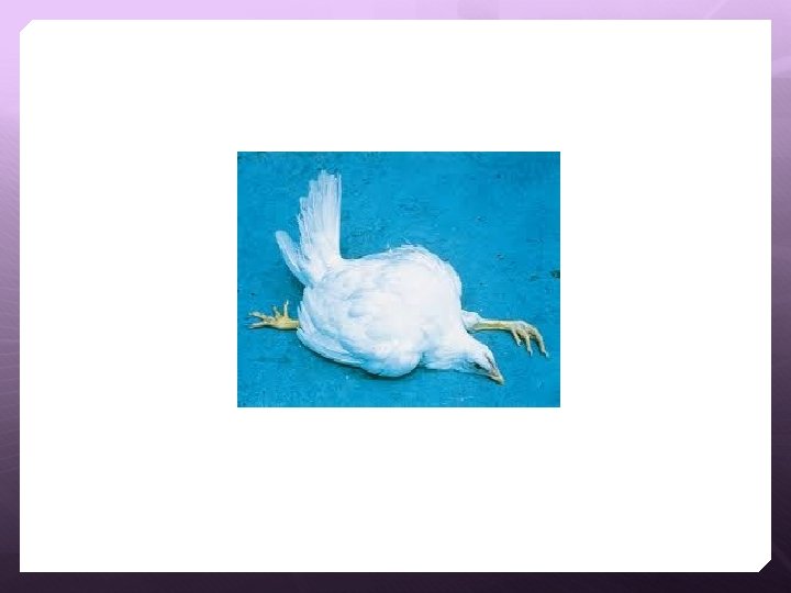
Syndrome There are six syndromes known to occur after infection with Marek's disease. These syndromes may overlap 1). Classical Marek's disease or neurolymphomatosis causes asymmetric paralysis of one or more limbs With vagus nerve involvement, difficulty breathing or dilation of the crop may occur. Besides lesions in the peripheral nerves, there are frequently lymphomatous infiltration/tumours in the skin, skeletal muscle, visceral organs. Organs that are commonly affected include the ovary, spleen, liver, kidneys, lungs, heart, proventriculus and adrenals.
")
Leg paresis (chicken Marek’s disease)
. Acute Marek's disease is an epidemic in a previously uninfected or unvaccinated flock,")
2). Acute Marek's disease is an epidemic in a previously uninfected or unvaccinated flock, causing depression, paralysis, and death in a large number of birds (up to 80 percent). • The age of onset is much earlier than the classic form; birds are 4 -8 weeks old when affected. • Infiltration into multiple organs/tissue is observed.
. Ocular lymphomatosis causes lymphocyte infiltration of the iris (making the iris turn grey),")
3). Ocular lymphomatosis causes lymphocyte infiltration of the iris (making the iris turn grey), unequal size of the pupils, and blindness. 4). Cutaneous Marek's disease causes round, firm lesions at the feather follicles 5). Atherosclerosis is induced in experimentally infected chickens
, unequal")
Ocular lymphomatosis causes lymphocyte infiltration of the iris (making the iris turn grey), unequal size of the pupils, and blindness. Left - normal chicken eye. Right - Eye of a chicken with Marek's disease

Cutaneous Marek's disease
 may be noted in broilers after defeathering")
Enlarged feather follicles (commonly termed skin leukosis) may be noted in broilers after defeathering during processing Marek's disease, skin involvement, chicken
. Immunosuppression – Impairment of the Tlymphocytes prevent competent immunological response against pathogenic challenge")
6). Immunosuppression – Impairment of the Tlymphocytes prevent competent immunological response against pathogenic challenge and the affected birds become more susceptible to disease conditions such as coccidiosis and "Escherichia coli" infection. Furthermore, without stimulation by cell-mediated immunity, the humoral immunity conferred by the Bcell lines from the Bursa of Fabricius also shuts down, thus resulting in birds that are totally immunocompromised.

Clinical Findings The incidence of Marek's disease is quite variable. Depends on: strain and dose of virus, age at exposure, maternal antibody, host gender and genetics, strain and dose of vaccine virus, and several environmental factors, including stress.

Diagnosis of lymphoid tumors in poultry is complicated due to multiple etiological agents capable of causing very similar tumors. More than one avian tumor virus can be present in a chicken Consider both the diagnosis of the disease/tumors (pathological diagnosis) and of the virus (etiological diagnosis).

A step-wise process has been proposed for diagnosis of Marek’s disease which includes (1) history, epidemiology, clinical observations and gross necropsy, (2) characteristics of the tumor cell, and (3) virological characteristics Peripheral nerve enlargement along with suggestive clinical signs in a bird, + 3 -4 months old (with or without visceral tumors) is highly suggestive of Marek's Disease. Histological nerves: infiltration of pleomorphic neoplastic and inflammatory lymphocytes. Peripheral neuropathy in young chickens, with paralysis and nerve enlargement, especially in nerves with interneuritic edema and infiltration of plasma cells

Nodules on the internal organs, but further testing is required for confirmation. Lymphomatous infiltration into the affected tissue. The T-cells are involved in the malignancy, showing neoplastic changes with evidence of mitosis.

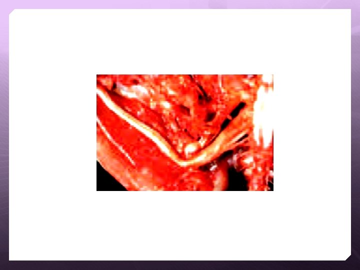
Enlarged nerves are one of the most consistent gross lesions in affected birds. Various peripheral nerves, but particularly the vagus, brachial, and sciatic, become enlarged and lose their striations Marek's disease, peripheral nerve enlargement, chicken

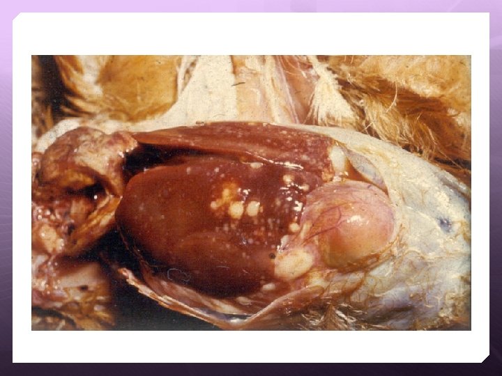
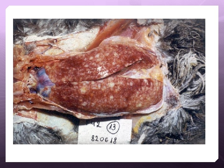
Diffuse or nodular lymphoid tumors may be seen in various organs, particularly the liver, spleen, gonads, heart, lung, kidney, muscle, and proventriculus. Marek's disease, liver and spleen involvement, chicken

In addition to gross pathology and histology, other advanced procedures used for a definitive diagnosis of Marek’s disease include immunohistochemistry to identify cell type and virus-specific antigens, standard and quantitative PCR for identification of the virus, virus isolation to confirm infections and serology to confirm/exclude infections.

Te. Ri. Ma Ka. Si. H
- Slides: 32