EYE MOVEMENTS EYE MOVEMENTS Eye is moved within


- Slides: 33
EYE MOVEMENTS
EYE MOVEMENTS �Eye is moved within orbit by six extraocular muscles. � 4 recti 1. 2. 3. 4. Superior rectus Inferior rectus Lateral rectus Medial rectus � 2 oblique muscles 1. 2. superior oblique Inferior oblique
Innervation of extraocular muscles �These are innervated by occulomotor, trochlear and abducent cranial nerves. �CN 3 innervates superior rectus, inferior rectus, medial rectus and inferior oblique. �Superior rectus is innervated by CN 4. �Lateral rectus is innervated by CN 6. �Superior and inferior recti moves eye upwards and downwards whereas medial rectus and lateral rectus moves eye from side to side. �Oblique muscles function mainly to rotate the eyeballs.
FIXATION MOVEMENTS OF EYE �Movements of eye that cause the eye to fix on a discrete portion of field of vision. �Controlled by 2 neuronal mechanisms: 1. Voluntary fixation mechanism Ø Allows a person to move the eye voluntarily to find the object on which he or she wants to fix the vision. Ø Controlled by cortical field located bilaterally in premotor cortical region of frontal lobe called voluntary fixation area.
Ø Bilateral dysfunction or destruction of these areas make it difficult for a person to unlock eye from point of fixation. It is usually necessary to blink the eyes or put a hand over eyes for short time which then allows the eye to be moved.
2. Involuntary fixation mechanism Ø It holds the eye firmly on object once it has been found. Ø Controlled by secondary visual area in occipital cortex. Ø When this area is destroyed bilaterally there is difficulty in keeping eyes directed towards a given fixation point.
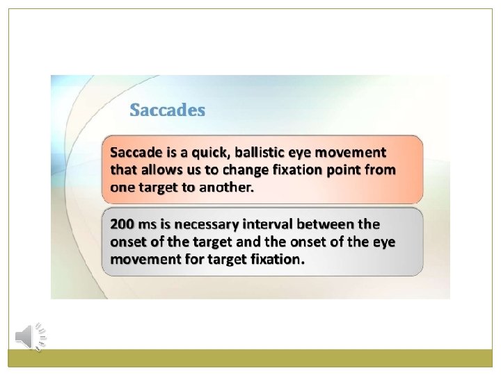
TYPES OF EYE MOVEMENTS 1. Sacccadic movement Ø sudden, jerky movements of eye. Ø Either eye or visual scene moves very rapidly from one locaion to another. Ø E. g during reading eyes move rapidly from word to word and line to line. Ø During driving the visual scenes change very rapidly and fixation occurs via saccadic movements of eyes.
� 2. Smooth persuit movements Ø Fixation on moving objects. Ø Eyes move steadily to track a moving object.
3. VESTIBULO OCULAR MOVEMENTS Ø Maintain visual fixation as the head moves. Ø The action of these movements can be appreciated by fixating an object and moving the head from side to side, the eyes automatically compensate for the head movement by moving eyes in opposite direction, thus keeping the image of object at more or less the same place on retina.
� 4. convergence movements Ø brings visual axis towaards each other as attention is focussed on near object.
�Smell is the least understood of our senses. This results partly from the fact that the sense of smell is a subjective phenomenon that cannot be studied with ease in lower animals. Another complicating problem is that the sense of smell is poorly developed in human beings in comparison with the sense of smell in many lower animals.
OLFACTORY EPITHELIUM
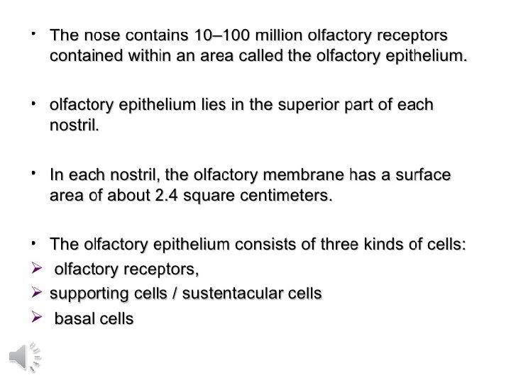
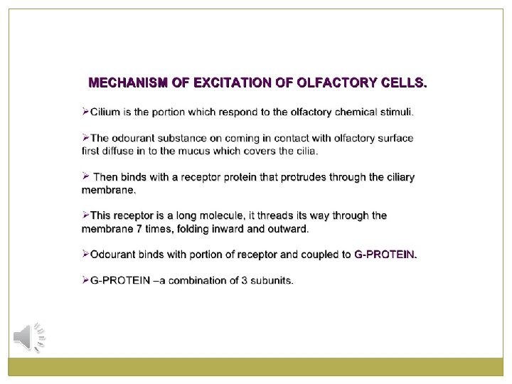
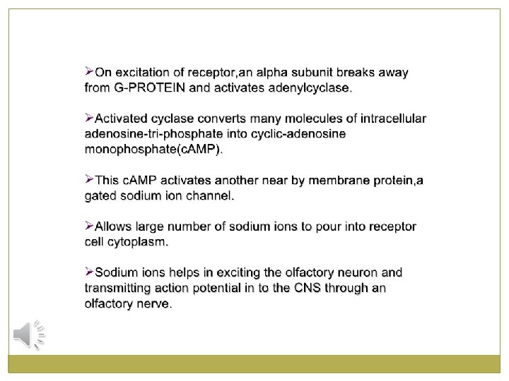
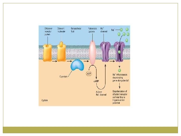
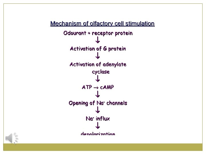
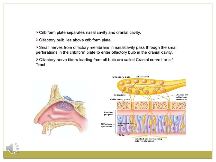
OLFACTORY RECEPTORS �The receptor cells for the smell sensation are olfactory cells. � they are actually bipolar cells derived from CNS. �There about 100 million of these cells in the olfactory epithelium interspersed among sustentacular cells. �The mucosal end of the olfactory cell forms a knob from which 4 to 25 olfactory hairs project into the mucus that coats the inner surface of the nasal cavity. �These projecting olfactory cilia form a dense mat in the mucus, and it is these cilia that react to odors in the air and stimulate the olfactory cells
�Spaced among the olfactory cells in the olfactory membrane are many small bowman’s gland, that secrete mucus onto the surface of olfactory membrane. �Mucus is carried to the surface of epithelium by ducts. �The secretion moisten the surface of olfactory epithelium and dissolves odourants so that transduction an occur.
SUPPORTING CELLS/SUSTENTACULAAR CELLS �The receptor cells in the olfactory epithelium are interspersed among sustentacular cells or supporting cells. �Supporting cells are columnar epithelial cells. �They provide physical support, nourishment and electrical insulation for the olfactory receptors. �They help detoxify chemicals that come in contact with the olfactory epithelium.
BASAL CELLS/ STEM CELLS �Basal cells are stem cells located between the bases of the supporting cells. �They continously undergo cell division to produce new olfactory receptors, which live for only a month or so before being replaced.
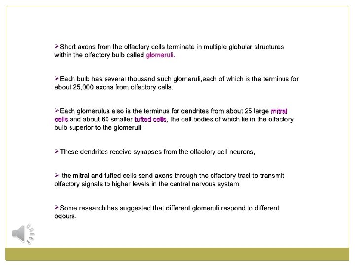
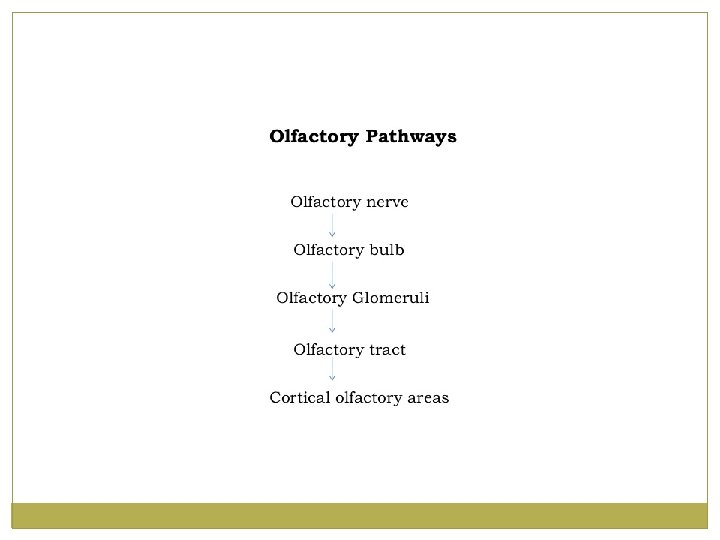
TRANSMISSION OF OLFACTORY SIGNALS TO OLFACTORY BULB
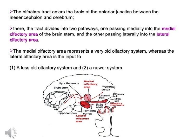
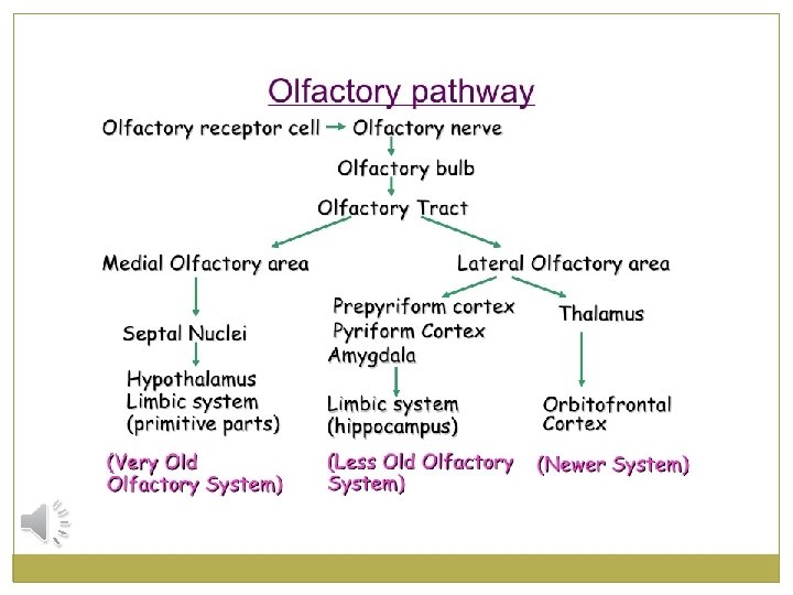
OLFACTORY PATHWAY