
Ophthalmology revision for MD finals Part II Dr






- Slides: 49
Ophthalmology revision for MD finals (Part II) Dr Haitham Al-Mahrouqi BMed. Sc (Hons)/MBCh. B Ophthalmology resident Oman Medical Specialty Board
Chronic visual loss
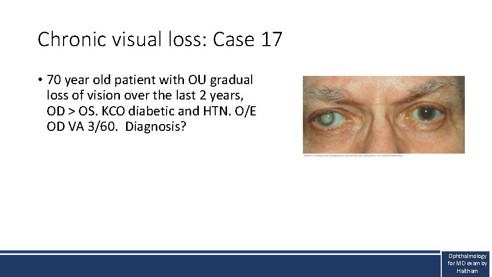
Chronic visual loss: Case 17 • 70 year old patient with OU gradual loss of vision over the last 2 years, OD > OS. KCO diabetic and HTN. O/E OD VA 3/60. Diagnosis? Ophthalmology for MD exam by Haitham
Chronic visual loss: Cataract High yield information • Etiology: Opacification of the lens proteins due to oxidative stress (age), polyol pathway (diabetic), traumatic or steroid induced. • History: Gradual loss of vision. • Examination: - Opacification of the crystalline lens • Management: - Surgery when visually significant.
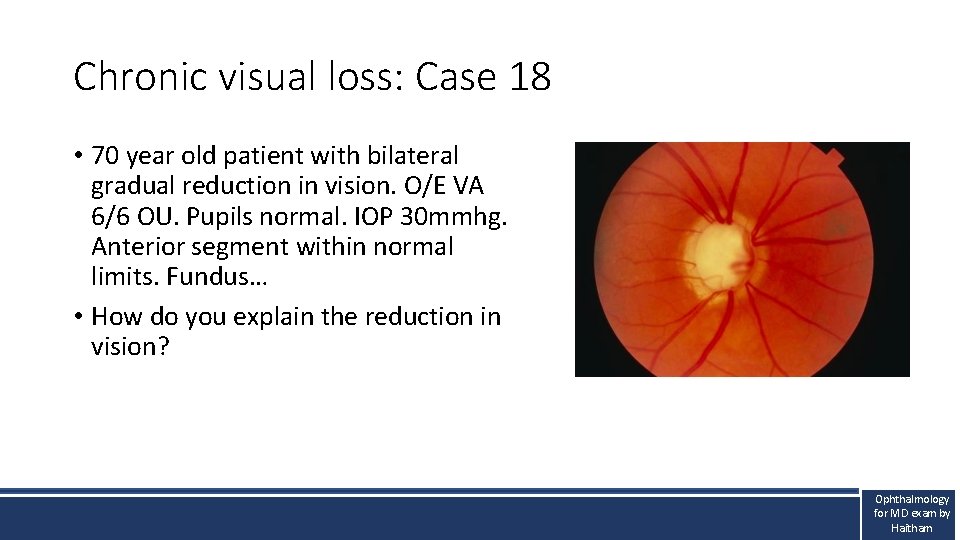
Chronic visual loss: Case 18 • 70 year old patient with bilateral gradual reduction in vision. O/E VA 6/6 OU. Pupils normal. IOP 30 mmhg. Anterior segment within normal limits. Fundus… • How do you explain the reduction in vision? Ophthalmology for MD exam by Haitham
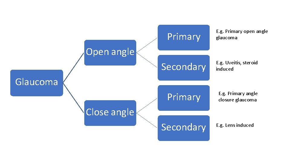
Chronic visual loss: Chronic glaucoma High yield information • Definition: Optic neuropathy where there is reduced retinal sensitivity and IOP is a risk factor • Etiology: Reduced aqueous outflow due to resistance of the trabecular meshwork (open angle) or closure (angle closure) leading to ganglion cell layer loss either due to pressure or ischaemia. • History: Gradual loss of peripheral vision (The silent thief) • Examination: - RAPD (if secondary glaucoma), angle abnormality, cupping • Management: - VF and optic disc OCT. - Anti-glaucoma medications or filtration surgery.
Primary E. g. Primary open angle glaucoma Open angle Secondary E. g. Uveitis, steroid induced Primary E. g. Primary angle closure glaucoma Glaucoma Close angle Secondary E. g. Lens induced
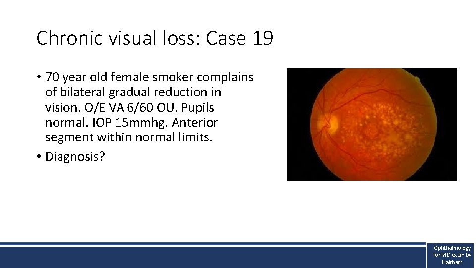
Chronic visual loss: Case 19 • 70 year old female smoker complains of bilateral gradual reduction in vision. O/E VA 6/60 OU. Pupils normal. IOP 15 mmhg. Anterior segment within normal limits. • Diagnosis? Ophthalmology for MD exam by Haitham
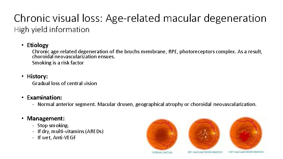
Chronic visual loss: Age-related macular degeneration High yield information • Etiology Chronic age-related degeneration of the bruchs membrane, RPE, photoreceptors complex. As a result, choroidal neovascularization ensues. Smoking is a risk factor • History: Gradual loss of central vision • Examination: - Normal anterior segment. Macular drusen, geographical atrophy or choroidal neovascularization. • Management: - Stop smoking. - If dry, multi-vitamins (AREDs) - If wet, Anti-VEGF
Neuro-ophthalmic emergencies
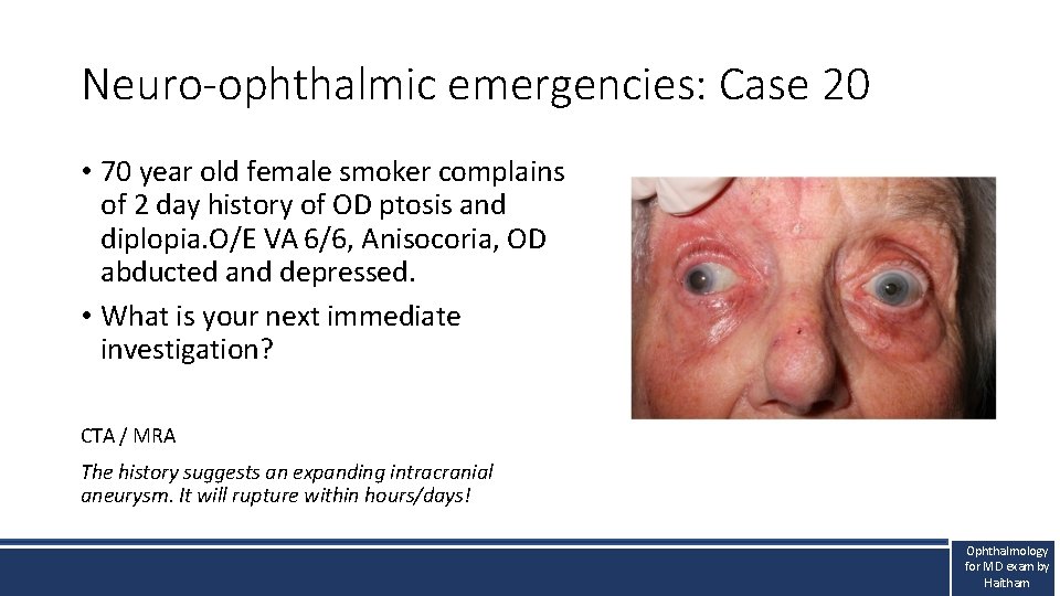
Neuro-ophthalmic emergencies: Case 20 • 70 year old female smoker complains of 2 day history of OD ptosis and diplopia. O/E VA 6/6, Anisocoria, OD abducted and depressed. • What is your next immediate investigation? CTA / MRA The history suggests an expanding intracranial aneurysm. It will rupture within hours/days! Ophthalmology for MD exam by Haitham
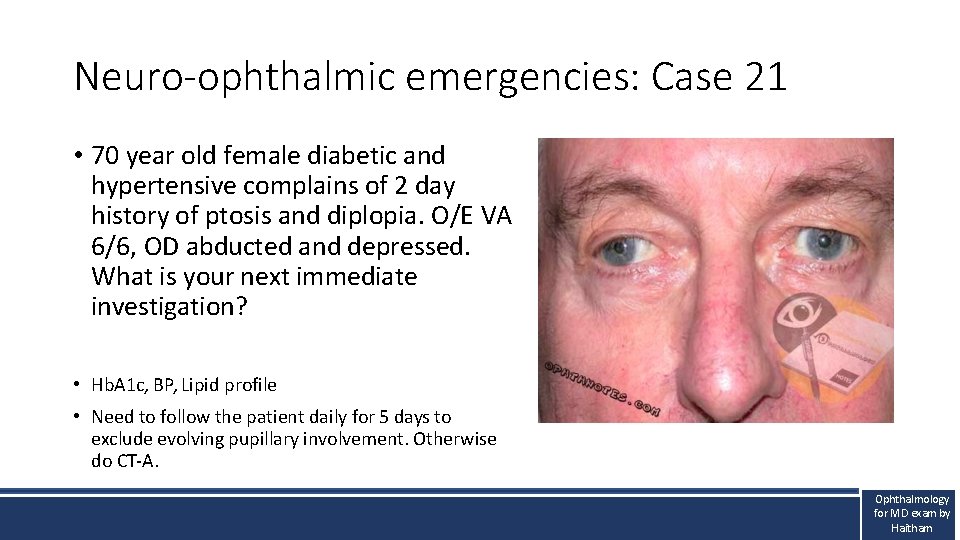
Neuro-ophthalmic emergencies: Case 21 • 70 year old female diabetic and hypertensive complains of 2 day history of ptosis and diplopia. O/E VA 6/6, OD abducted and depressed. What is your next immediate investigation? • Hb. A 1 c, BP, Lipid profile • Need to follow the patient daily for 5 days to exclude evolving pupillary involvement. Otherwise do CT-A. Ophthalmology for MD exam by Haitham
Neuro-ophthalmic emergencies: Oculomotor nerve palsy High yield information • Etiology (according to types): Pupillary involving: Compressive lesion, most commonly PCOM aneurysm Pupillary sparing: Ischaemia due to DM, HTN, hyperlipidaemia, smoking • History: Diplopia. Pain is subjective and can occur with both types. • Examination: - Ophthalmoplegia, +/- aniscoria, otherwise normal. • Management: - CTA/MRA to exclude an aneurysm. - Referral to neurosurgery for coiling/embolising aneurysms or GP to manage the ischaemic risk factors. - Resolves within 3 months
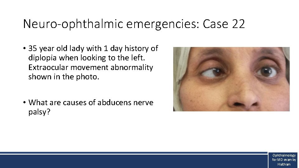
Neuro-ophthalmic emergencies: Case 22 • 35 year old lady with 1 day history of diplopia when looking to the left. Extraocular movement abnormality shown in the photo. • What are causes of abducens nerve palsy? Ophthalmology for MD exam by Haitham
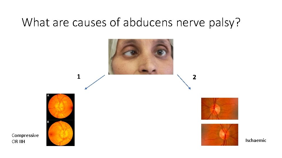
What are causes of abducens nerve palsy? 1 Compressive OR IIH 2 Ischaemic
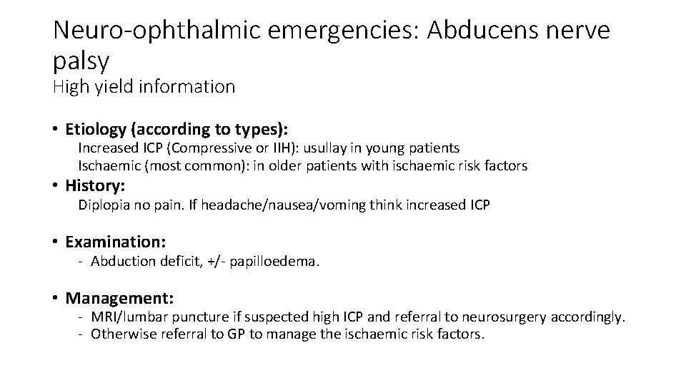
Neuro-ophthalmic emergencies: Abducens nerve palsy High yield information • Etiology (according to types): Increased ICP (Compressive or IIH): usullay in young patients Ischaemic (most common): in older patients with ischaemic risk factors • History: Diplopia no pain. If headache/nausea/voming think increased ICP • Examination: - Abduction deficit, +/- papilloedema. • Management: - MRI/lumbar puncture if suspected high ICP and referral to neurosurgery accordingly. - Otherwise referral to GP to manage the ischaemic risk factors.
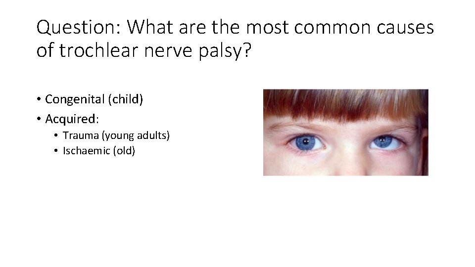
Question: What are the most common causes of trochlear nerve palsy? • Congenital (child) • Acquired: • Trauma (young adults) • Ischaemic (old)
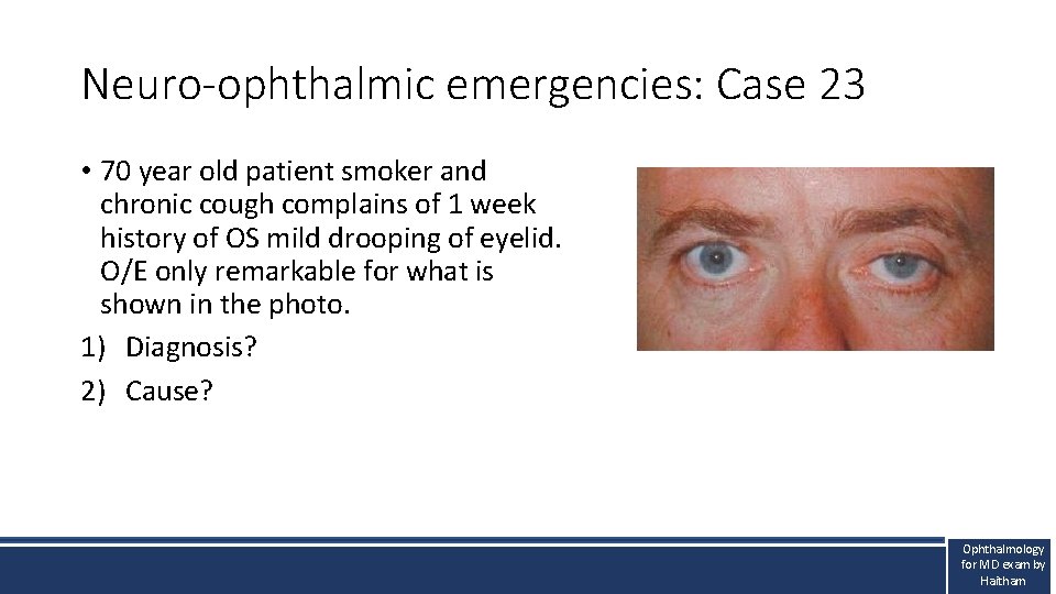
Neuro-ophthalmic emergencies: Case 23 • 70 year old patient smoker and chronic cough complains of 1 week history of OS mild drooping of eyelid. O/E only remarkable for what is shown in the photo. 1) Diagnosis? 2) Cause? Ophthalmology for MD exam by Haitham
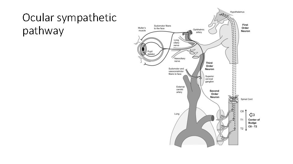
Ocular sympathetic pathway
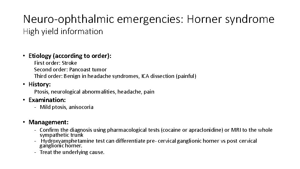
Neuro-ophthalmic emergencies: Horner syndrome High yield information • Etiology (according to order): First order: Stroke Second order: Pancoast tumor Third order: Benign in headache syndromes, ICA dissection (painful) • History: Ptosis, neurological abnormalities, headache, pain • Examination: - Mild ptosis, anisocoria • Management: - Confirm the diagnosis using pharmacological tests (cocaine or apraclonidine) or MRI to the whole sympathetic trunk - Hydroxyamphetamine test can differentiate pre- cervical ganglionic horner vs post cervical ganglionic horner. - Treat the underlying cause.
Trauma
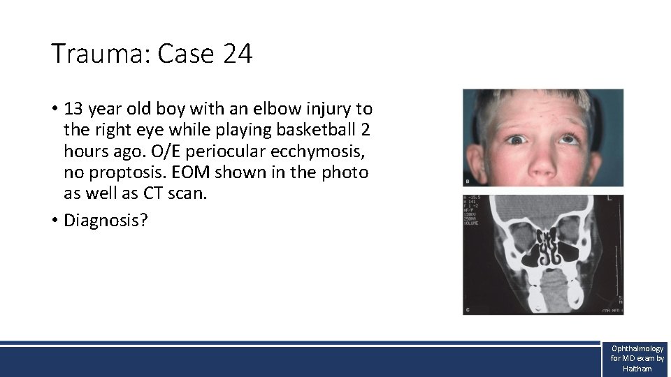
Trauma: Case 24 • 13 year old boy with an elbow injury to the right eye while playing basketball 2 hours ago. O/E periocular ecchymosis, no proptosis. EOM shown in the photo as well as CT scan. • Diagnosis? Ophthalmology for MD exam by Haitham
Ocular trauma: Orbital floor fracture High yield information • Etiology: Orbital floor is the weakest bone. The inferior rectus get trapped and the infraorbital nerve injured. Children are prone to greenstick # resulting in ischaemia and oculocardiac reflex. • History: Injury. No reduction in vision unless the globe or optic nerve are injured. • Examination: - ABC, exclude ruptured globe, retrobulbar injury, optic nerve injury ( RAPD) , ocular motility. • Management: - Stabilize ABC - Fracture to be fixed after 2 weeks once oedema subsides. In children, to be operated soon due to ischaemia of muscle and oculocardiac reflex.
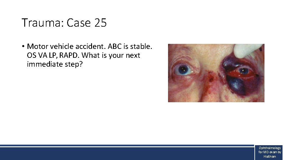
Trauma: Case 25 • Motor vehicle accident. ABC is stable. OS VA LP, RAPD. What is your next immediate step? Ophthalmology for MD exam by Haitham
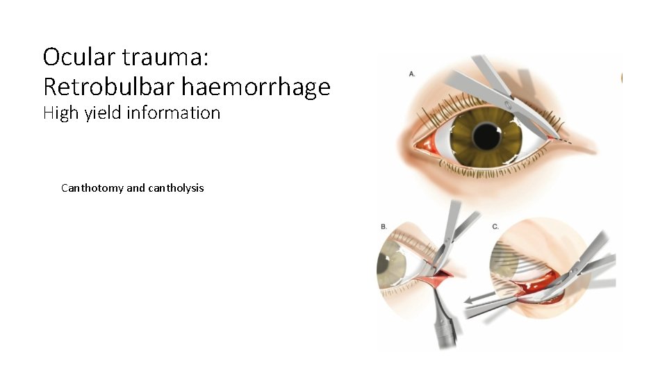
Ocular trauma: Retrobulbar haemorrhage High yield information Canthotomy and cantholysis
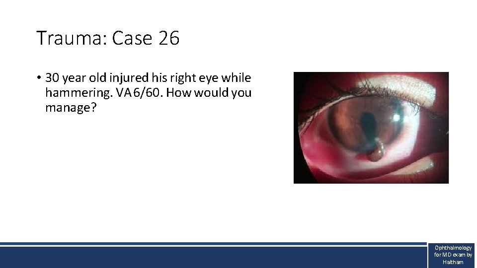
Trauma: Case 26 • 30 year old injured his right eye while hammering. VA 6/60. How would you manage? Ophthalmology for MD exam by Haitham
Ocular trauma: Open globe injury High yield information • Management: - Stabilize ABC and shield the eye (do not pressure patch) Tetanus toxoid IV Abx CT scan to exlude intraocular foreign body. Primary repair
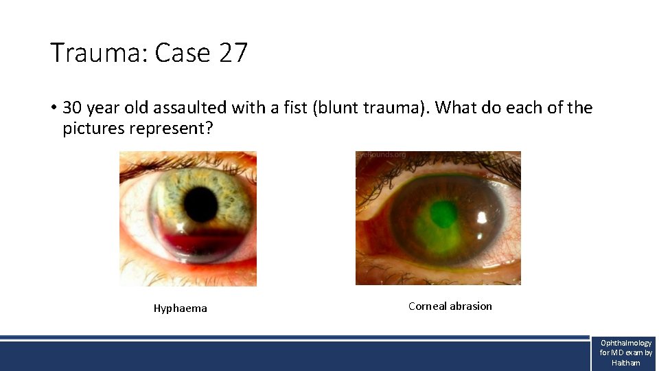
Trauma: Case 27 • 30 year old assaulted with a fist (blunt trauma). What do each of the pictures represent? Hyphaema Corneal abrasion Ophthalmology for MD exam by Haitham
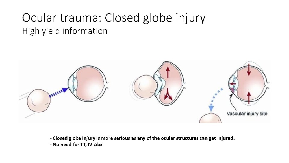
Ocular trauma: Closed globe injury High yield information - Closed globe injury is more serious as any of the ocular structures can get injured. - No need for TT, IV Abx
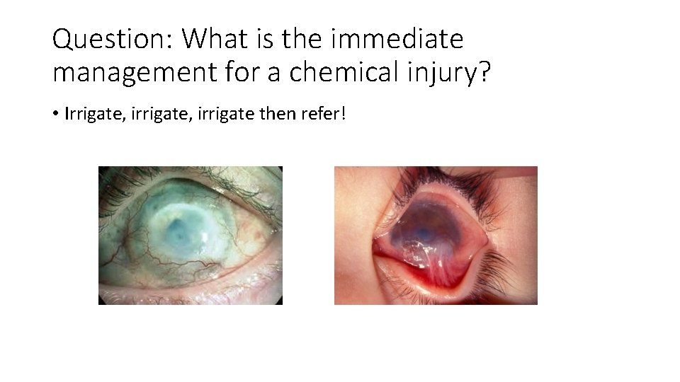
Question: What is the immediate management for a chemical injury? • Irrigate, irrigate then refer!
Proptosis
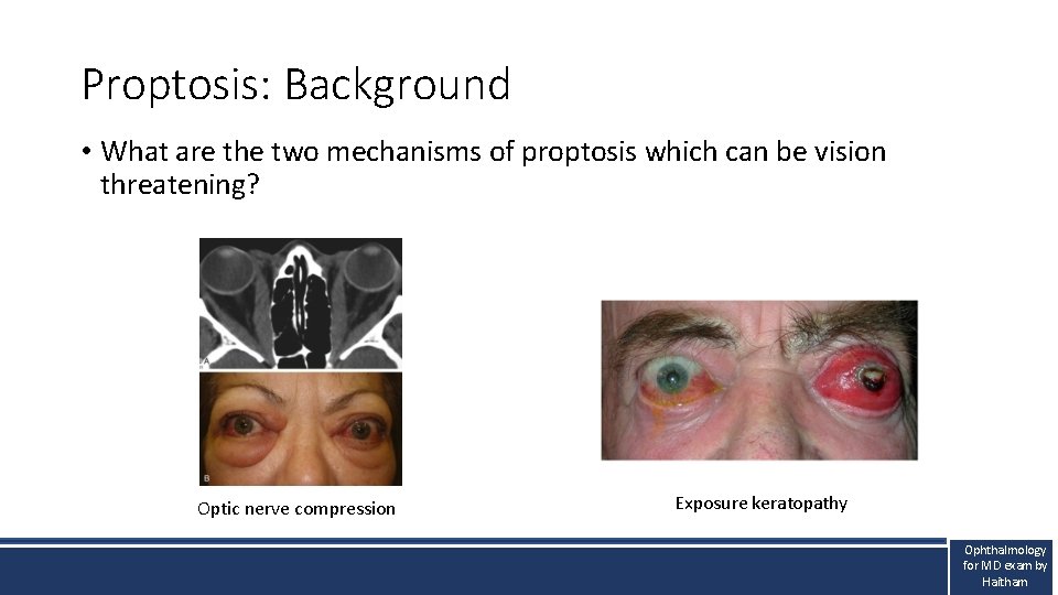
Proptosis: Background • What are the two mechanisms of proptosis which can be vision threatening? Optic nerve compression Exposure keratopathy Ophthalmology for MD exam by Haitham
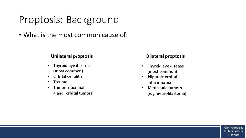
Proptosis: Background • What is the most common cause of: Unilateral proptosis • Thyroid eye disease (most common) • Orbital cellulitis • Trauma • Tumors (lacrimal gland, orbital tumors) Bilateral proptosis • Thyroid eye disease (most common) • Idipathic orbital inflammation • Metastatic tumors (e. g. neuroblastoma) Ophthalmology for MD exam by Haitham
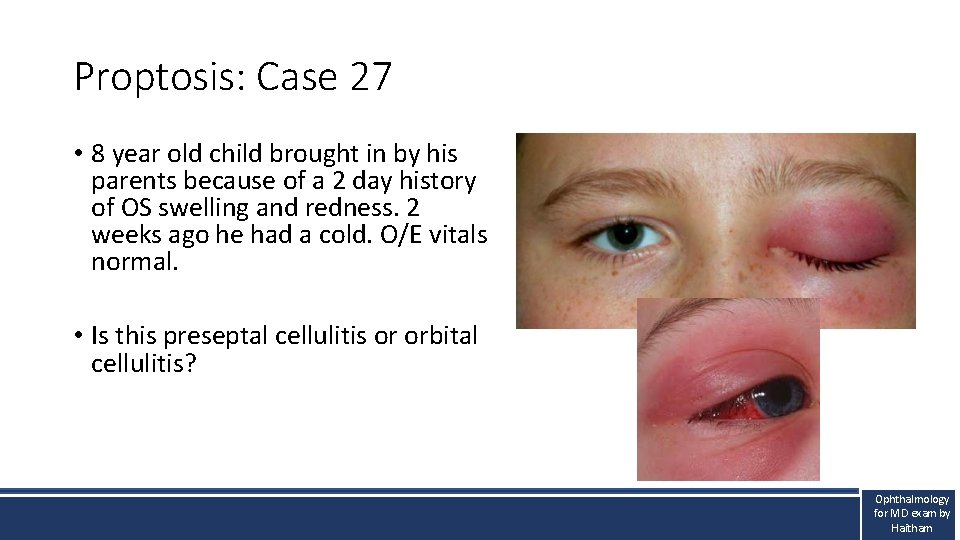
Proptosis: Case 27 • 8 year old child brought in by his parents because of a 2 day history of OS swelling and redness. 2 weeks ago he had a cold. O/E vitals normal. • Is this preseptal cellulitis or orbital cellulitis? Ophthalmology for MD exam by Haitham
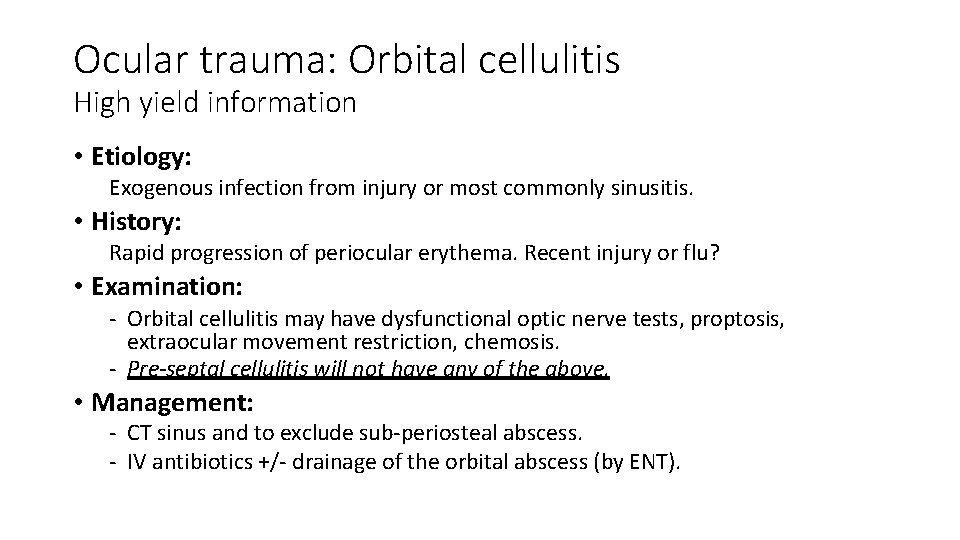
Ocular trauma: Orbital cellulitis High yield information • Etiology: Exogenous infection from injury or most commonly sinusitis. • History: Rapid progression of periocular erythema. Recent injury or flu? • Examination: - Orbital cellulitis may have dysfunctional optic nerve tests, proptosis, extraocular movement restriction, chemosis. - Pre-septal cellulitis will not have any of the above. • Management: - CT sinus and to exclude sub-periosteal abscess. - IV antibiotics +/- drainage of the orbital abscess (by ENT).
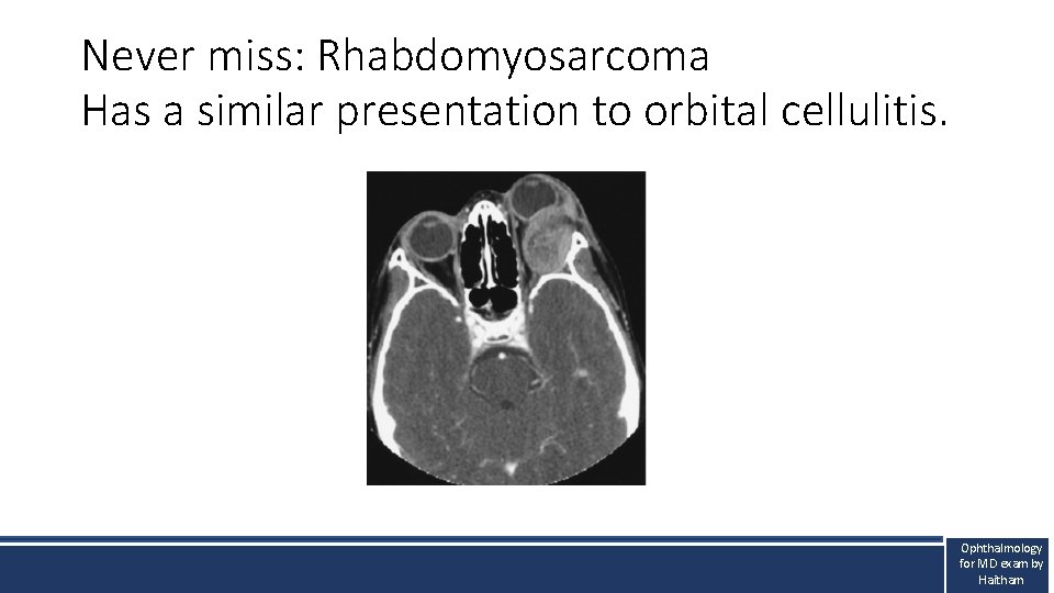
Never miss: Rhabdomyosarcoma Has a similar presentation to orbital cellulitis. Ophthalmology for MD exam by Haitham
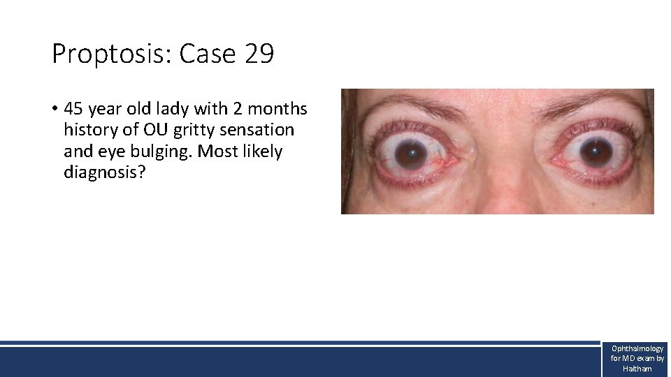
Proptosis: Case 29 • 45 year old lady with 2 months history of OU gritty sensation and eye bulging. Most likely diagnosis? Ophthalmology for MD exam by Haitham
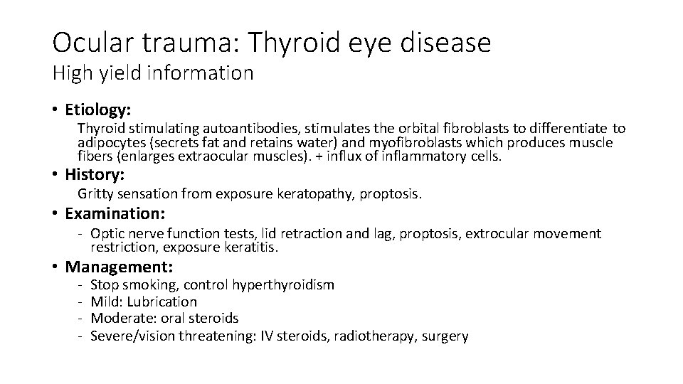
Ocular trauma: Thyroid eye disease High yield information • Etiology: Thyroid stimulating autoantibodies, stimulates the orbital fibroblasts to differentiate to adipocytes (secrets fat and retains water) and myofibroblasts which produces muscle fibers (enlarges extraocular muscles). + influx of inflammatory cells. • History: Gritty sensation from exposure keratopathy, proptosis. • Examination: - Optic nerve function tests, lid retraction and lag, proptosis, extrocular movement restriction, exposure keratitis. • Management: - Stop smoking, control hyperthyroidism Mild: Lubrication Moderate: oral steroids Severe/vision threatening: IV steroids, radiotherapy, surgery
Paediatric ophthalmology
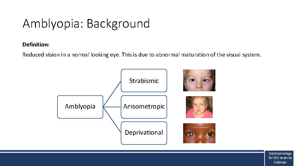
Amblyopia: Background Definition: Reduced vision in a normal looking eye. This is due to abnormal maturation of the visual system. Strabismic Amblyopia Anisometropic Deprivational Ophthalmology for MD exam by Haitham
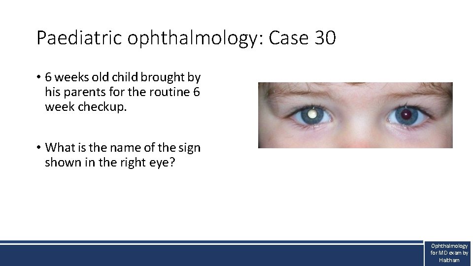
Paediatric ophthalmology: Case 30 • 6 weeks old child brought by his parents for the routine 6 week checkup. • What is the name of the sign shown in the right eye? Ophthalmology for MD exam by Haitham
Ocular trauma: Leukocoria (white pupil) High yield information • Etiology: Pathological lens, vitreous, retina (all behind the pupil). NEVER MISS retinoblastoma
Miscellaneous
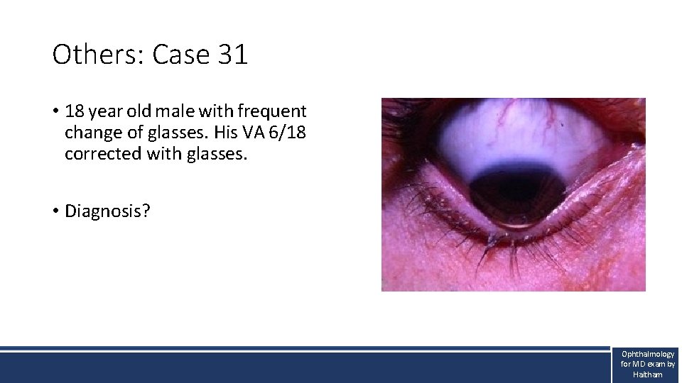
Others: Case 31 • 18 year old male with frequent change of glasses. His VA 6/18 corrected with glasses. • Diagnosis? Ophthalmology for MD exam by Haitham
Others: Keratoconus High yield information • Etiology: Genes, eye rubbing, allergic eye disease. Estimated prevalence in Oman : 5 -10% Onset 10 -20 years • History: Family history. Frequent change of glasses, glasses not giving full correction. • Examination: - high astigmatism, Papillae, corneal thinning with protrusion. • Management: - Stop the progression of the disease with corneal cross-linking - Rehabilitate vision with glasses, contact lenses, rings and last…. corneal transplantation.
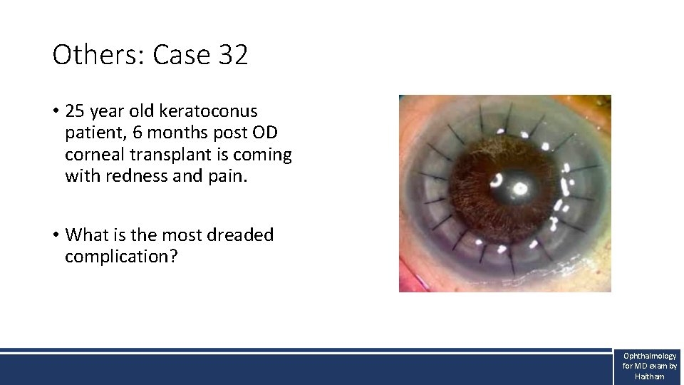
Others: Case 32 • 25 year old keratoconus patient, 6 months post OD corneal transplant is coming with redness and pain. • What is the most dreaded complication? Ophthalmology for MD exam by Haitham
Others: Corneal transplant High yield information • Must refer immediately to exclude rejection.
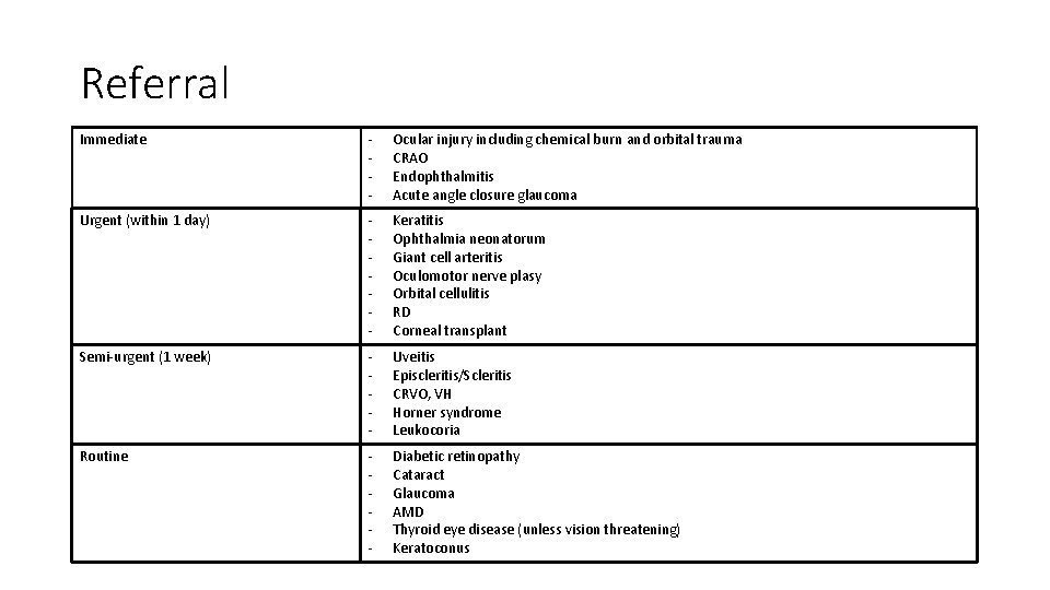
Referral Immediate - Ocular injury including chemical burn and orbital trauma CRAO Endophthalmitis Acute angle closure glaucoma Urgent (within 1 day) - Keratitis Ophthalmia neonatorum Giant cell arteritis Oculomotor nerve plasy Orbital cellulitis RD Corneal transplant Semi-urgent (1 week) - Uveitis Episcleritis/Scleritis CRVO, VH Horner syndrome Leukocoria Routine - Diabetic retinopathy Cataract Glaucoma AMD Thyroid eye disease (unless vision threatening) Keratoconus
Thank you