UVVis spectroscopy Electronic absorption spectroscopy spectroscopy The interactions

UV-Vis spectroscopy Electronic absorption spectroscopy

spectroscopy The interactions of radiation and matter are the subject of the science called spectroscopy.

Properties of Electromagnetic Radiation v Electromagnetic radiation is a form of energy that is transmitted through space at enormous velocities. v Electromagnetic radiation have properties of wavelength, frequency, velocity, and amplitude. v In contrast to sound waves, light requires no supporting medium for its transmission; thus, it readily passes through a vacuum.

ELECTROMAGNETIC SPECTRUM

Absorption and Emission Absorption: A transition from a lower level to a higher level with transfer of energy from the radiation field to an absorber, atom, molecule, or solid. Emission: A transition from a higher level to a lower level with transfer of energy from the emitter to the radiation field.

UV and Visible Spectroscopy Ultraviolet Spectroscopy: involves the measurement of absorption of light in the visible and ultraviolet regions (visible region 400 -800 nm; UV region 200 -400 nm) by the substances under investigation. Since the absorption of light involves the transition from one electronic energy level to another with in a molecule, UV spectroscopy is also known as electronic spectroscopy.

PRINCIPLE OF UV SPECTROSCOPY • Absorption of visible and ultraviolet light produces changes in the electronic states of molecules associated with the excitation of an electron from a lower to a higher energy level. • But it must be noted that each el electronic level in a molecule is associated with a no. of vibrational sub-levels and each vibrational energy level in turn is associated with a no. of rotational sub-levels.

Appearance of broad bands and not sharp peaks in the spectrum. • Due to the mixing of vibrational and rotational changes with electronic changes in the molecules, there will be a large no. of possible transitions requiring only slightly different energies. • As a result the absorption spectrum contains a large no. of lines which are too close together to be distinguished separately and are recorded in the form of broad bands in the spectrum obtained.

Electronic excitation from the ground state to the excited state accompanied By changes in vibrational and rotational sub-levels. Here E 0=Ground state E*=Excited state.

Beer-Lambert’s Law B eer ’ Law; “When a monochromatic light is passed through a substance dispersed in a non-absorbing solvent, the absorption of light is directly proportional to the molar concentration of the substance. ’’ Lambert ’s Law; “When a of monochromatic light is passed through a substance dispersed in a nonabsorbing solvent, the absorption of light is directly proportional to the path length of the sample. ’’ B eer- Lambert ’s Law ; B y combining above two Laws, " The absorption of light by a substance at a particular wavelength is directly proportional to the no. of molecules of the substance in the path of light. ”
 log I 0/I α ℓ (Lambert'Law)")
Thus log I 0/I α c (Beer's Law) log I 0/I α ℓ (Lambert'Law) log I 0/I α cℓ (Beer Lambert’s Law) log I 0/I =£cℓ log I 0/I =Optical Density or absorbance (A) Therefore, (A)=log I 0/I =£cℓ Where. A = Absorbance (optical density) I 0 = Intensity of light on the sample cell I = Intensity of light leaving the sample cell c = molar concentration of solute ℓ = length of sample cell (cm)

Limitation of Beer Lambert’s Law • High concentrations • Solute and solvent form complexes • Thermal equilibrium exist between the ground state and the excited state • Fluorescent compounds are present in solution
 Sources: Argon, Xenon, Deuterium, or Tungsten lamps • Deuterium Lamps-a")
Components of instrumentation: 1) Sources: Argon, Xenon, Deuterium, or Tungsten lamps • Deuterium Lamps-a truly continuous spectrum in the ultraviolet region is produced by electrical excitation of deuterium at low pressure. (160 nm~375 nm) • Tungsten Filament Lamps-the most common source of visible and near infrared radiation. 2) Sample Containers: Quartz, Borosilicate, Plastic.
 Monochromators: Quartz prisms and all gratings, Used as a filter: the monochromator will")
3) Monochromators: Quartz prisms and all gratings, Used as a filter: the monochromator will select a narrow portion of the spectrum (the band pass) of a given source Used in analysis: the monochromator will sequentially select for the detector to record the different components (spectrum) of any source or sample emitting light. 4) Detectors: which continuously measures the intensity ratio of the beams transmitted through the sample and the solvent respectively.

Instrumentation

General Instrument Designs Double Beam: Space resolved

Presentation of the spectrum p→p*

Origin of electronic spectra ØAbsorptions of UV-vis photons by molecule results in electronic excitation of molecule with chromophore. Ø The electronic transition involves promotion of electron from a electronic ground state to higher energy state, usually from a molecular orbital called HOMO to LUMO.

UV-Vis light causes electrons in lower energy molecular orbitals to be promoted to higher energy molecular orbitals. HOMO LUMO

Electronic transitions There are following types of electronic transition takes place in UV and visible region: 1. s ® s* Transitions 2. n ® s* Transitions 3. n ® p* transitions 4. p® p* transitions
 UV/VIS Vacuum UV or Far UV (λ <190 nm )")
(Important in organic chemist) UV/VIS Vacuum UV or Far UV (λ <190 nm )
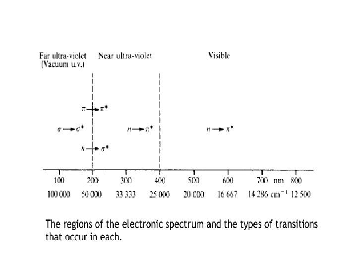

Electronic transition v s ® s* Transitions in which a s bonding electron is excited to an antibonding s* orbital are called s ® s* transitions. These transitions are shown by only saturated hydrocarbons. v For example, methane (which has only C-H bonds, and can only undergo s ® s* transitions) shows an absorbance maximum at 125 nm. Absorption maxima due to s ® s* transitions v These wavelengths are lesser than 200 nm and fall in the vacuum UV region.

n ® s* Transitions • n ® s* Transitions Saturated compounds containing atoms with lone pairs (non-bonding electrons) are capable of n ® s* transitions. • These transitions usually need less energy than s ® s * transitions. • They can be initiated by light whose wavelength is in the range 150 - 250 nm. • The number of organic functional groups with n ® s* peaks in the UV region is small. • E. g. , CH 3 Cl.

n ® p* Transitions • n ® p* transitions These are the transitions in which an electron in a non – bonding atomic orbital is promoted to an antibonding p* orbital. • Compounds having double bonds between heteroatoms, e. g. , C=O, C=S, and N=O. • For e. g. , the >C=O group of saturated aldehydes or ketones exhibit an absorption of low intensity at about 285 nm. • These transitions require only small amounts of energy and takes place with in the range of ordinary UV spectrophotometer. • These are generally forbidden transitions.

p ® p* Transitions p® p* transitions These are the transitions in which an electron in a p electron is promoted to an antibonding p* orbital. • These transitions require relatively higher amount of energy than n ® p* transitions. • For e. g. , the >C=O group of saturated aldehydes or ketones exhibit an absorption of high intensity at about 180 nm.

Selection Rules of electronic transition • Electronic transitions may be classified as intense or weak according to the magnitude of εmax that corresponds to allowed or forbidden transition : 1)Allowed transitions: These are transitions have εmax 104 or more and probability of their occurrence is very high. These are generally due to p® p* transition For e. g. , p® p* transition in 1, 3 -butadiene which shows absorption at 217 nm and εmax 20900 represents an allowed transition.

Forbidden Transition v. These are usually related to n ® p* transition. v. These are the transitions for which εmax is generally less than 104. v. For e. g. , n ® p* transition of a saturated aldehydes or ketones exhibit a weak absorption of low intensity near about 285 nm and having εmax less than 100 is a forbidden transition. .

Chromophores 1. But the term chromophore was originally used to denote a functional group or some other structural feature, the presence of which imparts a colour to a compound. 2. A functional group which exhibits absorption of electromagnetic radiations in the visible or ultraviolet region is called a chromophore (colour loving). 29

Chromophore Excitation lmax, nm Solvent C=C p→p* 171 hexane C=O n→p* p→p* 290 180 hexane N=O n→p* p→p* 275 200 ethanol

AUXOCHROME An auxochrome is a group which itself does not act as a chromophore but when attached to a chromophore it shifts the absorption maximum towards longer wavelength along with an increase in the intensity of absorption. NH 2 Benzene Aniline λmax= 254 nm λ max= 280 nm £max=203 nm £max=1430 nm
 Bathochromic shift or red shift: It")
Change in position and intensity of absorption 1) Bathochromic shift or red shift: It involves the shift of absorption maximum towards longer wavelength. It can be brought about by three different methods as given below; a) By an attachment of an auxochrome to the chromophore, b) By conjugation of two or more chromophoric groups, c) By using solvent of lower polarity.
Hypsochromic shift or blue shift: It involves the shift of absorption maximum towards shorter")
2)Hypsochromic shift or blue shift: It involves the shift of absorption maximum towards shorter wavelength. It may be brought about as follows; a) By removal of conjugation in a system, c) By using solvent of higher polarity. 3)Hyperchromic Effect: This effect involves an increase in the intensity of absorption. It is brought about by introduction of an auxochrome. 4)Hypochromic Effect: This effect involves a decrease in the intensity of absorption. It is brought about by groups which distort the geometry of the molecule.

HYPERCHROMIC HYPSOCHROMIC ABSORBANCE BATHOCHROMIC HYPOCHROMIC λ max λ Terminology of shifts in the position of an absorption band

Choice of solvents q They need to be transparent and do not affect the fine structure arising from the vibrational effects q. Polar solvents generally tend to cause this problem q. Same solvent must be used when comparing absorption spectra for identification purpose.
")
Solvents for the Ultraviolet and Visible regions Solvent Ethanol Lower Solvent wavelength limit (nm) 180 Diethyl ether 220 Hexane 195 Cyclohexane Carbon tetrachloride 195 260 Water Lower wavelength limit (nm) 210

Solvent effect
 (Hypsochromic shift) – Increasing polarity")
Effects of solvents • 1. Blue shift (n- p*) (Hypsochromic shift) – Increasing polarity of solvent better solvation of electron pairs (n level has lower E) – peak shifts to the blue (more energetic) – 30 nm (hydrogen bond energy) • 2. Red shift (n- p* and p –p*) (Bathochromic shift) – Increasing polarity of solvent, then increase the attractive polarization forces between solvent and absorber, thus decreases the energy of the unexcited and excited states with the later greater – peaks shift to the red – 5 nm

Effect of polar solvent and shift in n - π* transition π* π* ΔE 1 n ΔE 2 Non- polar solvent n Polar solvent ΔE 2> ΔE 1

Effect of polar solvent and shift in π - π* transition π* ΔE 1 ΔE 2 π Non- polar solvent π* π ΔE 1> ΔE 2 Polar solvent

Woodward-Fieser Rules This quantification is referred to as the Woodward-Fieser Rules which we will apply to three specific chromophores: Conjugated dienes Conjugated dienones

Woodward-Fieser Rules Dienes The rules begin with a base value for λ max of the chromophore being observed: acyclic butadiene = 217 nm The incremental contribution of substituents is added to this base value from the group tables: Group Extended conjugation Increment +30 Each exo-cyclic C=C +5 Alkyl +5 -OCOCH 3 +0 -OR +6 -SR +30 -Cl, -Br -NR 2 +5 +60

Woodward-Fieser Rules Dienes Isoprene - acyclic butadiene = 217 nm one alkyl subs. = + 5 nm = 222 nm Experimental value =220 nm Allylidene cyclohexane - acyclic butadiene =217 nm one exocyclic C=C =+ 5 nm 2 alkyl subs. = +10 nm = 232 nm Experimental value = 237 nm

Woodward-Fieser Rules – Cyclic Dienes There are two major types of cyclic dienes, with two different base values Heteroannular (transoid): e = 5, 000 – 15, 000 base λmax = 214 Homoannular (cisoid): e = 12, 000 -28, 000 base λmax = 253 The increment table is the same as for acyclic butadienes with a couple additions: Group Additional homoannular Where both types of diene are present, the one with the longer l becomes the base Increment +39

Woodward-Fieser Rules Cyclic Dienes In the pre-NMR era of organic spectral determination, the power of the method for discerning isomers is readily apparent Consider abietic vs. levopimaric acid : abietic acid levopimaric acid

1. Woodward-Fieser Rules – Cyclic Dienes heteroannular diene = 214 nm 4 alkyl subs. (4 x 5) +20 nm 1 exo C=C + 5 nm 239 nm homoannular diene = 253 nm 4 alkyl subs. (4 x 5) +20 nm 1 exo C=C + 5 nm 278 nm

3. Woodward-Fieser Rules – Cyclic Dienes Be careful with your assignments – three common errors: This compound has three exocyclic double bonds; the indicated bond is exocyclic to two rings This is not a heteroannular diene; you would use the base value for an acyclic diene Likewise, this is not a homoannular diene; you would use the base value for an acyclic diene

Woodward-Fieser Rules - Enones Group Increment 6 -membered ring or acyclic enone Base 215 nm 5 -membered ring parent enone Base 202 nm Acyclic dienone Base 245 nm Double bond extending conjugation 30 Alkyl group or ring residue a, b, g and higher 10, 12, 18 -OH a, b, g and higher 35, 30, 18 -OR a, b, g, d 35, 30, 17, 31 a, b, d 6 -Cl a, b 15, 12 -Br a, b 25, 30 b 95 -O(C=O)R -NR 2 Exocyclic double bond Homocyclic diene component 5 39

1. Woodward-Fieser Rules – Enones Aldehydes, esters and carboxylic acids have different base values than ketones Unsaturated system Base Value Aldehyde 208 With a or b alkyl groups 220 With a, b or b, b alkyl groups 230 With a, b, b alkyl groups 242 Acid or ester With a or b alkyl groups 208 With a, b or b, b alkyl groups 217 Group value – exocyclic a, b double bond +5 Group value – endocyclic a, b bond in 5 or 7 membered ring +5

2. Woodward-Fieser Rules – Enones cyclic enone = 215 nm 2 x b- alkyl sub (2 x 12) +24 nm 239 nm Experimental value 238 nm cyclic enone = extended conj. b-ring residue d-ring residue exocyclic double bond 215 nm +30 nm +12 nm +18 nm + 5 nm 280 nm Experimental 280 nm

- Slides: 51