Ultrasonographic anatomy of the pelvis including its 3


")


 Uterine volume (cm 3)")


 PROLIFERATIVE PHASE 4")


")


 benign adnexal")


- Slides: 72
Ultrasonographic anatomy of the pelvis including its 3 D aspects Frédéric Chantraine, ULg
Introduction • Anatomy of the pelvis • Ultrasound techniques • Female organs in the pelvis • Uterus • Ovaries • Fallopian Tubes
The female pelvis
And in ultrasound? • Different approaches: – Transvaginal • Bladder is empty – Transabdominal • Bladder is filled – Transrectal – Transperineal
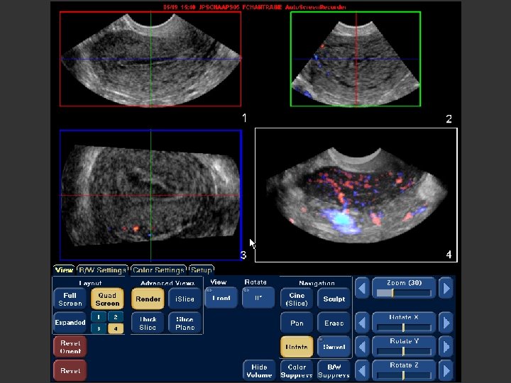
And in ultrasound? • Different techniques – Classic “ 2 D” – Colour and Pulsed Doppler – Volumetric “ 3 D”
Uterus
Uterus • Organ of reference in the pelvis • Different orientation – Ante-, retroflexion – Ante-, retroversion • Three main parts – Corpus – Isthmus – Cervix
Uterus • Classic plans Longitudinal Transversal
Uterus – Coronal plan by 3 D
Uterus – Coronal plan by 3 D
Uterus – Completed by transabdominal scan
Transabdominal scan – Check if possible the kidneys • Dilation by ureter-compression (Myomas, …) • In combination with congenital uterine malformations
Uterus • Volume depends – Patient age – Number of gestations – Endocrinal level
Uterus
Main uterine diameters during different stages of life LONG. DIAMETER ANT-POST DIAMETER TRANS. DIAMETER VOLUME PREPUBERTAL 1 -3 cm 0, 5 -1 cm 10 -20 ml MULTIPAROUS 8 cm 4 cm 5 cm 60 -80 ml NULLIPAROUS 6 cm 3 -4 cm 30 -40 ml POSTMENOPA USAL 4 -6 cm 2 -3 cm 14 -17 ml Platt, 1990. Modified
Normal dimensions of the uterus before puberty Uterine diameter (cm) Uterine volume (cm 3) Age N°. Pts. DL DAP cervix 2 7 33. 1 4. 4 7. 0 3. 4 8. 3 2. 0 1. 98 1. 58 3 8 32. 4 4. 3 6. 4 1. 3 7. 6 2. 2 1. 63 0. 81 4 15 32. 9 3. 3 7. 6 1. 8 8. 6 1. 8 2. 10 0. 57 5 7 33. 1 5. 5 8. 0 2. 8 8. 4 1. 8 2. 36 1. 39 6 9 33. 2 4. 1 6. 7 2. 9 7. 5 1. 80 1. 57 7 9 32. 3 3. 9 8. 0 2. 2 7. 7 2. 5 2. 32 1. 07 8 11 35. 8 7. 3 9. 0 2. 8 8. 4 1. 7 3. 12 1. 52 9 11 37. 1 4. 4 9. 7 3. 0 8. 8 2. 0 3. 70 1. 62 10 13 40. 3 6. 4 12. 8 5. 3 10. 7 2. 6 6. 54 3. 78 11 13 42. 2 5. 1 12. 8 3. 1 10. 7 2. 6 6. 66 2. 87 12 6 54. 3 8. 4 17. 3 5. 3 14. 3 5. 2 16. 18 9. 15 13 5 53. 8 11. 4 15. 8 4. 5 15. 0 2. 4 13. 18 5. 64 Orsini, 1984, Modified
Uterus – Myometrium • Three different layers
Uterus – Myometrium • Three different layers
Uterus – Myometrium • Contractile • bjl
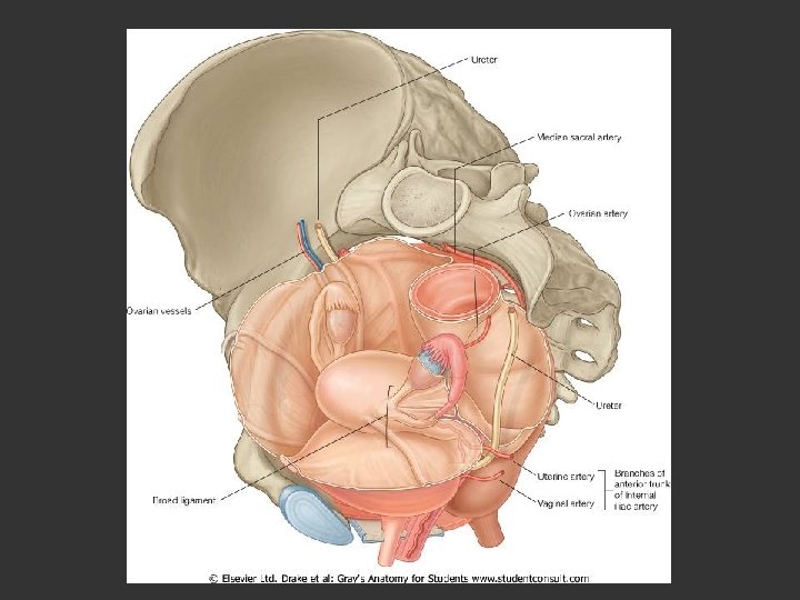
Uterus – Vascularisation • Uterine artery • Arcuate artery • Radial artery • Spiral artery
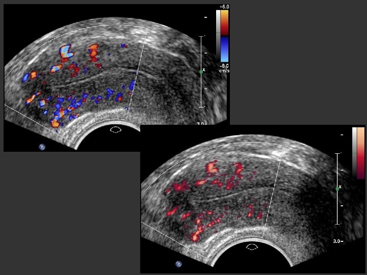
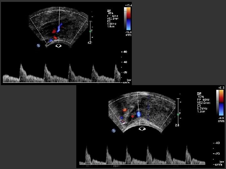
Uterus – Doppler of the uterine artery • Systole: high velocity peak • Diastole: lower flow • Telesystolic Notch • Changes during menstrual cycle
Uterus – Endometrium 1. Thickness: technique of measurement
Uterus
Uterus
Uterus – Endometrium 2. Echostructure – Patients age – Moment of the spontaneous cycle – Eventual hormonal treatment
Uterus –Endometrium • Prepubertal age – Newborn: thickened and hyperechogenic – Up to menarch: thin, <1 mm – Useful in diagnosis of early or delayed puberty • Reproductive age
Uterus – Endometrium • Range of normal endometrial thicknesses THICKNESS (mm) PROLIFERATIVE PHASE 4 -8 PERIOVULATORY PHASE 6 -10 SECRETIVE PHASE 7 -14 POSTMENOPAUSAL PHASE <6
Appearance of the endometrial mucosa during the course of a physiological cycle Early proliferative phase Late proliferative phase Early luteal phase Late luteal phase L. Savelli, ISUOG
Early proliferative phase
Late proliferative phase
Early luteal phase
Late luteal phase
And the next step?
Postmenopausal
To complete the uterus examination Check the cervix • Polyps • Myomas • Pregnancy location
IUD-Fibre Nabothian cyst
Cervical pregnancy
Ovary
Ovary • Location – Between uterus and pelvic wall – Behind uterus, Douglas pouch
Ovary • Dimension – Pré-ménopause: 3, 5 x 2 x 1, 5 cm – Post-ménopause: 1, 5 x 0, 7 cm • Morphology – Patient age – Moment of the menstrual cycle
Ovary • Prepubertal – Small hypoechogenic masse – Years before puberty: • Increase in volume • Follicles development
Ovary • Reproductive age – Classic multifollicular form
Ovary • Reproductive age – Mid-cycle: dominant follicle (10 to 20 -25 mm)
Ovary • Reproductive age – After ovulation: corpus luteum
Ovary • Reproductive age – After ovulation: free fluid in the Douglas
Ovary • Reproductive age – Functional cysts
Ovary • Reproductive age – Functional cysts with Mirena® IUD
Ovary • Reproductive age – Polycystic ovary
Ovary and 3 D
Ovary and 3 D
Ovary and 3 D
Ovary and 3 D
Ovary and 3 D
Ovary • Postmenopausal – Small masses, difficult to visualize
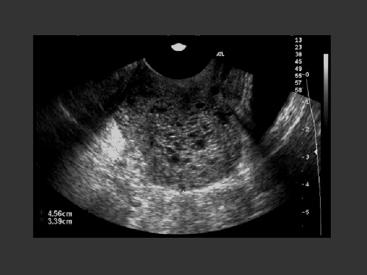
Ovary • Post-menopausal – Unilocular cysts “Small (< or = 50 mm) benign adnexal cysts and small benign solid tumors are so common in postmenopausal women that their presence may be regarded as normal” L. Valentin UOG 2003
Fallopian Tubes
Fallopian Tubes • Anatomy – Interstitielle – Isthmus – Ampulla • Difficult to visualize – Fluid around – Hydrosalpinx
Fallopian Tubes © Luca Savelli
And what else?
Cervix after subtotal hysterectomy
Pelvic kidney
Bladder filling
Heterotopic pregnancy
Dank uw ! Questions?
Don’t worry, they normally look like little humans during the early scans