CHAPTER 21 THE THIGH HIP GROIN AND PELVIS


 and Retroversion (B) – Relationship between neck and shaft of femur")
 • Galenslen’s test •")

 – Detects")


 strains • Trochanter Bursitis: inflam")


- Slides: 61
CHAPTER 21 THE THIGH, HIP, GROIN, AND PELVIS
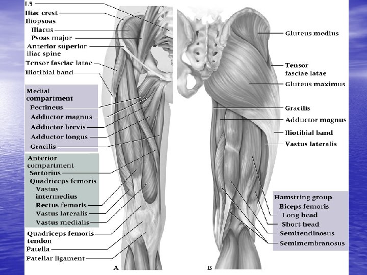
ANATOMY OF THE THIGH • Bones: Femur
THIGH MUSCLES • ANTERIOR THIGH: -Quadriceps: 4 muscles: -Rectus femoris, Vastus Medialis, Vastus Lateralis and Vastus intermedius. -Sartorius ** Action: Extension of leg
THIGH MUSCLES • POSTERIOR: -Hamstring: Biceps femoris, semitendonosus and semimembranosus **Action: Flexion of the leg
THIGH MUSCLES • MEDIAL: - Adductors - Pectineus - Gracilis Action: Adducts and laterally rotates the thigh
NERVE AND BLOOD SUPPLY • Nerves: Tibial, common Peroneal, and greater Sciatic nerve. • Blood: Femoral artery
HOPS OF THE THIGH • History: How, What and why • Observation: swelling, bruising, deformation, pain and ability to walk • Palpation: Bones-Greater Trochanter -Lesser Trochanter -Lateral and medial femoral condyle -Anterior superior iliac spine (ASIS)
PALPATION • SOFT TISSUE: - ANTERIOR: sartorius, Quad - POSTERIOR: Hamstring - MEDIAL: Adductors - LATERAL: Abuctors, Iliotibial band (ITB), Gluteus medius and tensor fasciae latae
SPECAL TESTS • ROM: Active, Passive, Resistive • Muscle weakness • Xrays: if fx is suspected
PREVENTION OF THIGH INJURIES • Conditioning • Strength • Endurance • Extensibility: ability to withstand strain • Collision sports: pads
THIGH INJURIES • Contusions: bruising • Myositis Ossificans Traumatica: blow to the thigh that causes ectopic bone production • Quadriceps Muscle strain • Hamstring Muscle strain • Acute Femoral Fracture • Femoral stress Fractures
THIGH CONTUSIONS
MYOSITIS OSSIFICANS TRAUMATICA
QUAD STRAIN HAMSTRING STRAIN
FEMORAL FRACTURE
FEMORAL STRESS FRACTURE
ANATOMY OF THE HIP, GROIN AND PELVIC REGION • BONES: Pelvis, Pelvic girdle, sacrum and coccyx.
LATERAL VIEW OF THE HIP
HIP JOINT • Ball in socket joint • Articulates with the femur and hip bone • Acetabulum: socket part of the joint • Greater trochanter: the head of the femur and is the ball of the joint
BALL AND SOCKET JOINT
LIGAMENTS OF THE HIP
MUSCLES OF THE HIP • ANTERIOR MUSCLES: - Iliacus: action: Assists in flexion - Psoas: action: Flexes thigh and trunk on the femur - iliopsoas: action: flexes trunk and hip • POSTERIOR MUSCLES: - Tensor fasciae latae: action: flexion, abduction and rotates thigh
MUSCLES OF THE HIP • POSTERIOR: - 3 Gluteal muscles: Maximus, medius and minimus. Action: Extends, abducts and medially rotates the thigh
BURSAE • Has many bursae. • Examples: iliopsoas bursae, greater trochanter bursae, and many others • Bursae are used to cushion the joint
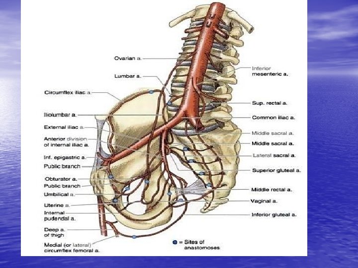
BLOOD SUPPLY AND NERVES • NERVES: lumbar plexus, 4 th and 5 th lumbar nerves, 1 -3 sacral nerves, sciatic nerves, tibial and common peroneal nerves • BLOOD: Arteries: 2 common iliac arteries, internal and external iliac arteries • Veins: 3 major: found in the hip, groin and pelvis. Common iliac vein, internal iliac vein and external iliac vein
FUNCTIONAL ANATOMY OF THE HIP, GROIN AND PELVIS • Pelvis and hip: pelvic girdle • Articulates with the femoral head. -At the acetabulum (hip) with greater trochanter (femur) * Muscles include: Glutes, Quads, Hamstrings, Hip flexors and so on
HOPS • History: the how, what and why • Observation: Observe for postural asymmetry and while standing on one leg and during ambulation (recovery) • Palpation: -Boney: Iliac crest, ASIS, AIIS, Greater trochanter, Pubic symphysis, Ischial tuberosity, PIIS and PSIS
HOPS • Palpation: soft tissue - Quads, Ham, Gracilis, Abductors, Sartorius, Iliopsoas, Glutes, ITB, and Tensor fasciae latae. • Special Tests: -ROM -Tests for hip flexor tightness: Kendall test Thomas test Femoral Anteversion and retroversion
KENDALL TEST
Thomas Test
Femoral Anteversion (A) and Retroversion (B) – Relationship between neck and shaft of femur – Normal angle is 15 degrees anterior to the long axis of the femur and condyles – Internal rotation in excess of 35 degrees is indicative of anteversion, 45 degrees of external rotation is an indicator of retroversion
TESTS FOR HIP AND SACROILIAC JOINT • Patrick test (FABER) • Galenslen’s test • • TESTIN THE TENSOR FASCIAE LATAE AND ITB: Renne’s Test Nobel’s Test Ober’s test Trendlelenburg’s test Piriformis test Ely’s test
PATRICK’S TEST
• Test for Hip and Sacroiliac Joint • Patrick Test (FABER) – Detects pathological conditions of the hip and SI joint – Pain may be felt in the hip or SI joint
• Gaenslen’s Test – Test works to push SI joint into extension – Test is positive if hyperextension on affected side increases pain
Testing the Tensor Fasciae Latae and Iliotibial Band • Renne’s test – Athlete stands w/ knee bent at 30 40 degrees – Positive response of TFL tightness occurs when pain is felt at lateral femoral condyle
• Nobel’s Test – Lying supine the athlete’s knee is flexed to 90 degrees – Pressure is applied to lateral femoral condyle while knee is extended – Pain at 30 degrees at lateral femoral condyle indicates a positive test
• Ober’s Test – Used to determine presence of contracted TFL or IT-band – Thigh will remain in abducted position, not falling into adduction
OBER’S TEST
Trendelenburg’s Test - Iliac crest on unaffected side should be higher when standing on one leg - Test is positive when affected side is higher indicating weak abductors (glut medius)
• Piriformis Test – Hip is internally rotated – Tightness or pain is indicative of piriformis tightness
MEASURING LEG-LENGTH DISCREPANCY • Leg length discrepancies of more than 1 inch can cause symptoms • 1/8” may cause symptoms in highly active athletes. • Areas that may be affected: lower limbs (shins), hip and pelvis or lower back. • 2 types of leg length discrep: 1. true or anatomical shortening 2. apparent or functional shortening.
TYPES OF LEG LENGTH DISCREPANCY • Anatomical: may be equal throughout the lower limbs. • Functional: can occur as a result of lateral pelvic tilt or from flexion or adduction deformity.
Leg Length Discrepancy Measures
HIP, GROIN AND PELVIC INJURIES • Groin strain: muscle(s) strains • Trochanter Bursitis: inflam of burseas • Sprains of the hip joint • Dislocated hip joint: Greater trochanter out of • • • acetabulum Avascular Necrosis: loss of blood flow Legg-Calve-Perthes Disease (coxa plana) Snapping Hip Phenomenon: instablility of ligaments in the hip
GROIN STRAIN
TROCHANTER BURSITIS
DISLOCATION OF THE HIP JOINT
AVASCULAR NECROSIS
LEGG-CALVE-PERTHES DISEASE
PELVIC CONDITIONS • Hip Pointer: contusion to the iliac crest • Osteitis Pubis: Pull of muscles from the pubis. Common in runners • Athletic Pubalgia: Chronic pubic region pain. • Stress Fx • Avulsion Fx and apophysitis: Inflam of the apophyses of the hip
HIP POINTER
OSTEITIS PUBIS
AVULSION FRACTURES AND APOPHYSITIS
THIGH AND HIP REHAB • General conditioning • Flexibility • Mobilization • Strength • Neuromuscular Control • Functional Progression • Return to Activity
STRETCHING
STRENGTHENING EXERCISE
CORE STRENGTH EXCERISES
Balance Shoe for Neuromuscular Control