
Hip Region Dr S Nishan Silva MBBS Regional
Hip Region Dr. S. Nishan Silva (MBBS)
Regional anatomy of the lower limb
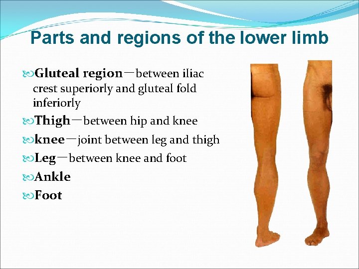
Parts and regions of the lower limb Gluteal region-between iliac crest superiorly and gluteal fold inferiorly Thigh-between hip and knee-joint between leg and thigh Leg-between knee and foot Ankle Foot
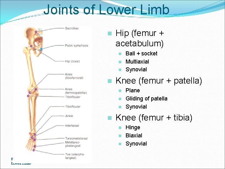
Joints of Lower Limb n Hip (femur + acetabulum) n n Knee (femur + patella) n n Plane Gliding of patella Synovial Knee (femur + tibia) n n n Frolich, Human Anatomy, Lower LImb Ball + socket Multiaxial Synovial Hinge Biaxial Synovial
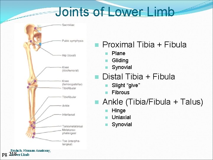
Joints of Lower Limb n Proximal Tibia + Fibula n n Distal Tibia + Fibula n n n pg Slight “give” Fibrous Ankle (Tibia/Fibula + Talus) n Frolich, Human Anatomy, 218 Lower LImb Plane Gliding Synovial Hinge Uniaxial Synovial
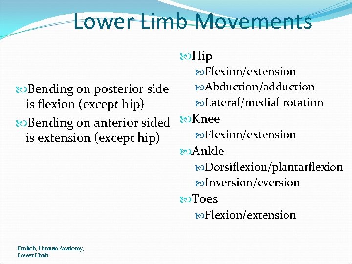
Lower Limb Movements Hip Flexion/extension Abduction/adduction Bending on posterior side Lateral/medial rotation is flexion (except hip) Bending on anterior sided Knee Flexion/extension is extension (except hip) Ankle Dorsiflexion/plantarflexion Inversion/eversion Toes Flexion/extension Frolich, Human Anatomy, Lower LImb
Surface anatomy of lower limb Gluteal region and thigh anterior superior and inferior iliac spines tubercle of iliac crest ischial tuberosity greater trochanter pubic tubercle pubic crest superior border of pubic symphysis
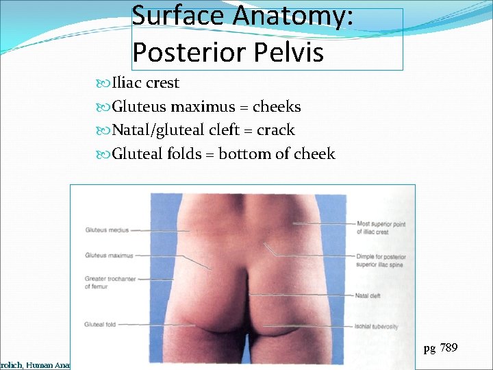
Surface Anatomy: Posterior Pelvis Iliac crest Gluteus maximus = cheeks Natal/gluteal cleft = crack Gluteal folds = bottom of cheek Frolich, Human Anatomy, Lower LImb pg 789
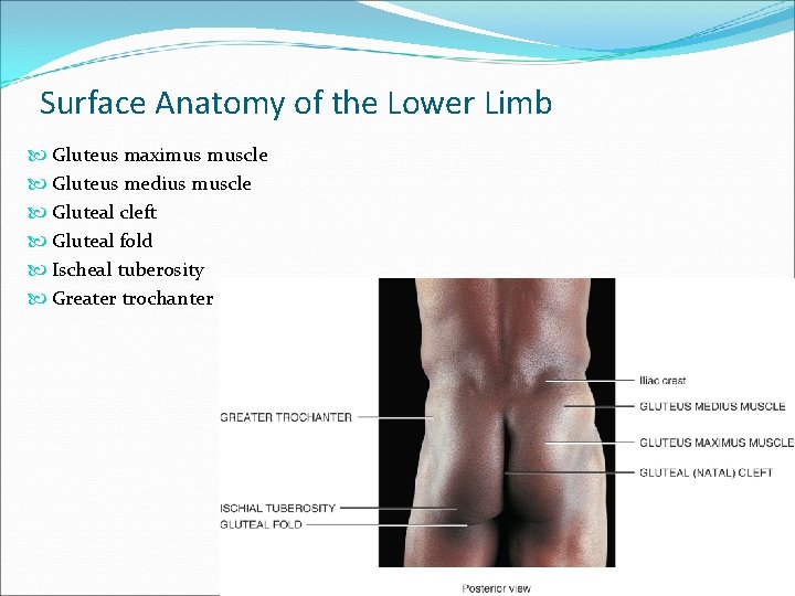
Surface Anatomy of the Lower Limb Gluteus maximus muscle Gluteus medius muscle Gluteal cleft Gluteal fold Ischeal tuberosity Greater trochanter 12 -9
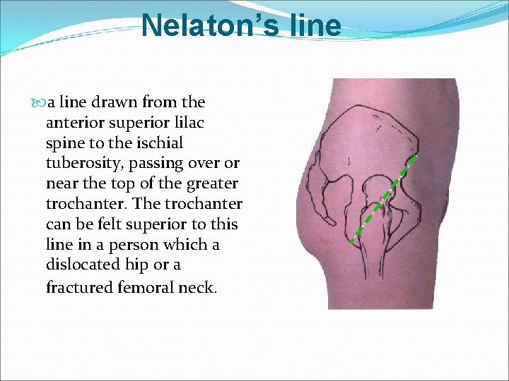
Nelaton’s line a line drawn from the anterior superior lilac spine to the ischial tuberosity, passing over or near the top of the greater trochanter. The trochanter can be felt superior to this line in a person which a dislocated hip or a fractured femoral neck.
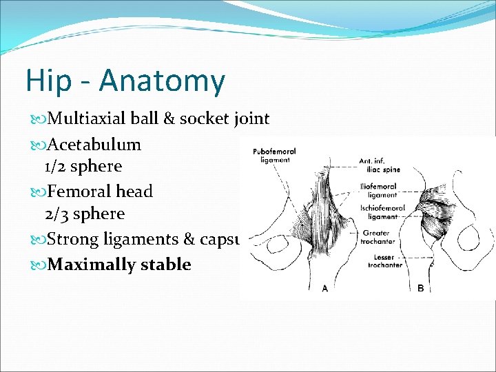
Hip - Anatomy Multiaxial ball & socket joint Acetabulum 1/2 sphere Femoral head 2/3 sphere Strong ligaments & capsule Maximally stable
Anatomy Forces Standing - 0. 3 times body weight Standing on 1 leg - 2. 5 times body weight Walking - 1. 3 to 5. 8 times body weight Walking up stairs - 3 times body weight Running - 4. 5+ times body weight
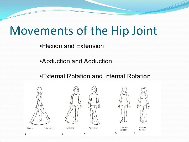
Movements of the Hip Joint • Flexion and Extension • Abduction and Adduction • External Rotation and Internal Rotation.
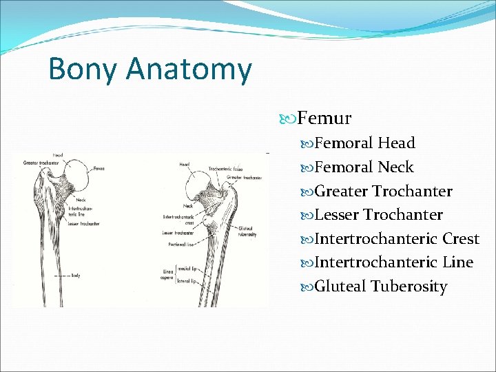
Bony Anatomy Femur Femoral Head Femoral Neck Greater Trochanter Lesser Trochanter Intertrochanteric Crest Intertrochanteric Line Gluteal Tuberosity
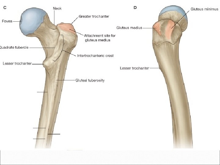
BONY ANATOMY OF THE HIP
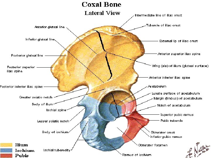
BONY ANATOMY OF THE HIP
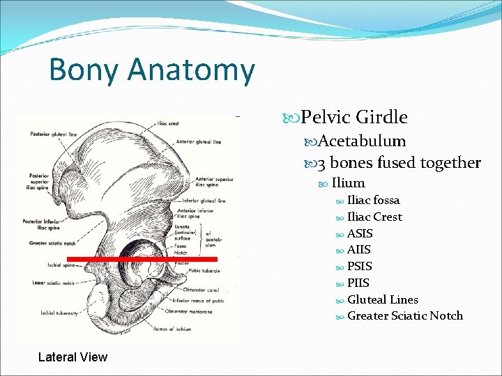
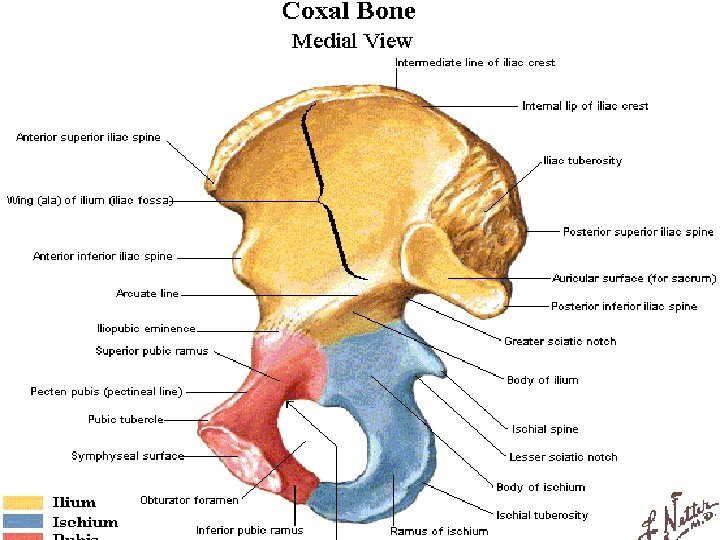
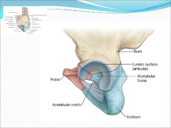
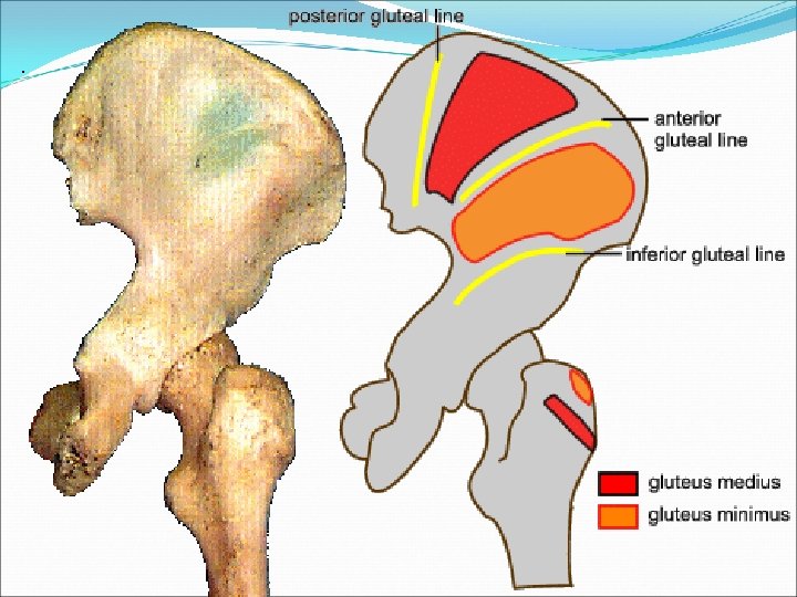
Bony Anatomy Pelvic Girdle Acetabulum 3 bones fused together Ilium Iliac fossa Iliac Crest ASIS AIIS PSIS PIIS Gluteal Lines Greater Sciatic Notch Lateral View
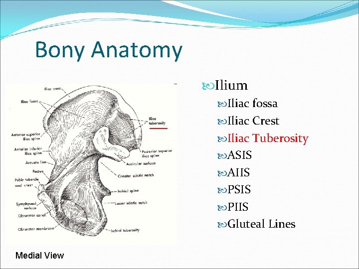
Bony Anatomy Ilium Iliac fossa Iliac Crest Iliac Tuberosity ASIS AIIS PSIS PIIS Gluteal Lines Medial View
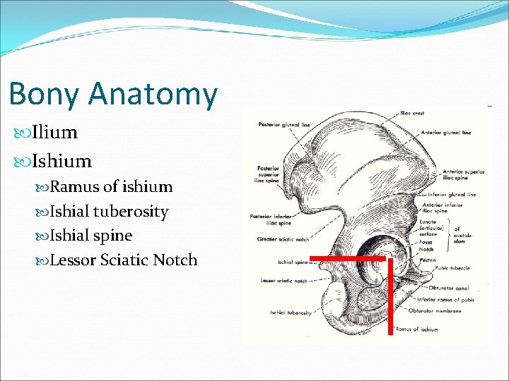
Bony Anatomy Ilium Ishium Ramus of ishium Ishial tuberosity Ishial spine Lessor Sciatic Notch

Bony Anatomy Ilium Ishium Pubis Superior Ramus of Pubis Inferior Ramus of Pubis Pubic Crest Pubic Tubercle Pectin Symphyseal Surface
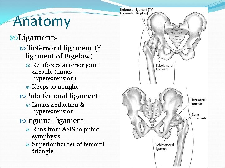
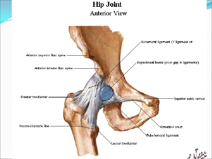
Anatomy Ligaments Iliofemoral ligament (Y ligament of Bigelow) Reinforces anterior joint capsule (limits hyperextension) Keeps us upright Pubofemoral ligament Limits abduction & hyperextension Inguinal ligament Runs from ASIS to pubic symphysis Superior border of femoral triangle
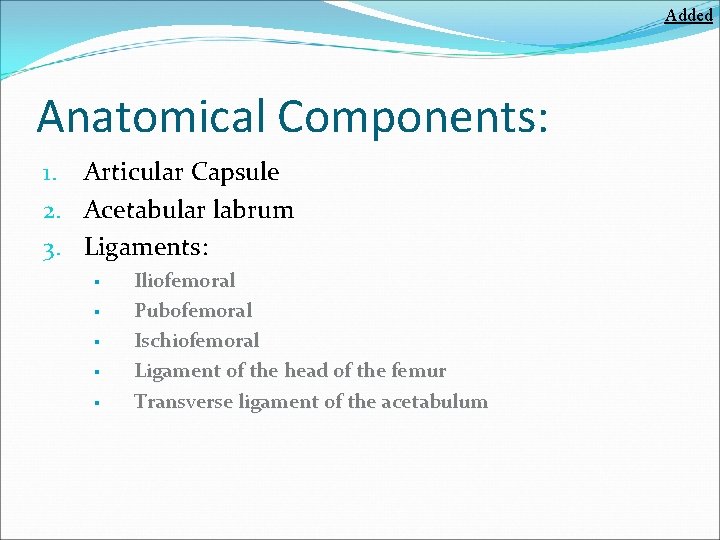
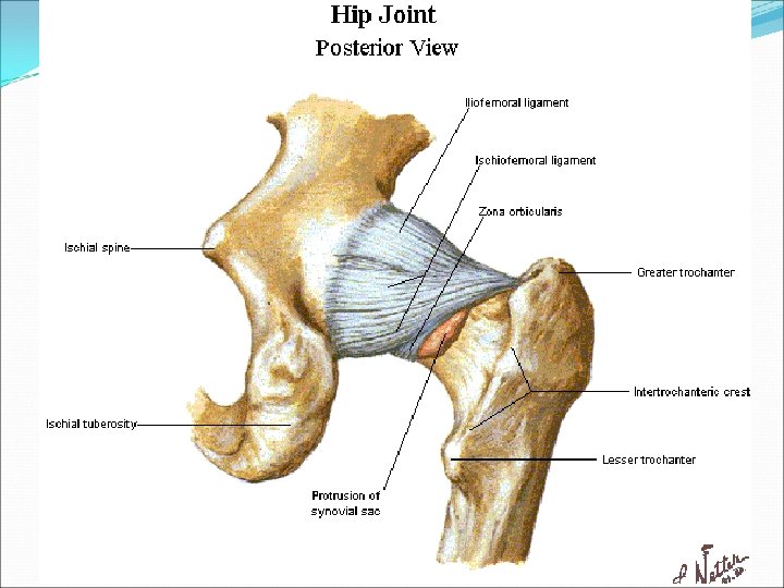
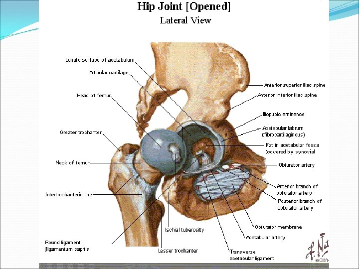
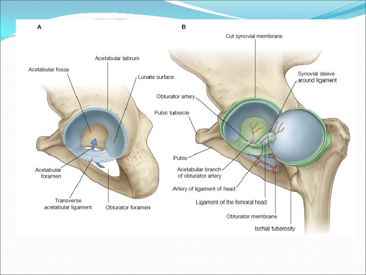
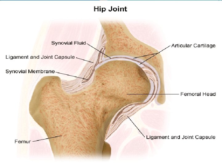
Added Anatomical Components: 1. Articular Capsule 2. Acetabular labrum 3. Ligaments: § § § Iliofemoral Pubofemoral Ischiofemoral Ligament of the head of the femur Transverse ligament of the acetabulum
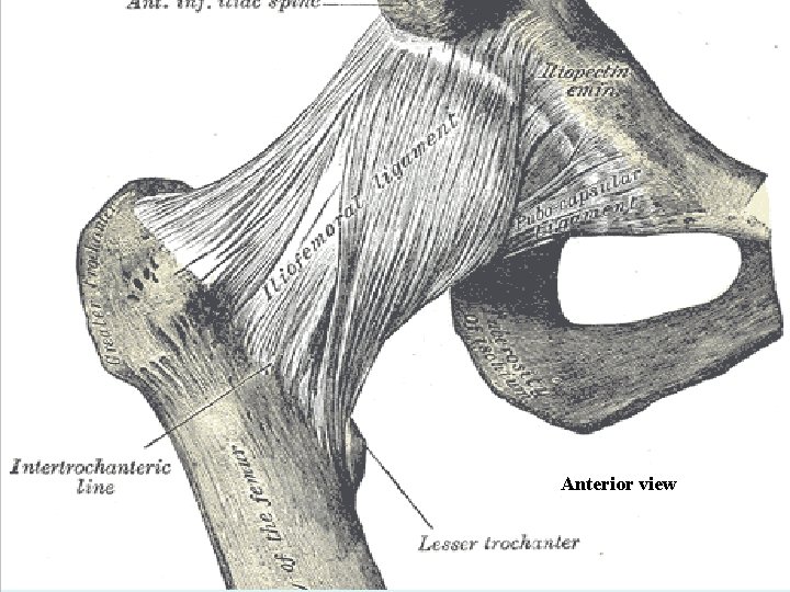
Anterior view
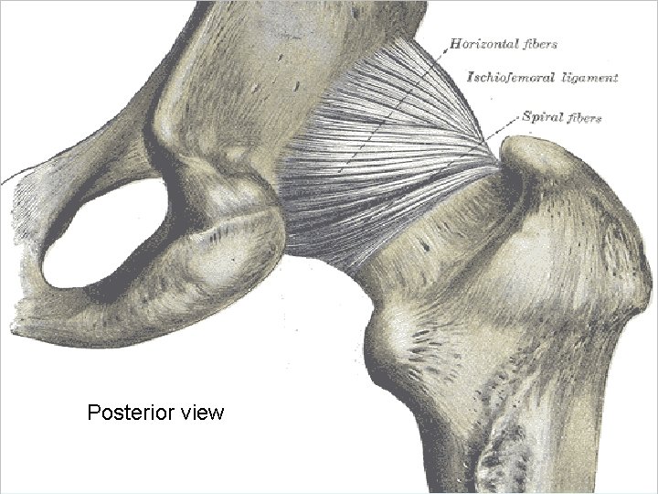
Posterior view
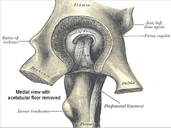
Medial view with acetabular floor removed
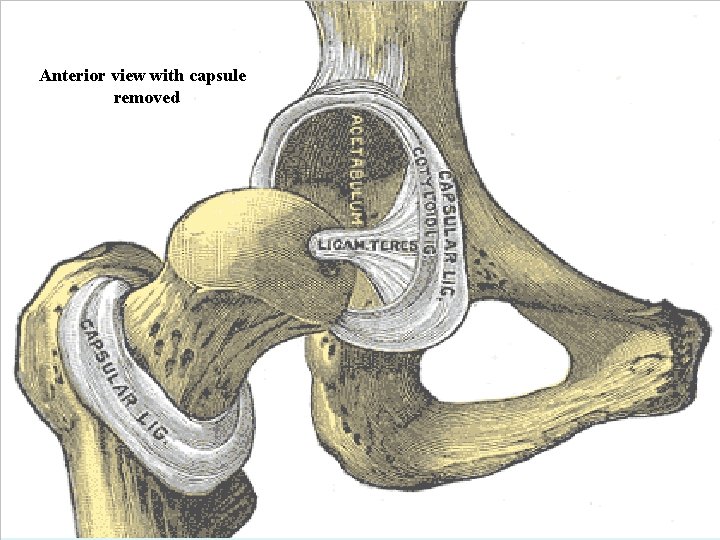
Anterior view with capsule removed
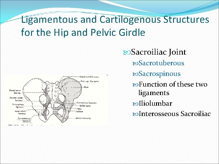
Ligamentous and Cartilogenous Structures for the Hip and Pelvic Girdle Sacroiliac Joint Sacrotuberous Sacrospinous Function of these two ligaments Iliolumbar Interosseous Sacroiliac
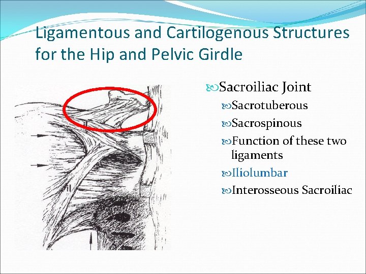
Ligamentous and Cartilogenous Structures for the Hip and Pelvic Girdle Sacroiliac Joint Sacrotuberous Sacrospinous Function of these two ligaments Iliolumbar Interosseous Sacroiliac
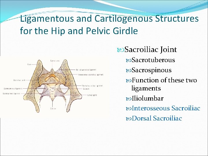
Ligamentous and Cartilogenous Structures for the Hip and Pelvic Girdle Sacroiliac Joint Sacrotuberous Sacrospinous Function of these two ligaments Iliolumbar Interosseous Sacroiliac Dorsal Sacroiliac
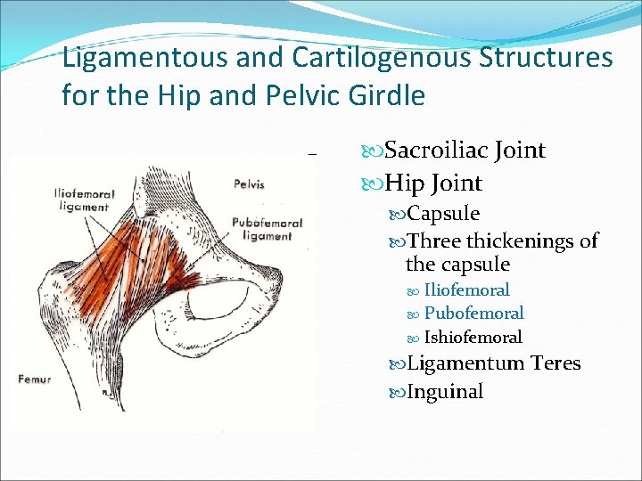
Ligamentous and Cartilogenous Structures for the Hip and Pelvic Girdle Sacroiliac Joint Hip Joint Capsule Three thickenings of the capsule Iliofemoral Pubofemoral Ishiofemoral Ligamentum Teres Inguinal
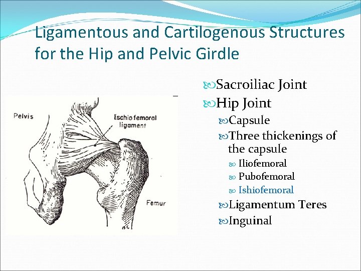
Ligamentous and Cartilogenous Structures for the Hip and Pelvic Girdle Sacroiliac Joint Hip Joint Capsule Three thickenings of the capsule Iliofemoral Pubofemoral Ishiofemoral Ligamentum Teres Inguinal
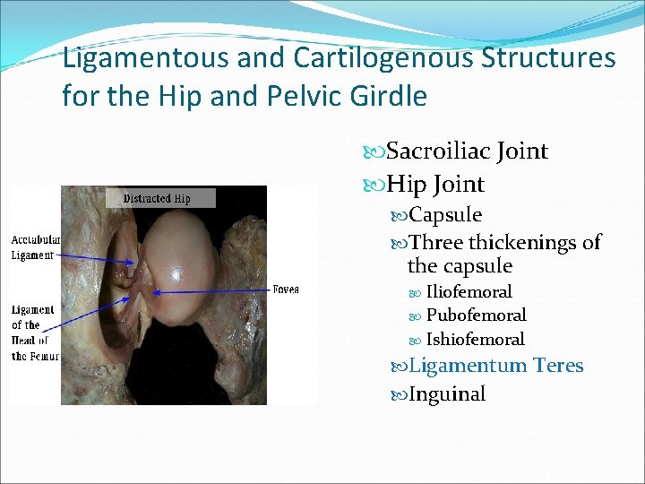
Ligamentous and Cartilogenous Structures for the Hip and Pelvic Girdle Sacroiliac Joint Hip Joint Capsule Three thickenings of the capsule Iliofemoral Pubofemoral Ishiofemoral Ligamentum Teres Inguinal
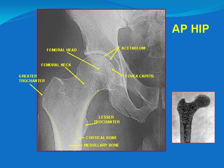
AP HIP ACETABLUM FEMORAL HEAD FEMORAL NECK GREATER TROCHANTER FOVEA CAPITIS LESSER TROCHANTER CORTICAL BONE MEDULLARY BONE
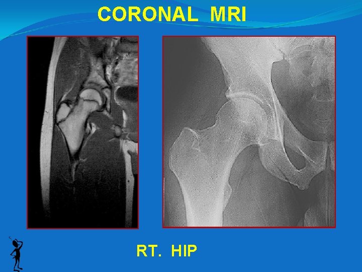
CORONAL MRI RT. HIP
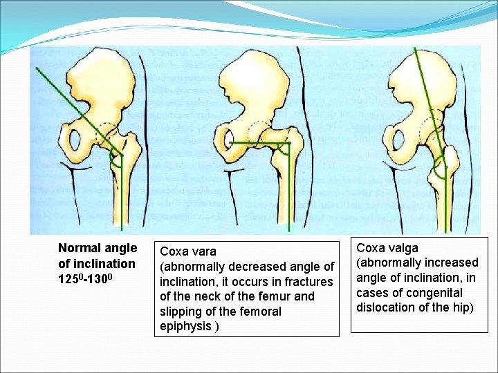
Normal angle of inclination 1250 -1300 Coxa vara (abnormally decreased angle of inclination, it occurs in fractures of the neck of the femur and slipping of the femoral epiphysis ) Coxa valga (abnormally increased angle of inclination, in cases of congenital dislocation of the hip)
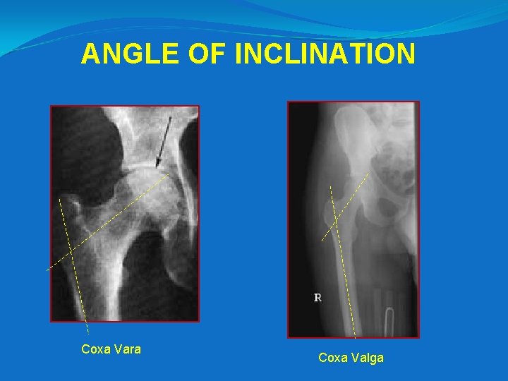
ANGLE OF INCLINATION Coxa Vara Coxa Valga
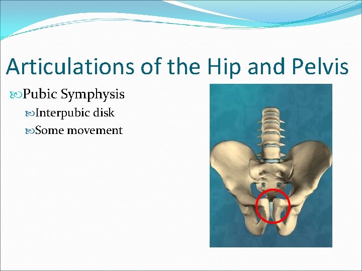
Articulations of the Hip and Pelvis Pubic Symphysis Interpubic disk Some movement
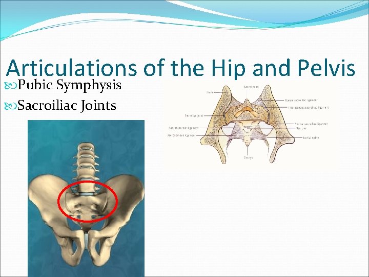
Articulations of the Hip and Pelvis Pubic Symphysis Sacroiliac Joints
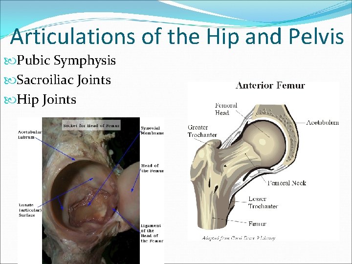
Articulations of the Hip and Pelvis Pubic Symphysis Sacroiliac Joints Hip Joints
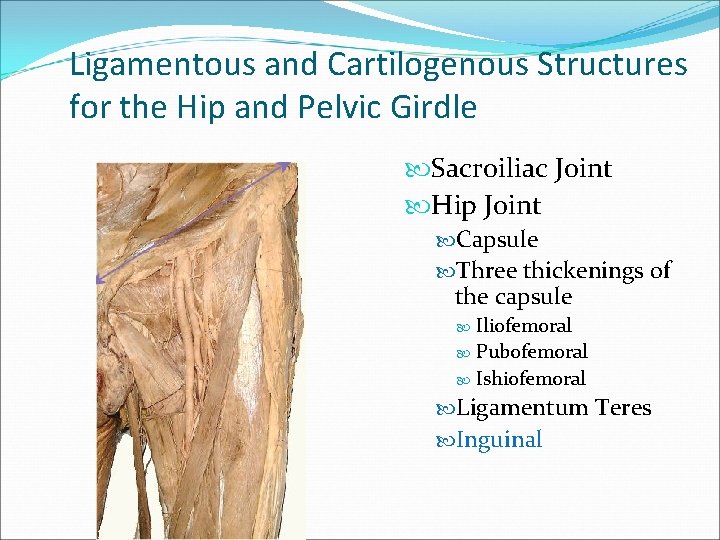
Ligamentous and Cartilogenous Structures for the Hip and Pelvic Girdle Sacroiliac Joint Hip Joint Capsule Three thickenings of the capsule Iliofemoral Pubofemoral Ishiofemoral Ligamentum Teres Inguinal
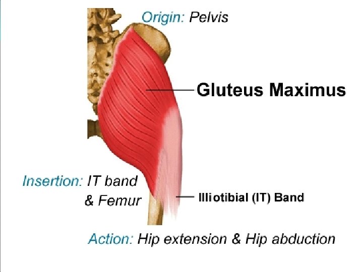
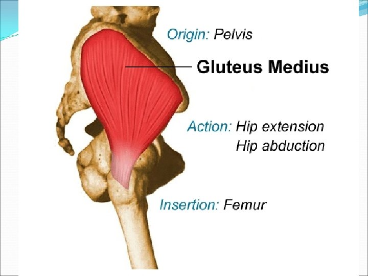
Buttock Supericial Gluteus Maximus Gluteus Medius – reverse action Gluteus Minimus Tensor Fascia Latae – iliotibial band, functional considerations “Gower’s” Sign Positive Trendelenburg
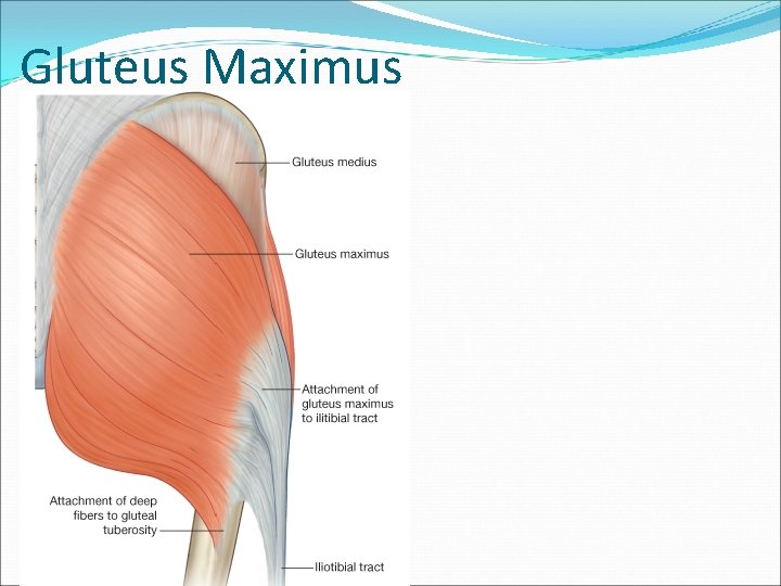
Gluteus Maximus
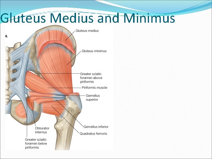
Gluteus Medius and Minimus
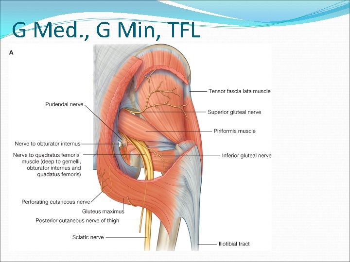
G Med. , G Min, TFL
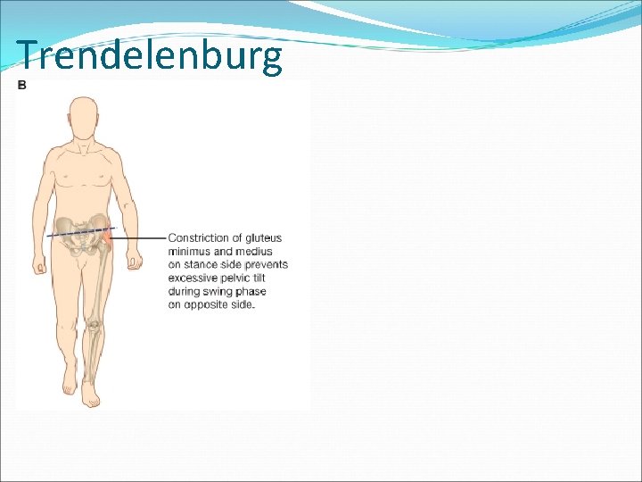
Trendelenburg
Deep Buttocks “External Rotators of the Hip” Small Muscles Mostly attach near or on greater trochanter Excellent mechanical advantage for 1) producing external rotation and 2) to help maintain stability of the hip All but one innervated by named nerves specific to one or two of them The exception is the obturator externus – innervated by posterior brach of obturator n.
Deep Buttock Muscles: Piriformis Superior Gemellus Obturator Internus Inferior Gemellus Quadratus Femoris Obturator Externus
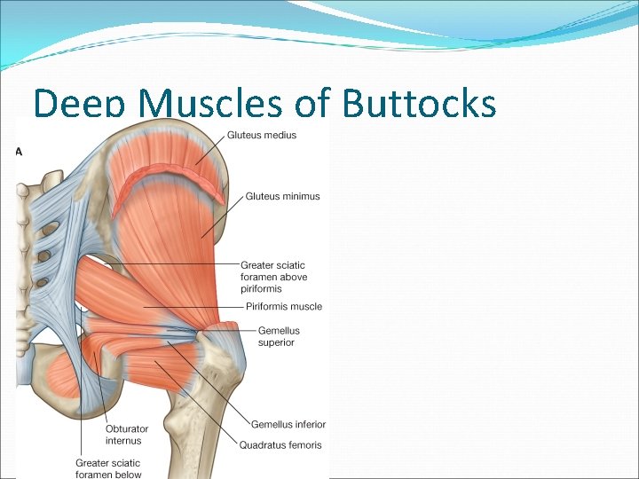
Deep Muscles of Buttocks
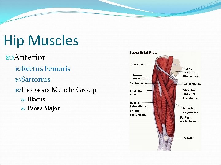
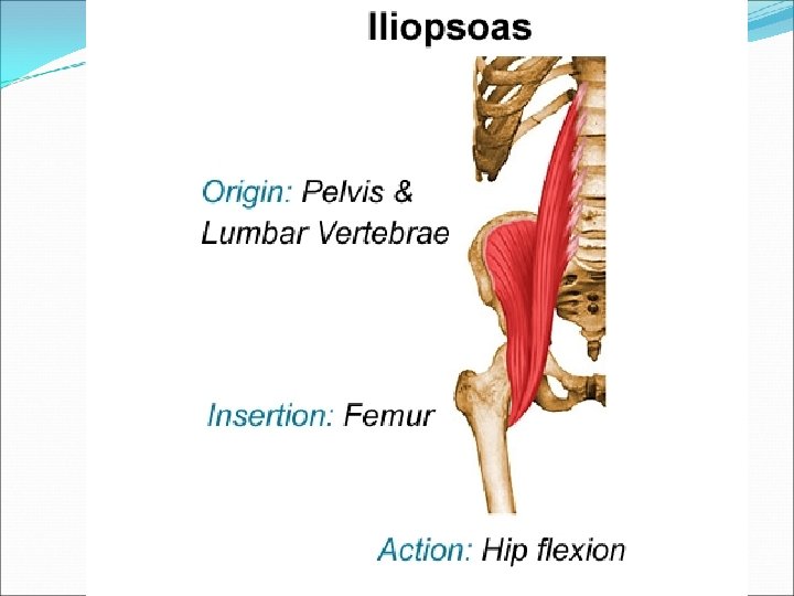
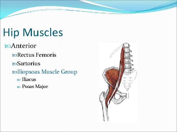
Hip Muscles Anterior Rectus Femoris Sartorius Iliopsoas Muscle Group Iliacus Psoas Major
Hip Muscles Anterior Rectus Femoris Sartorius Iliopsoas Muscle Group Iliacus Psoas Major
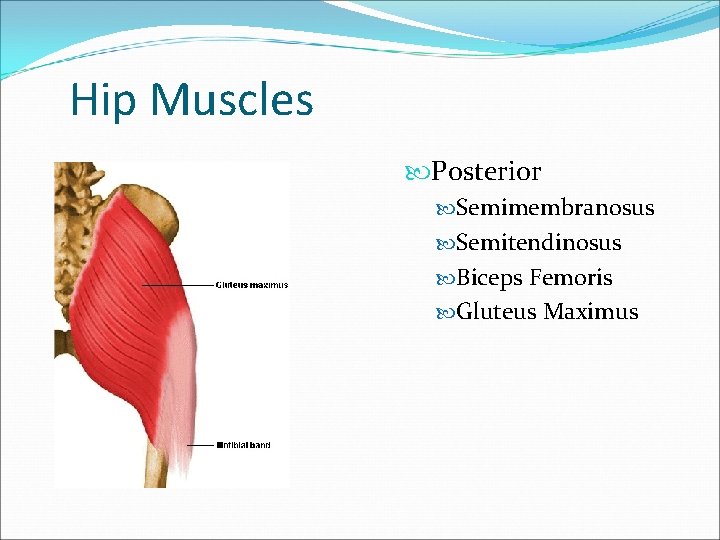
Hip Muscles Posterior Semimembranosus Semitendinosus Biceps Femoris Gluteus Maximus
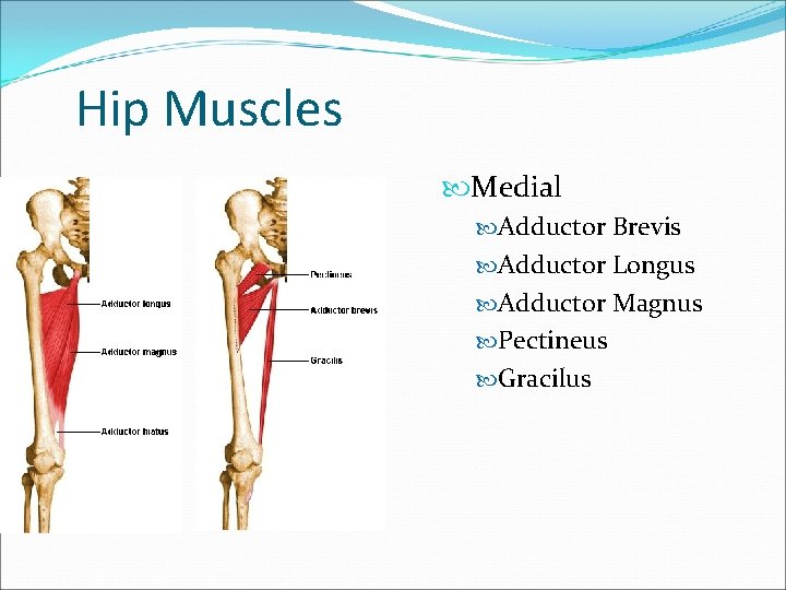
Hip Muscles Medial Adductor Brevis Adductor Longus Adductor Magnus Pectineus Gracilus
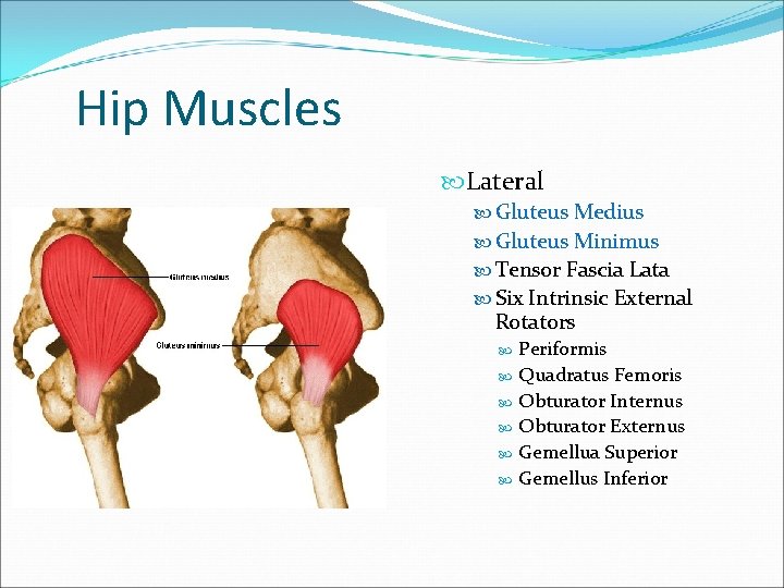
Hip Muscles Lateral Gluteus Medius Gluteus Minimus Tensor Fascia Lata Six Intrinsic External Rotators Periformis Quadratus Femoris Obturator Internus Obturator Externus Gemellua Superior Gemellus Inferior
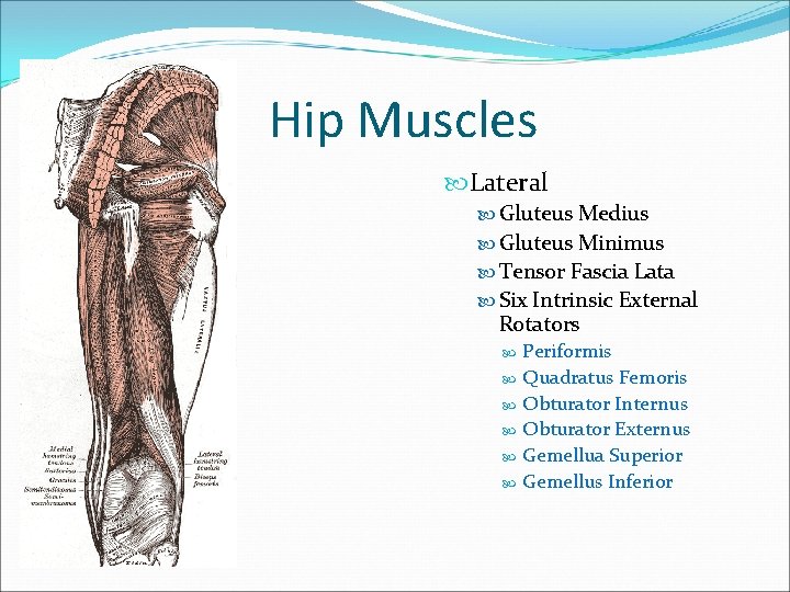
Hip Muscles Lateral Gluteus Medius Gluteus Minimus Tensor Fascia Lata Six Intrinsic External Rotators Periformis Quadratus Femoris Obturator Internus Obturator Externus Gemellua Superior Gemellus Inferior
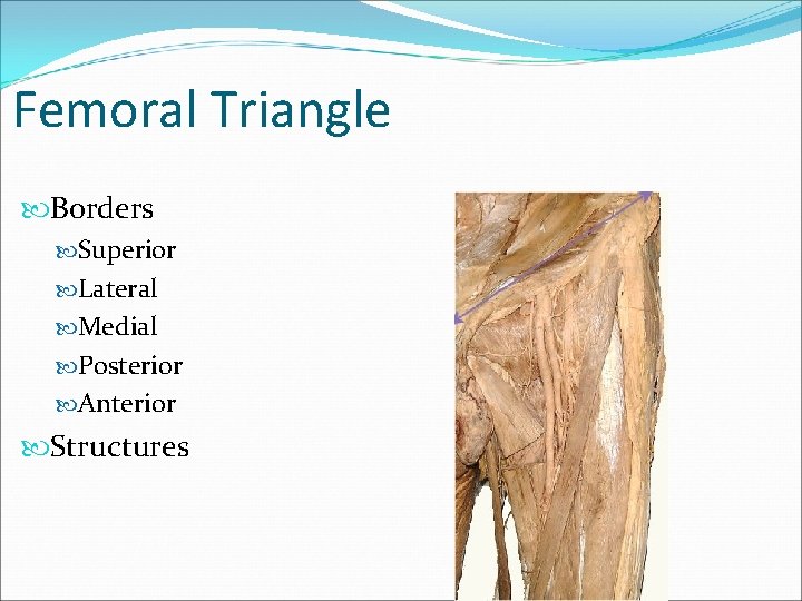
Femoral Triangle Borders Superior Lateral Medial Posterior Anterior Structures
Movements of the Pelvis Forward and Backward Tilt Left and right Lateral Tilt Left and Right Rotation
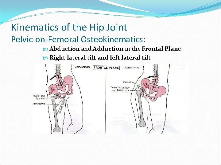
Kinematics of the Hip Joint Pelvic-on-Femoral Osteokinematics: Abduction and Adduction in the Frontal Plane Right lateral tilt and left lateral tilt
The Hip and Pelvic Girdle A. B. C. D. E. General Structure & Function of Specific Joints Muscular Considerations Specific Functional Considerations Common Injuries
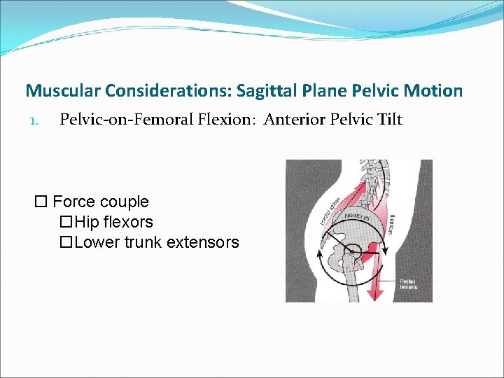
Muscular Considerations: Sagittal Plane Pelvic Motion 1. Pelvic-on-Femoral Flexion: Anterior Pelvic Tilt o Force couple o. Hip flexors o. Lower trunk extensors
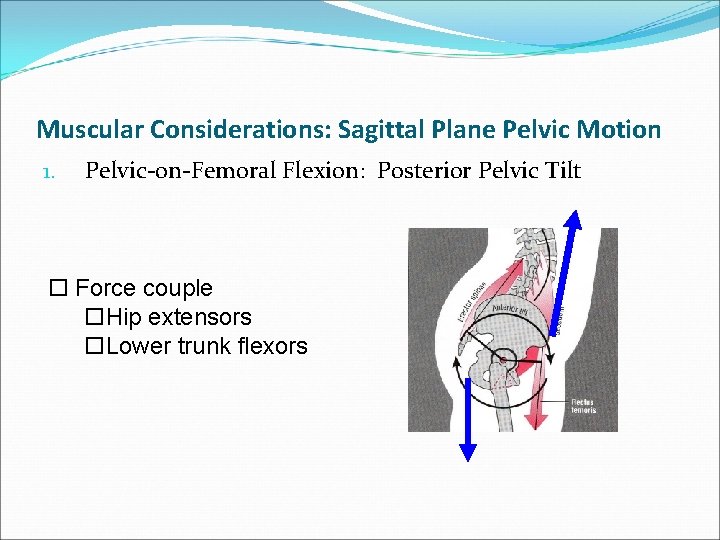
Muscular Considerations: Sagittal Plane Pelvic Motion 1. Pelvic-on-Femoral Flexion: Posterior Pelvic Tilt o Force couple o. Hip extensors o. Lower trunk flexors
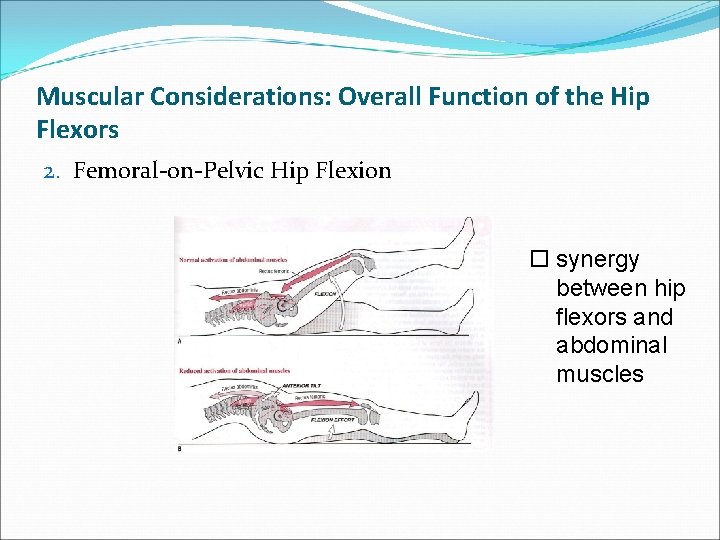
Muscular Considerations: Overall Function of the Hip Flexors 2. Femoral-on-Pelvic Hip Flexion o synergy between hip flexors and abdominal muscles
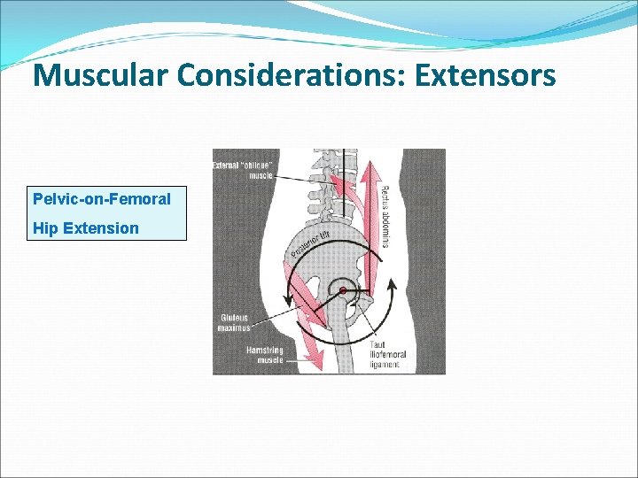
Muscular Considerations: Extensors Pelvic-on-Femoral Hip Extension
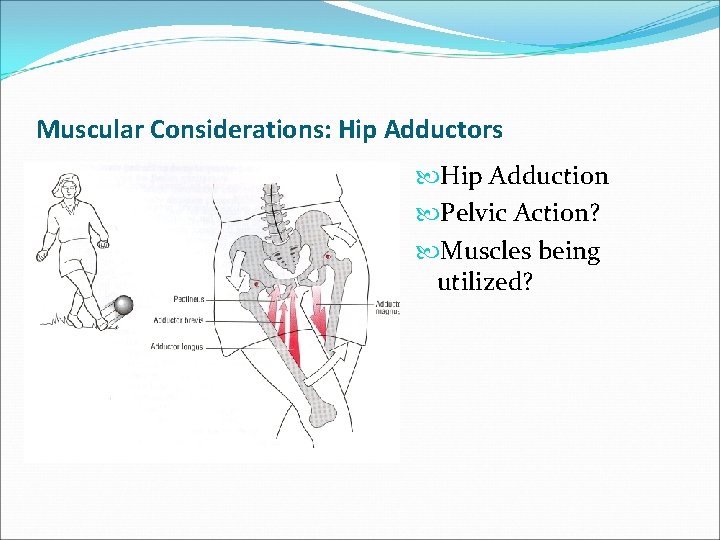
Muscular Considerations: Hip Adductors Hip Adduction Pelvic Action? Muscles being utilized?
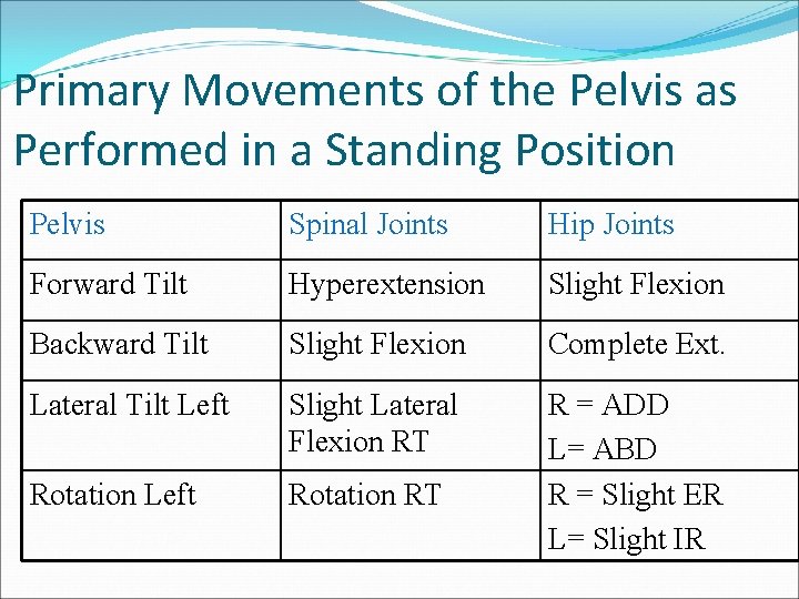
Primary Movements of the Pelvis as Performed in a Standing Position Pelvis Spinal Joints Hip Joints Forward Tilt Hyperextension Slight Flexion Backward Tilt Slight Flexion Complete Ext. Lateral Tilt Left Slight Lateral Flexion RT Rotation Left Rotation RT R = ADD L= ABD R = Slight ER L= Slight IR
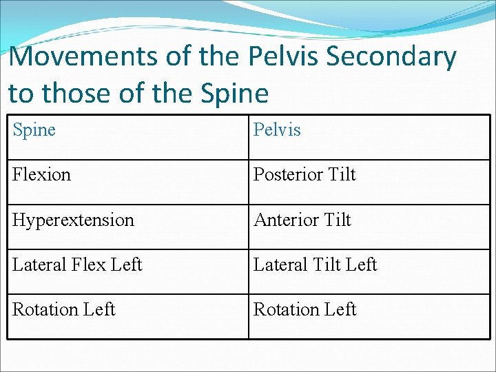
Movements of the Pelvis Secondary to those of the Spine Pelvis Flexion Posterior Tilt Hyperextension Anterior Tilt Lateral Flex Left Lateral Tilt Left Rotation Left
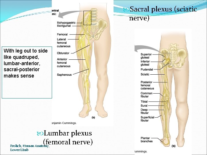
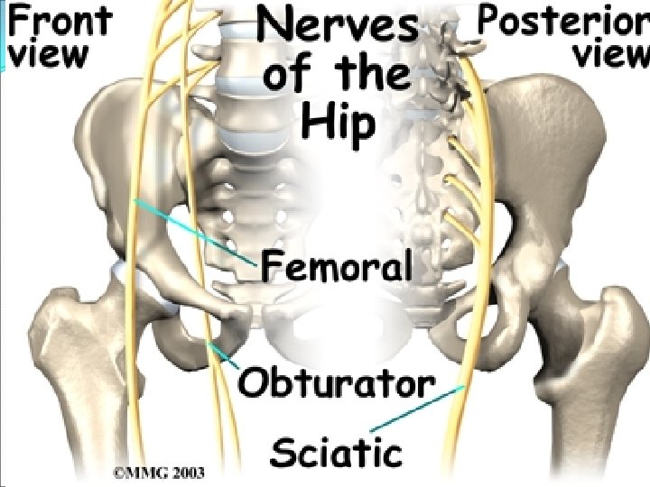
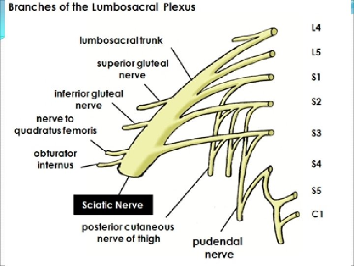
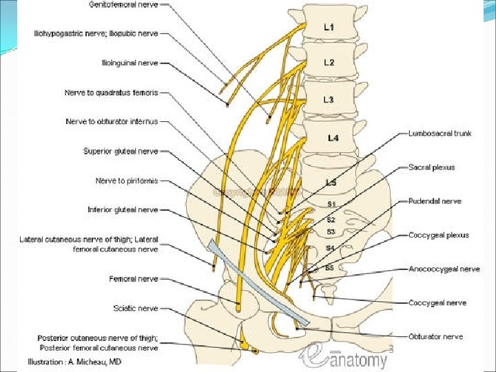
Sacral plexus (sciatic nerve) With leg out to side like quadruped, lumbar-anterior, sacral-posterior makes sense Lumbar plexus (femoral nerve) Frolich, Human Anatomy, Lower LImb
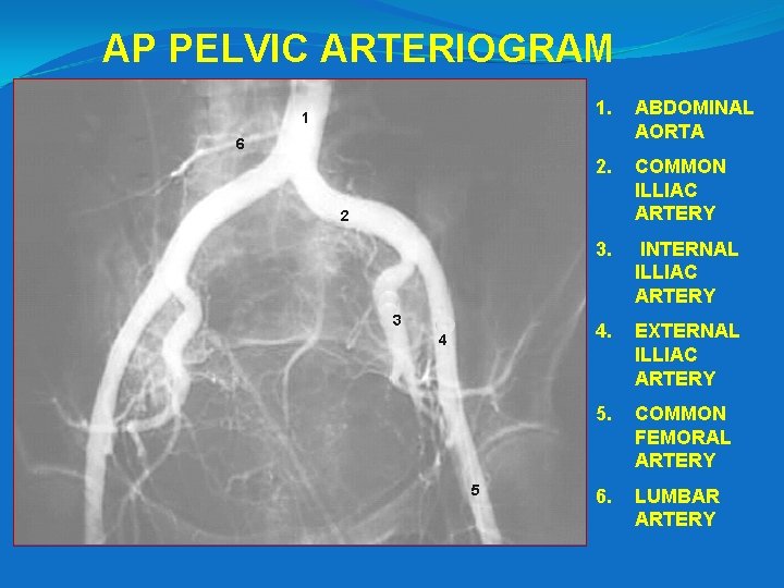
AP PELVIC ARTERIOGRAM 1 1. ABDOMINAL AORTA 2. COMMON ILLIAC ARTERY 3. INTERNAL ILLIAC ARTERY 4. EXTERNAL ILLIAC ARTERY 5. COMMON FEMORAL ARTERY 6. LUMBAR ARTERY 6 2 3 4 5
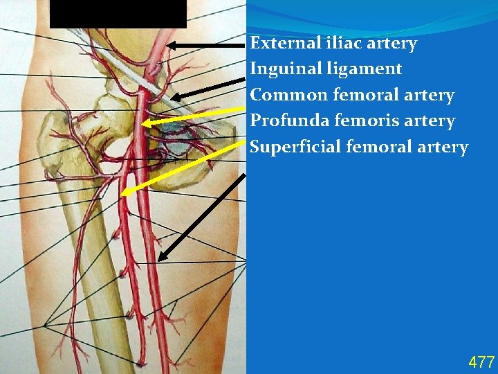
Anterior Thigh External iliac artery Inguinal ligament Common femoral artery Profunda femoris artery Superficial femoral artery 477
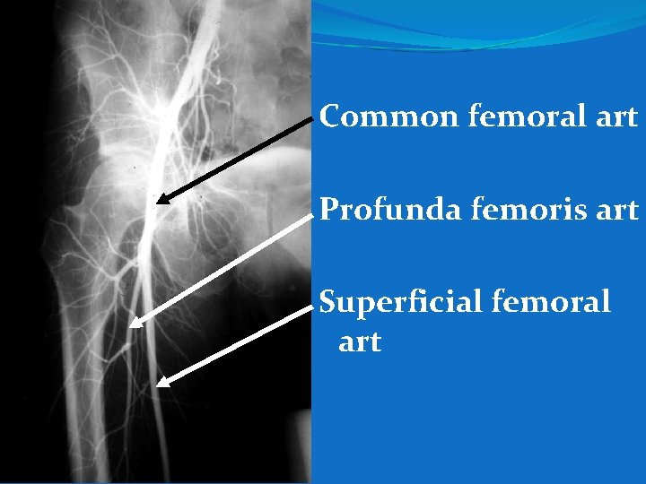
Common femoral art Profunda femoris art Superficial femoral art
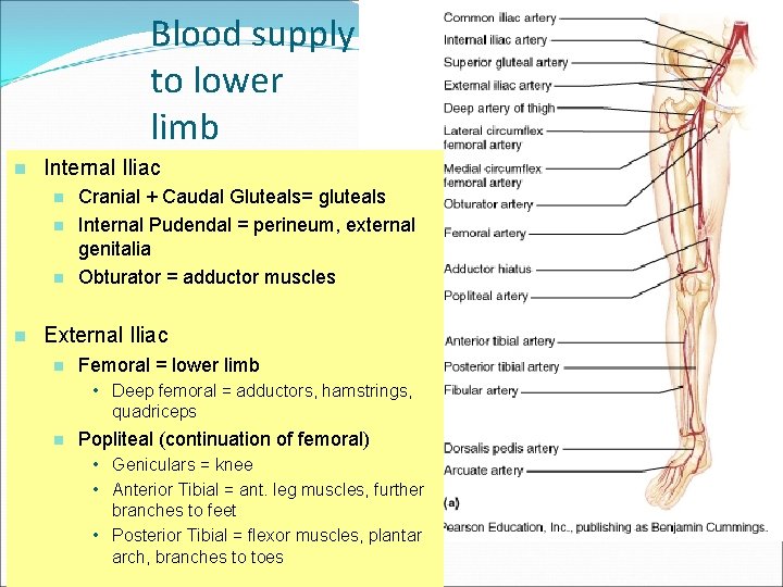
Blood supply to lower limb n Internal Iliac n n Cranial + Caudal Gluteals= gluteals Internal Pudendal = perineum, external genitalia Obturator = adductor muscles External Iliac n Femoral = lower limb • Deep femoral = adductors, hamstrings, quadriceps n Popliteal (continuation of femoral) • Geniculars = knee • Anterior Tibial = ant. leg muscles, further branches to feet • Posterior Tibial = flexor muscles, plantar Frolich, Human Anatomy, arch, branches to toes Lower LImb
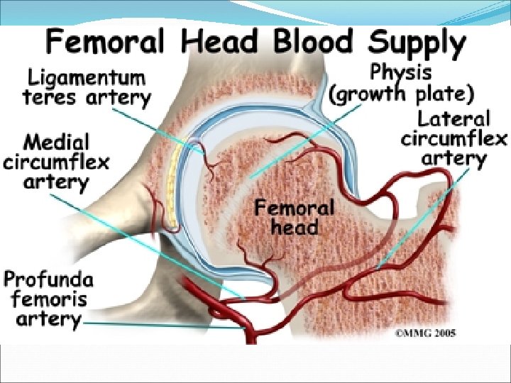
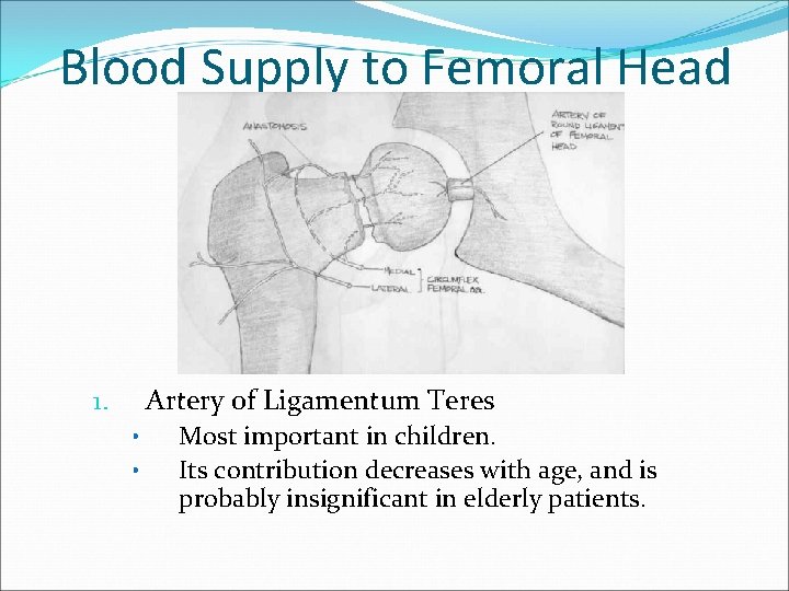
Blood Supply to Femoral Head Artery of Ligamentum Teres 1. • • Most important in children. Its contribution decreases with age, and is probably insignificant in elderly patients.
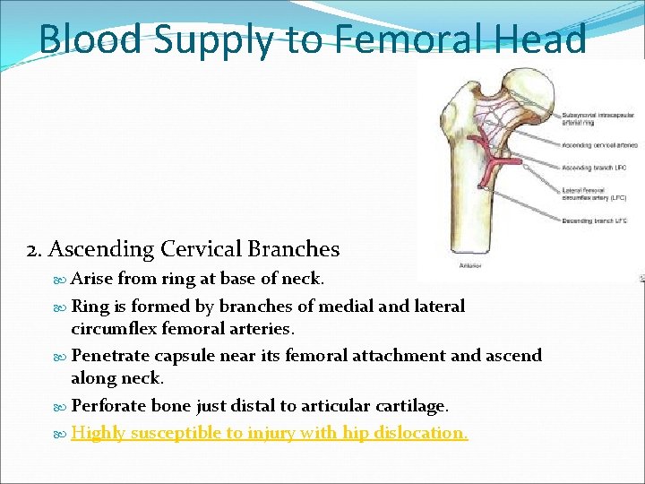
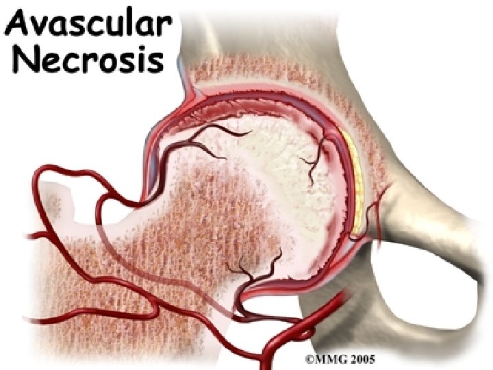
Blood Supply to Femoral Head 2. Ascending Cervical Branches Arise from ring at base of neck. Ring is formed by branches of medial and lateral circumflex femoral arteries. Penetrate capsule near its femoral attachment and ascend along neck. Perforate bone just distal to articular cartilage. Highly susceptible to injury with hip dislocation.
Some pathologies…
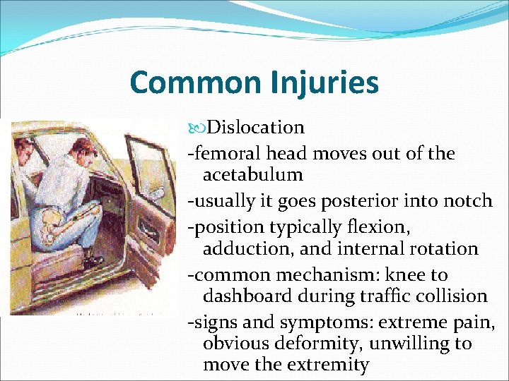
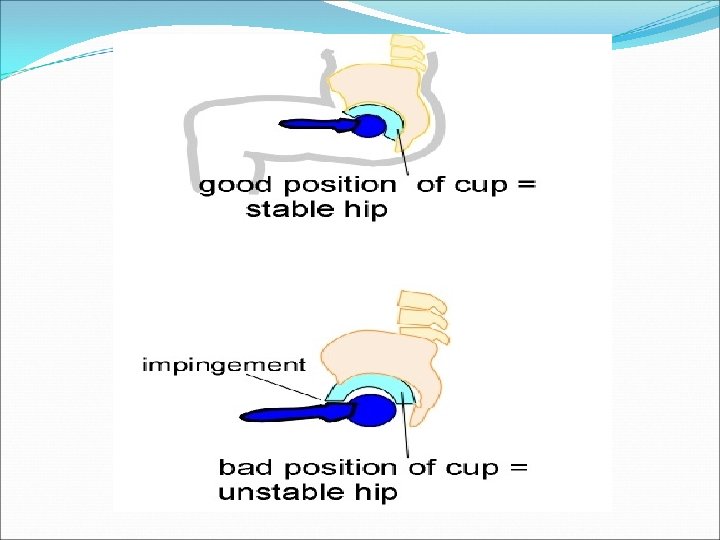
Common Injuries Dislocation -femoral head moves out of the acetabulum -usually it goes posterior into notch -position typically flexion, adduction, and internal rotation -common mechanism: knee to dashboard during traffic collision -signs and symptoms: extreme pain, obvious deformity, unwilling to move the extremity
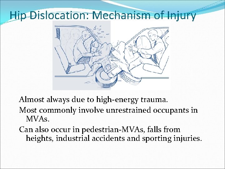
Hip Dislocation: Mechanism of Injury Almost always due to high-energy trauma. Most commonly involve unrestrained occupants in MVAs. Can also occur in pedestrian-MVAs, falls from heights, industrial accidents and sporting injuries.
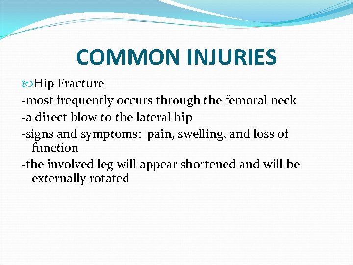
COMMON INJURIES Hip Fracture -most frequently occurs through the femoral neck -a direct blow to the lateral hip -signs and symptoms: pain, swelling, and loss of function -the involved leg will appear shortened and will be externally rotated
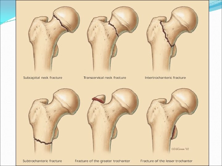
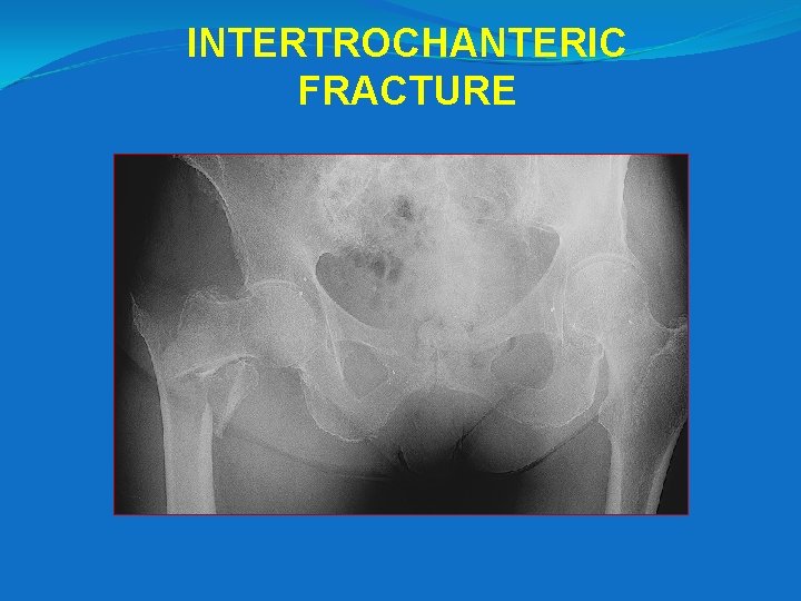
INTERTROCHANTERIC FRACTURE
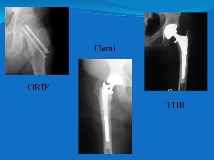
COMMON INJURIES Avascular Necrosis of the Femoral Head -blood supply to the femur head is severed or is occluded for a prolonged period of time. -this is a common complication following hip dislocations, fractures, and chronic synovitis and often necessitates a hip replacement
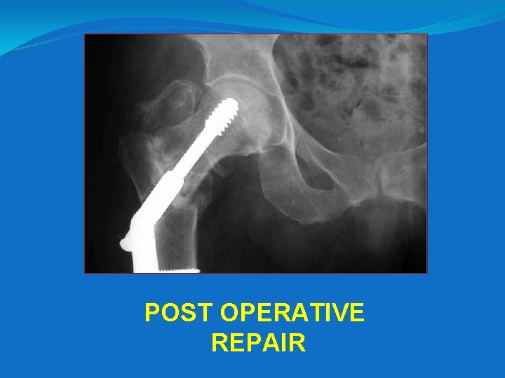
POST OPERATIVE REPAIR
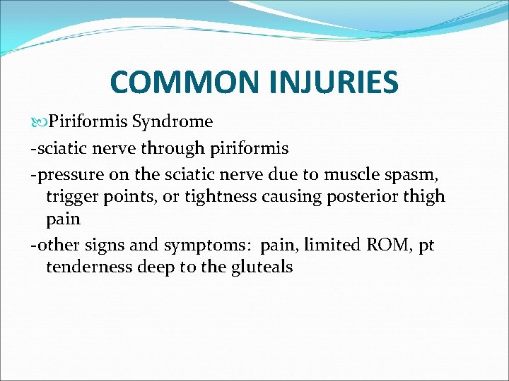
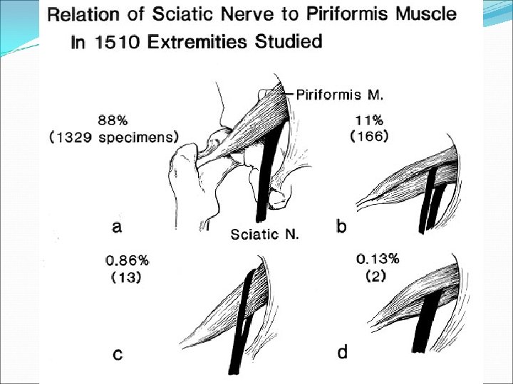
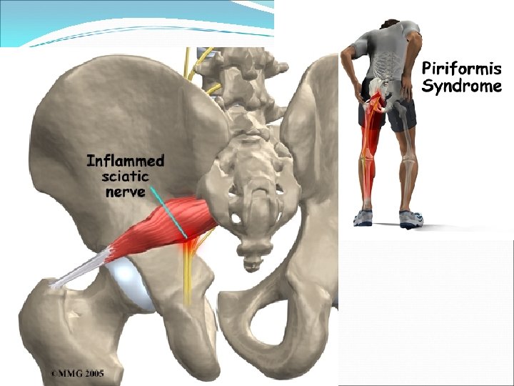
COMMON INJURIES Piriformis Syndrome -sciatic nerve through piriformis -pressure on the sciatic nerve due to muscle spasm, trigger points, or tightness causing posterior thigh pain -other signs and symptoms: pain, limited ROM, pt tenderness deep to the gluteals
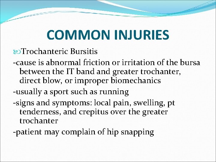
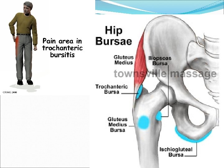
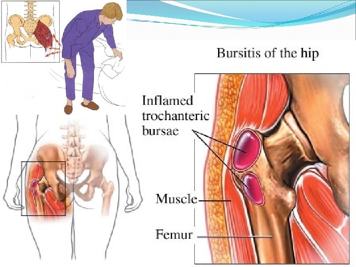
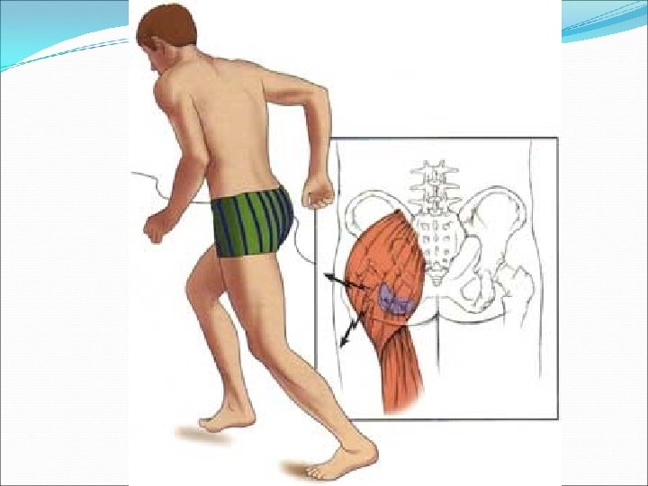
COMMON INJURIES Trochanteric Bursitis -cause is abnormal friction or irritation of the bursa between the IT band greater trochanter, direct blow, or improper biomechanics -usually a sport such as running -signs and symptoms: local pain, swelling, pt tenderness, and crepitus over the greater trochanter -patient may complain of hip snapping
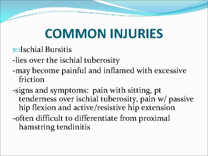
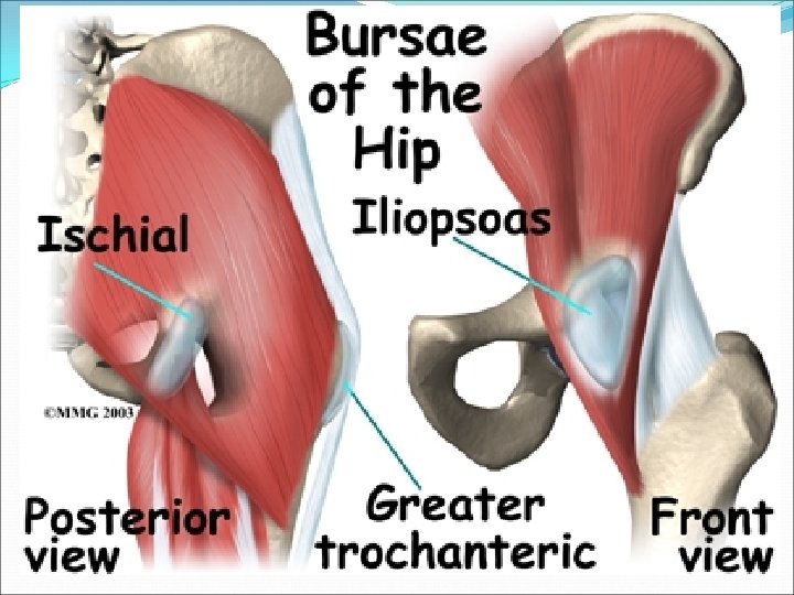
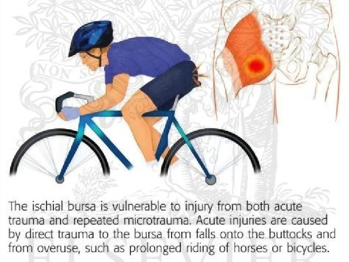
COMMON INJURIES Ischial Bursitis -lies over the ischial tuberosity -may become painful and inflamed with excessive friction -signs and symptoms: pain with sitting, pt tenderness over ischial tuberosity, pain w/ passive hip flexion and active/resistive hip extension -often difficult to differentiate from proximal hamstring tendinitis
COMMON INJURIES Hip Joint Sprain -less common -excessive forcible exertion of the extremity that stretch or tear the surrounding ligaments -signs and symptoms: pain and decrease ROM
COMMON INJURIES Hip Joint Strains -resulting from overstretching or from a rapid, forceful contraction of the muscle -explosive starts and slipping of the foot during cutting are common mechanisms for hip flexor and adductor strains -these injuries frequently occur during the beginning of practice and preseason training -signs and symptoms: pain, pt tenderness, muscle spasm, swelling, ecchymosis , and decreased ROM
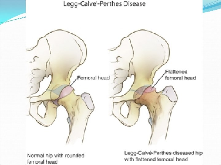
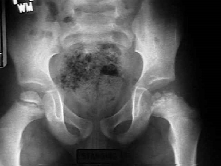
COMMON INJURIES Legg-Calve-Perthes Disease -characterized by avascular necrosis of the proximal femoral epiphysis -a chronic condition that develops slowly in children -more often in males than in females -signs and symptoms: pain in the hip or groin that radiates to the knee, limping, decreased ROM, and hip flexor tightness may be noted -physician should be consulted to rule out serious pathologies such as this
COMMON INJURIES Chronic Synovitis -inflammatory process at the hip that is characterized by chronic irritation and excess secretion of synovial fluid within the capsule -this condition is very difficult to detect -may lead to avascular necrosis of the femoral head
Hemi ORIF THR

Hips The End

- Slides: 117