THYROIDITIS Inflammatory diseases of the thyroid gland with



- Slides: 72
THYROIDITIS Inflammatory diseases of the thyroid gland with different etiologic, biologic, histologic and clinical aspects CLASSIFICATION • ACUTE: bacterial, viral • SUBACUTE: de Quervain’s thryoiditis • CHRONIC: • chronic autoimmune thyroidits • Tuberculous • Mycotic • Riedel’s thyroidits
ACUTE BACTERIAL THYROIDITIS Signs and symptoms • Fever • Pain profound and severe • Dysfagia - 90 % din cazuri • Dyspnea – 50 % • Spasmodic cough Laboratory data • increased ESR • leukocytosis with neutrophilia • Ultrasound: small or large hypoechoic areas • FNB: isolation of germs Treatment : antibiotics
ACUTE BACTERIAL THYROIDITIS
Acute thyroiditis - histology
SUBACUTE ”DE QUERVAIN’S” THYROIDITIS PREVALENTA Sex ratio F/M: 3, 6/1 – 10, 6 /1 1 caz TS for 5 cases of Graves disease and for 20 cases of AIT • 0, 01 % of all hospitalized patients • 1, 89 % of all patiens hospitalized for thyroid diseases • 9, 9 % of subjects presenting with thyrotoxicosis • 1, 52 % of patients investigated by FNB Szabolosz I. Subacute thyroidits Budapesta 2000
SUBACUTE THYROIDITIS ETIOLOGY • probably the disease is a response to a viral infection GENETICS • those with HLA-Bw 35 have a risk to develop the disease of 8 -56. 6 % • HLA-Bw 35 allows the development of clincal symtoms • it has no relatioship with the evolution of the disease
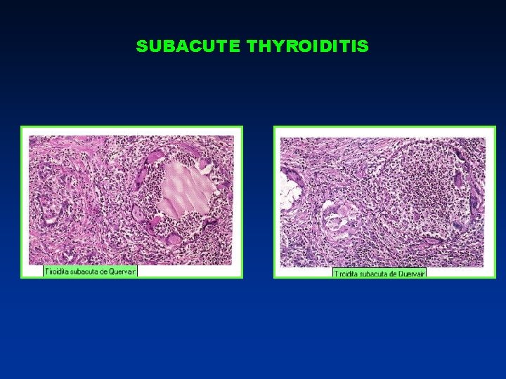
SUBACUTE THYROIDITIS PATOGENICITY • interleukine 6 produced by monocytes si macrophages determine inflammation • interleukine 2 +TNF + interferon determine destructive thyroiditis in 10 % of cases • VEGF, basic FGF, PDGF determine granulomatous reaction • EGF determines by mitogenic effect the regeneration of the follicles PATHOLOGY • Follicular disruption with thyroglobulin liberation is responsible for the initial phase of thyrotoxicosis • granuloma: • a center of giant cells surrounded by macrophages • epithelial cells surrounded by a crown of macrophages involved with antigen presentation
SUBACUTE THYROIDITIS
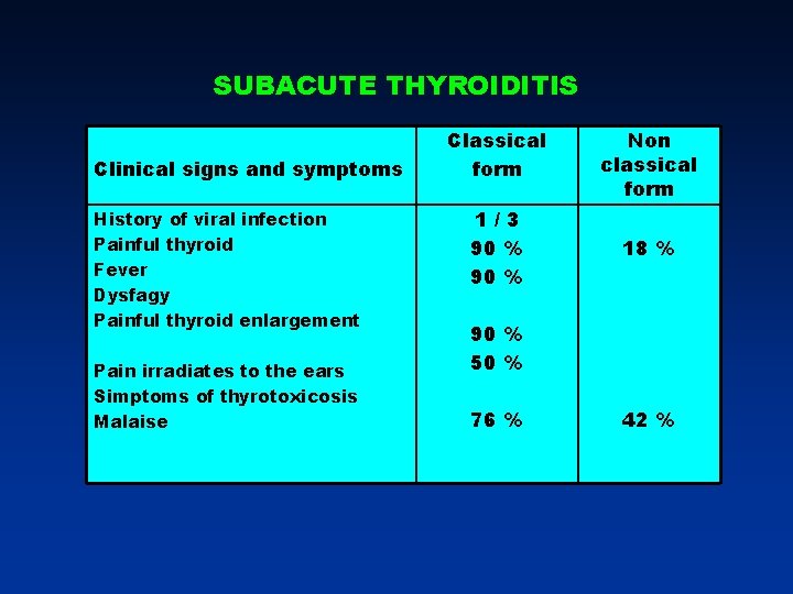
SUBACUTE THYROIDITIS Clinical signs and symptoms History of viral infection Painful thyroid Fever Dysfagy Painful thyroid enlargement Pain irradiates to the ears Simptoms of thyrotoxicosis Malaise Classical form Non classical form 1/3 90 % 18 % 90 % 50 % 76 % 42 %
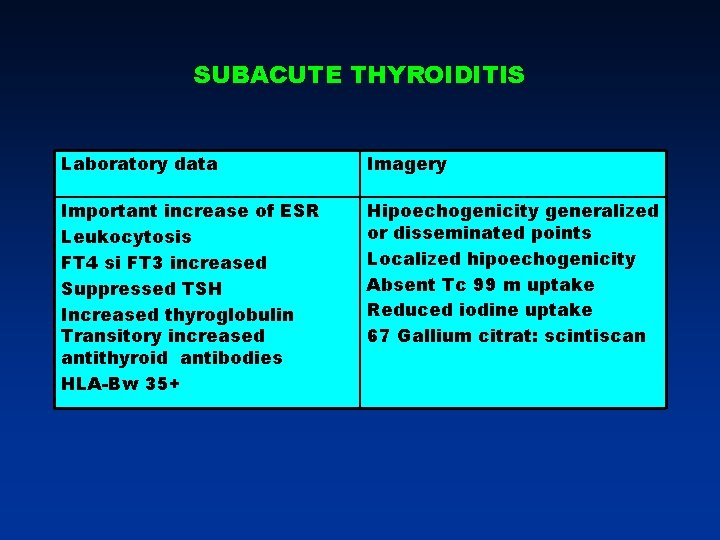
SUBACUTE THYROIDITIS Laboratory data Imagery Important increase of ESR Leukocytosis FT 4 si FT 3 increased Suppressed TSH Increased thyroglobulin Transitory increased antithyroid antibodies HLA-Bw 35+ Hipoechogenicity generalized or disseminated points Localized hipoechogenicity Absent Tc 99 m uptake Reduced iodine uptake 67 Gallium citrat: scintiscan
SUBACUTE THYROIDITIS Differential diagnosis Evolution and complications Cyst with intracystic hemorrhage Tirotoxicosis induced by iodine loading (amiodarone) Interpheron induced thyroiditis Thyroid cancer: FNB Transient hypothyroidism second phase of evolution Recurrent disease is unpredictable Heeling Definitive hypothyroidism <1/10 Painless forms
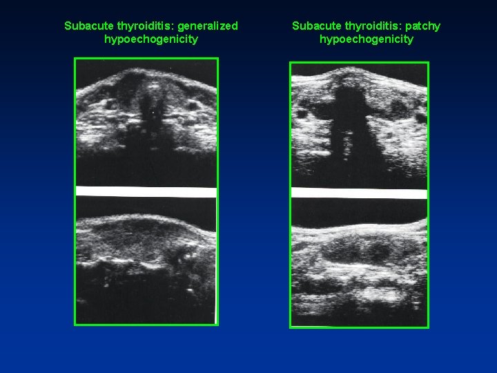
Subacute thyroiditis: generalized hypoechogenicity Subacute thyroiditis: patchy hypoechogenicity
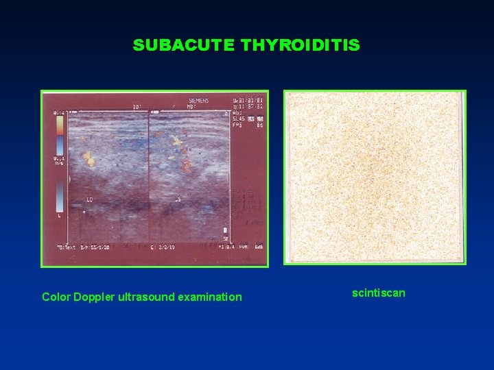
SUBACUTE THYROIDITIS Color Doppler ultrasound examination scintiscan
SUBACUTE THYROIDITIS – TREATMENT FORME SEVERE: GLUCOCORTICODS: • Prednisone: 30 -40 mg / day at the beginning of the disease with further reduction of the dosage • Dexametazone: 3 -4 mg /zi FORME USOARE: Nonsteroidal anti inflammatory drugs: indometacin
AUTOIMMUNE THYROIDITIS INCIDENCE • 3, 5 – 4, 5 % of population present autoimmune thyroid diseases • 4, 6 % of women and 1, 23 %of men have antithyroid antibodies • 15 % of women over 60 years • lymphocytic infiltrations: 6, 8 5 of women and 2, 7 % of men • 50 % of those with antithyroid antibodies have TSH > 6 U. I. /ml • 60 % of those with TSH > 6 U. I. /ml have antithyroid antibodies • 80 % of those with TSH > 10 6 U. I. /ml have antithyroid antibodies • 5 % of those with TSH > 6 U. I. /ml develop overt hypothyroidism each year
AUTOIMMUNE THYROIDITIS PATOGENY • genetic predisposition • Viral aggression • excessive iodine supply GENETIC PREDISPOSITION • relatives with autoimmune thyroid diseases • patients with genetic abnormalities : Turner, Klinefelter, Down syndrome • association with other autoimmune diseases: • multiple autoimune endocrine diseases type I and II (ICSR, ovarian failure with precocious menopause ) autoimmune hypophysitis Biermer disease , sd, Sjogren, lupus, rheumatoid arthritis , miastenia gravis, interstitial lung disease • HLA-DR 3 si HLA-DR 4
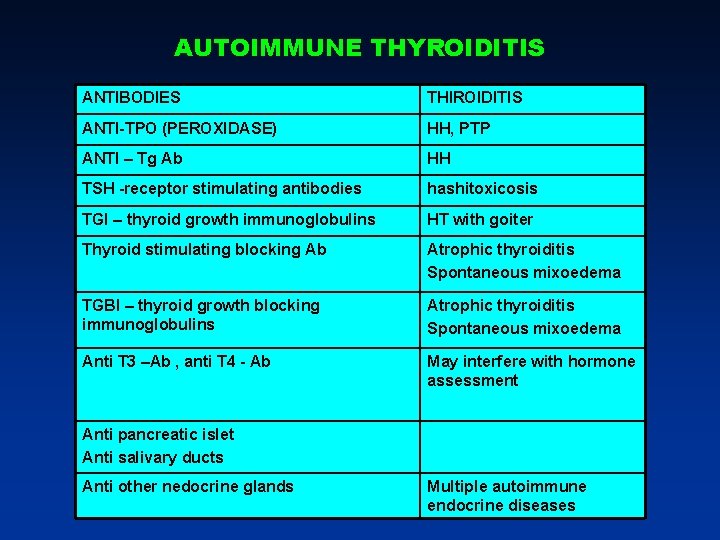
AUTOIMMUNE THYROIDITIS ANTIBODIES THIROIDITIS ANTI-TPO (PEROXIDASE) HH, PTP ANTI – Tg Ab HH TSH -receptor stimulating antibodies hashitoxicosis TGI – thyroid growth immunoglobulins HT with goiter Thyroid stimulating blocking Ab Atrophic thyroiditis Spontaneous mixoedema TGBI – thyroid growth blocking immunoglobulins Atrophic thyroiditis Spontaneous mixoedema Anti T 3 –Ab , anti T 4 - Ab May interfere with hormone assessment Anti pancreatic islet Anti salivary ducts Anti other nedocrine glands Multiple autoimmune endocrine diseases
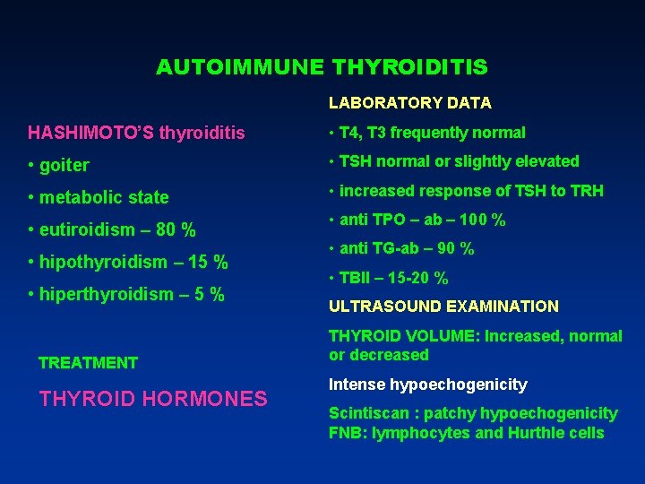
AUTOIMMUNE THYROIDITIS LABORATORY DATA HASHIMOTO’S thyroiditis • T 4, T 3 frequently normal • goiter • TSH normal or slightly elevated • metabolic state • increased response of TSH to TRH • eutiroidism – 80 % • hipothyroidism – 15 % • hiperthyroidism – 5 % TREATMENT THYROID HORMONES • anti TPO – ab – 100 % • anti TG-ab – 90 % • TBII – 15 -20 % ULTRASOUND EXAMINATION THYROID VOLUME: Increased, normal or decreased Intense hypoechogenicity Scintiscan : patchy hypoechogenicity FNB: lymphocytes and Hurthle cells
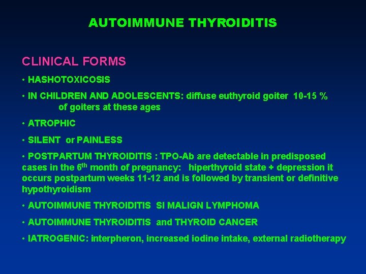
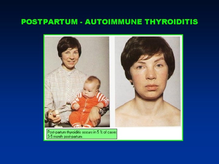
AUTOIMMUNE THYROIDITIS CLINICAL FORMS • HASHOTOXICOSIS • IN CHILDREN AND ADOLESCENTS: diffuse euthyroid goiter 10 -15 % of goiters at these ages • ATROPHIC • SILENT or PAINLESS • POSTPARTUM THYROIDITIS : TPO-Ab are detectable in predisposed cases in the 6 th month of pregnancy: hiperthyroid state + depression it occurs postpartum weeks 11 -12 and is followed by transient or definitive hypothyroidism • AUTOIMMUNE THYROIDITIS SI MALIGN LYMPHOMA • AUTOIMMUNE THYROIDITIS and THYROID CANCER • IATROGENIC: interpheron, increased iodine intake, external radiotherapy
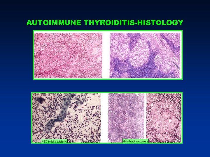
AUTOIMMUNE THYROIDITIS-HISTOLOGY
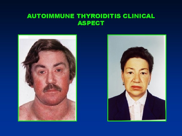
AUTOIMMUNE THYROIDITIS CLINICAL ASPECT
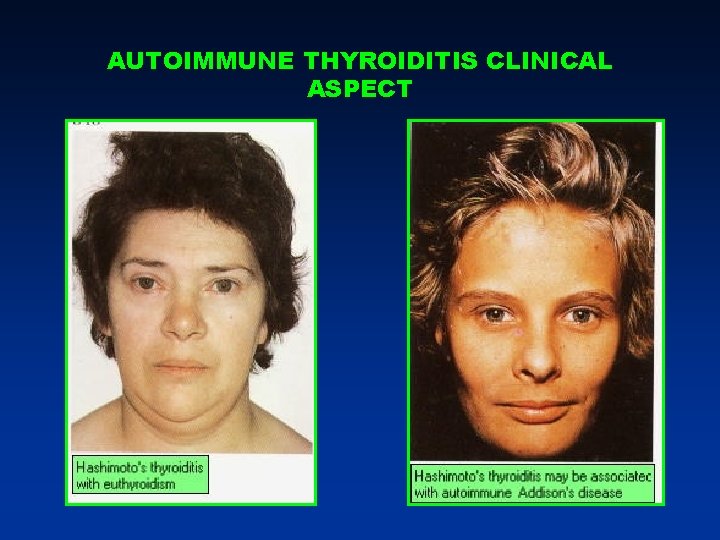
AUTOIMMUNE THYROIDITIS CLINICAL ASPECT
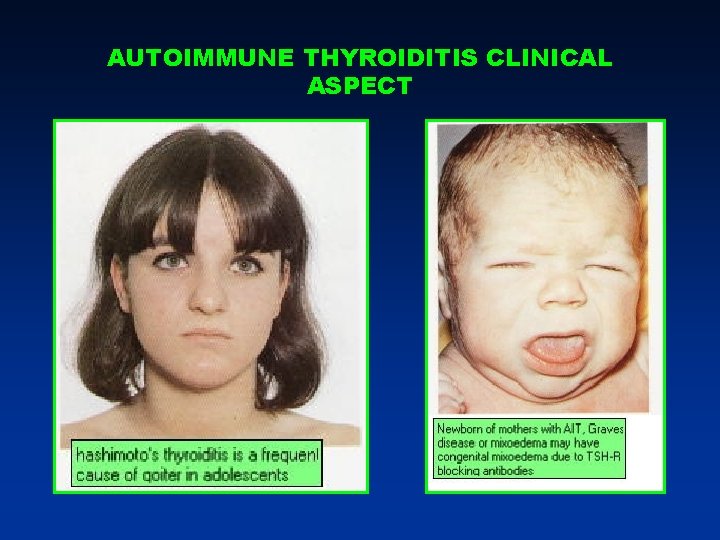
AUTOIMMUNE THYROIDITIS CLINICAL ASPECT
POSTPARTUM - AUTOIMMUNE THYROIDITIS
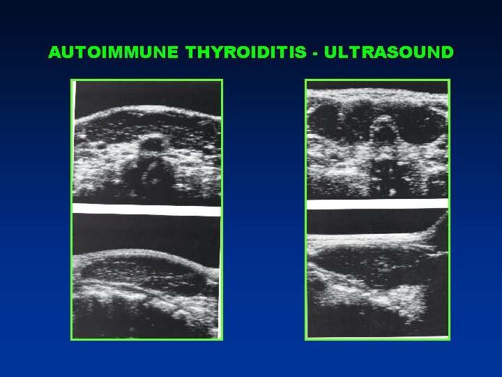
AUTOIMMUNE THYROIDITIS - ULTRASOUND
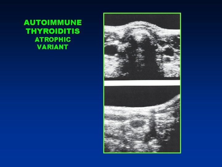
AUTOIMMUNE THYROIDITIS ATROPHIC VARIANT
THYROID NODULES
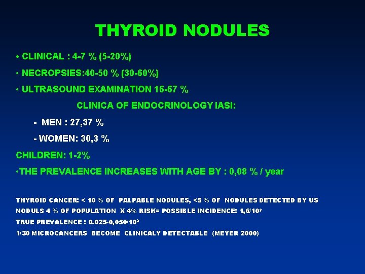
THYROID NODULES • CLINICAL : 4 -7 % (5 -20%) • NECROPSIES: 40 -50 % (30 -60%) • ULTRASOUND EXAMINATION 16 -67 % CLINICA OF ENDOCRINOLOGY IASI: - MEN : 27, 37 % - WOMEN: 30, 3 % CHILDREN: 1 -2% • THE PREVALENCE INCREASES WITH AGE BY : 0, 08 % / year THYROID CANCER: < 10 % OF PALPABLE NODULES, <5 % OF NODULES DETECTED BY US NODULS 4 % OF POPULATION X 4% RISK= POSSIBLE INCIDENCE: 1, 6/103 TRUE PREVALENCE : 0. 025 -0, 050/103 1/30 MICROCANCERS BECOME CLINICALY DETECTABLE (MEYER 2000)
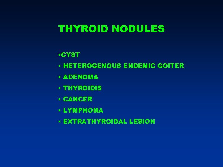
THYROID NODULES • CYST • HETEROGENOUS ENDEMIC GOITER • ADENOMA • THYROIDIS • CANCER • LYMPHOMA • EXTRATHYROIDAL LESION
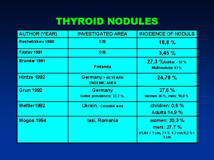
THYROID NODULES AUTHOR (YEAR) INVESTIGATED AREA INCIDENCE OF NODULS Reshetnikov 1990 CIS 18, 8 % Filatov 1991 CIS 3, 45 % Brander 1991 Finlanda Hintze 1992 Germany > 60 YEARS 27, 3 %Solitar – 57 % Multinodular 43 % 24, 78 % ENDEMIC AREA Grun 1992 Mettler 1992 Mogos 1994 Germany 27, 6 % Goiter prevalence: 37, 7 %, women 36 %, men: 18, 8 % Ukrain, Cernobil area Iasi, Romania children: 0, 5 % Adults 14, 9 % women: 30, 3 % meni: 27, 7 % 61, 84 < 1 cm, 21 % 1 -2 cm. 9, 2 %> 3 cm
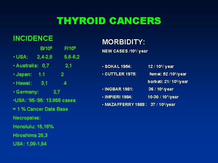
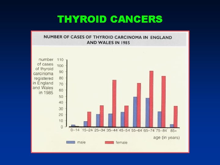
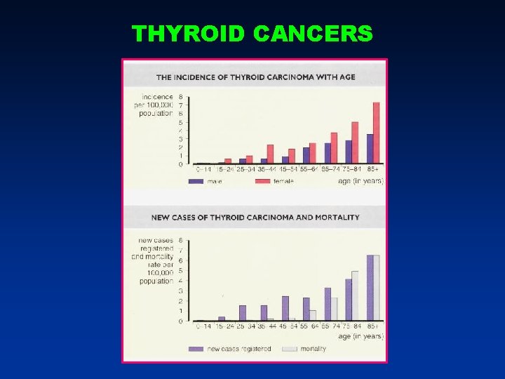
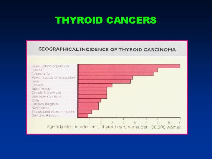
THYROID CANCERS INCIDENCE B/106 • USA: F/106 2, 4 -2, 8 2, 1 1. 1 • Hawai: 2 3, 1 • Germany: 2, 7 = 1 % Cancer Data Base Honolulu: 15, 16% Hiroshima 25, 3 USA: 1, 09 -1, 84 • SOKAL 1954: • CUTTLER 1975: 12 / 106/ year femei: 52 /106/year barbati: 21/ 10 6/year 4 • USA: ’ 85 -’ 95: 13. 856 cases Necropsies: NEW CASES /106/ year 5, 6 -6, 2 • Australia: 0, 7 • Japan: MORBIDITY: • INGBAR 1981: 36 / 106/year • IMPIERI 1984: 10 -30 / 106/year • MAZAFFERRY 1988 : 37 / 106/year
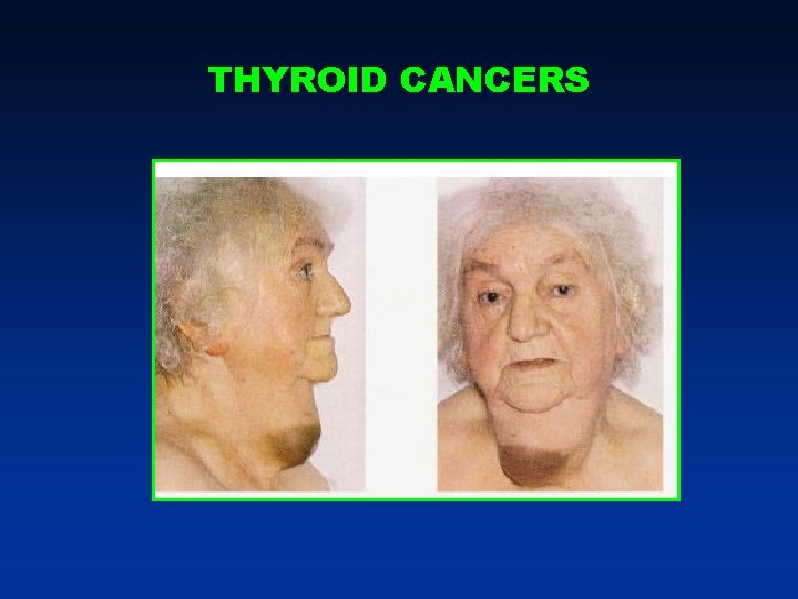
THYROID CANCERS
THYROID CANCERS
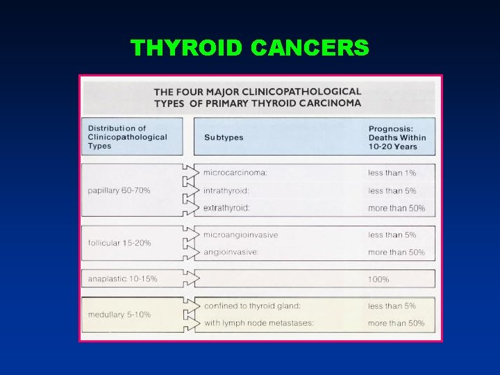
THYROID CANCERS
THYROID CANCERS
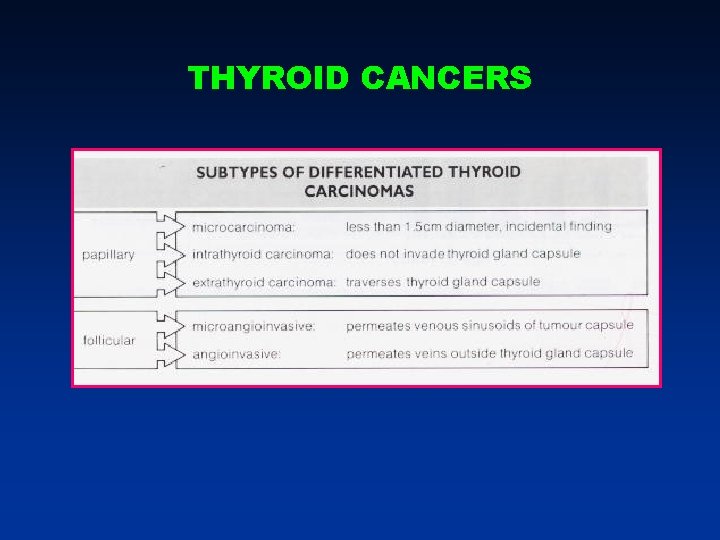
THYROID CANCERS
THYROID CANCERS
THYROID CANCERS
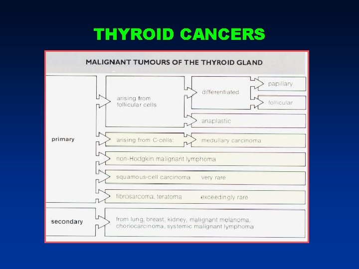
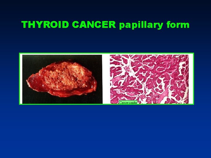
THYROID CANCER papillary form
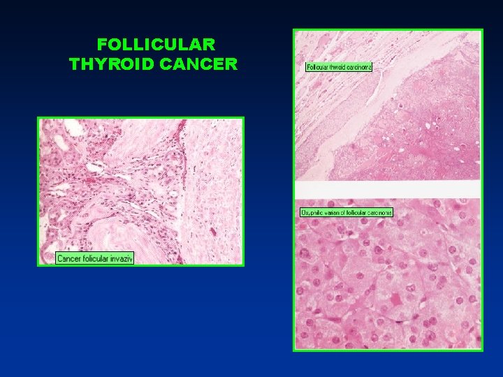
FOLLICULAR THYROID CANCER
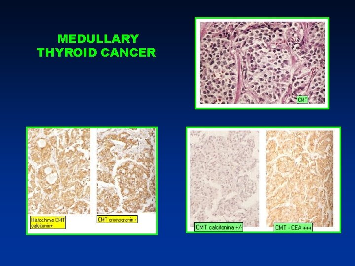
MEDULLARY THYROID CANCER
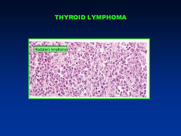
THYROID LYMPHOMA
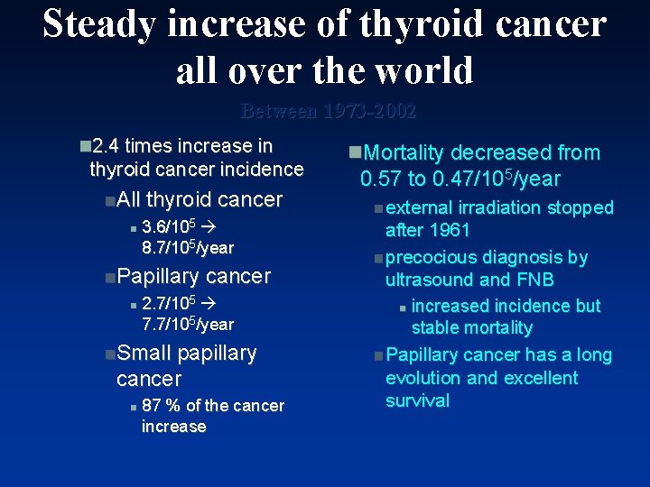
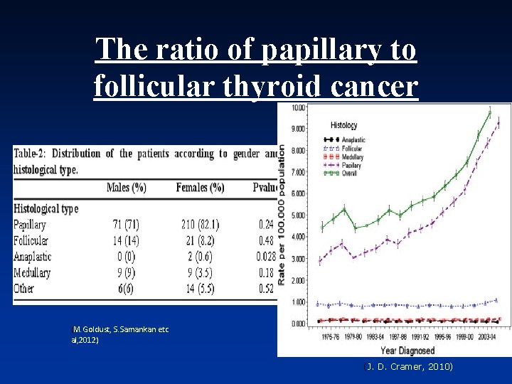
Steady increase of thyroid cancer all over the world Between 1973 -2002 n 2. 4 times increase in thyroid cancer incidence n. All n thyroid cancer 3. 6/105 8. 7/105/year n. Papillary n cancer 2. 7/105 7. 7/105/year n. Small papillary cancer n 87 % of the cancer increase n. Mortality decreased from 0. 57 to 0. 47/105/year n external irradiation stopped after 1961 n precocious diagnosis by ultrasound and FNB n increased incidence but stable mortality n Papillary cancer has a long evolution and excellent survival
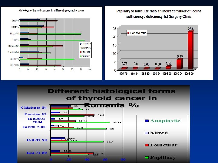
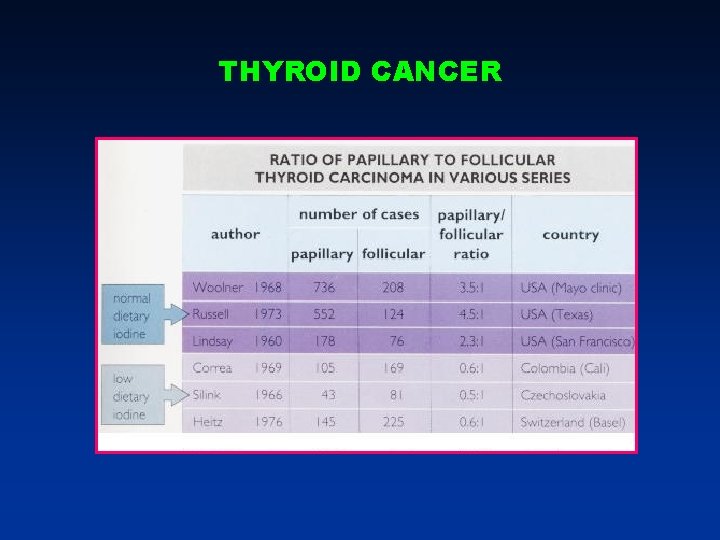
Trends in thyroid cancer n. There was noticed steady increase of thyroid cancer all over the world n. External irradiation is the only well documented cause in papillary thyroid cancer leading to RET/PTC re-arrangements n. Iodine deficiency may play a role in the development of follicular cancer and may favor the development of anaplastic carcinoma n. Iodine repletion is associated with increased incidence of papillary carcinoma with excellent prognosis
The ratio of papillary to follicular thyroid cancer (M. Goldust, S. Samankan etc al, 2012) (J. D. Cramer, 2010)
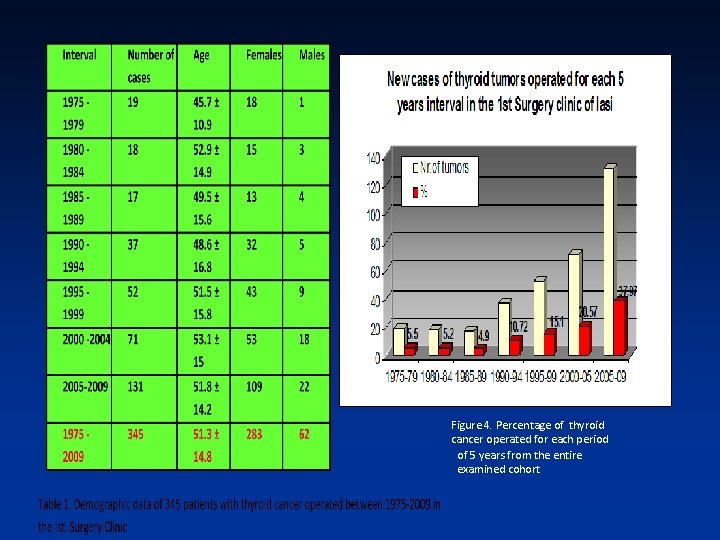
Figure 4. Percentage of thyroid cancer operated for each period of 5 years from the entire examined cohort
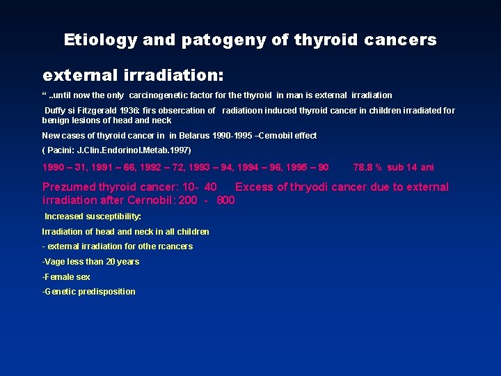
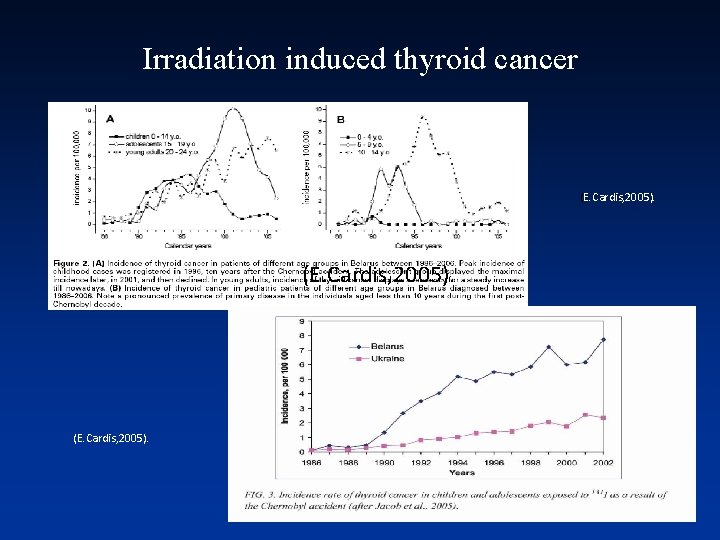
Etiology and patogeny of thyroid cancers external irradiation: “. . until now the only carcinogenetic factor for the thyroid in man is external irradiation Duffy si Fitzgerald 1936: firs obsercation of radiatioon induced thyroid cancer in children irradiated for benign lesions of head and neck New cases of thyroid cancer in in Belarus 1990 -1995 –Cernobil effect ( Pacini: J. Clin. Endorinol. Metab. 1997) 1990 – 31, 1991 – 66, 1992 – 72, 1993 – 94, 1994 – 96, 1995 – 90 78. 8 % sub 14 ani Prezumed thyroid cancer: 10 - 40 Excess of thryodi cancer due to external irradiation after Cernobil: 200 - 800 Increased susceptibility: Irradiation of head and neck in all children - external irradiation for othe rcancers -Vage less than 20 years -Female sex -Genetic predisposition
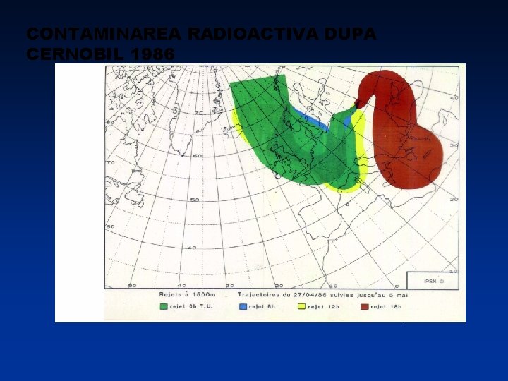
CONTAMINAREA RADIOACTIVA DUPA CERNOBIL 1986
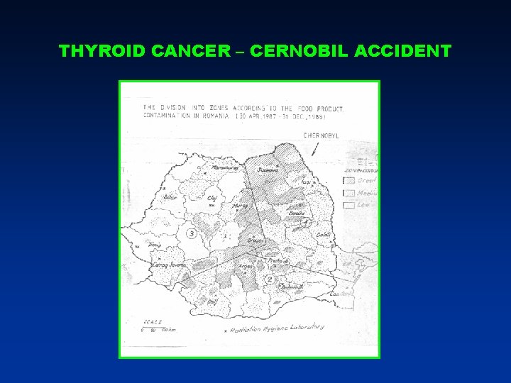
THYROID CANCER – CERNOBIL ACCIDENT
Irradiation induced thyroid cancer (E. Cardis, 2005).
THYROID CANCER
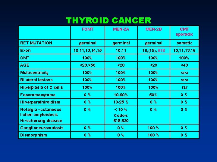
THYROID CANCER FCMT MEN-2 A MEN-2 B CMT sporadic germinal somatic Exon 10, 11, 13, 14, 15 10, 11 16, (15), 918 10, 11, 13, 16 CMT 100% AGE <20, >50 <20 <40 Multicentricity 100% rara Bilateral lesions 100% rara Hiperplasia of C cells 100% rar Feocromocytoma 0% 10 -60% 50% 0% Hiperparathiroidism 0% 10 -25 % 0% 0% Notalgia –cutaneous lichen amyloidosis Hirschprung disease 0% < 10 % Codon: 618, 620 0% 0% Ganglioneuromatosis 0% 0% 100 % 0% Dismorphism 0% 0% 100 % 0% RET MUTATION
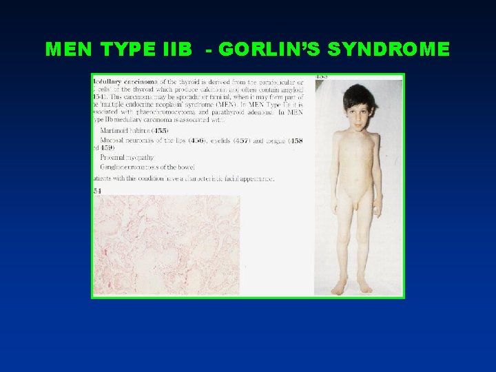
MEN TYPE IIB - GORLIN’S SYNDROME
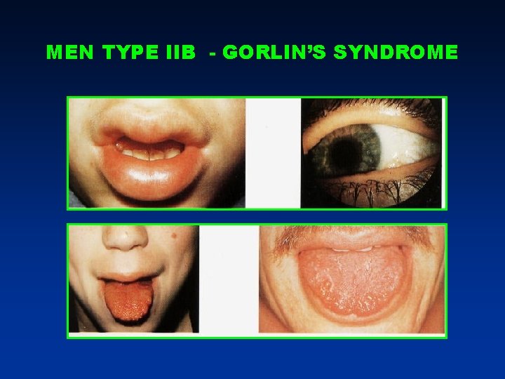
MEN TYPE IIB - GORLIN’S SYNDROME
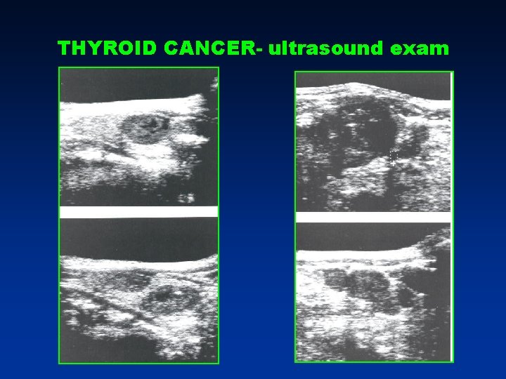
THYROID CANCER- ultrasound exam
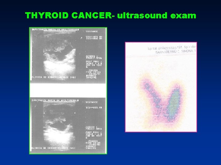
THYROID CANCER- ultrasound exam
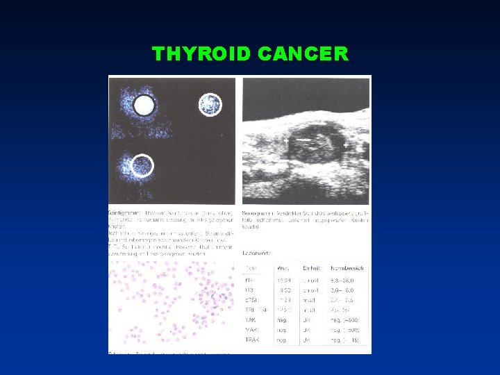
THYROID CANCER
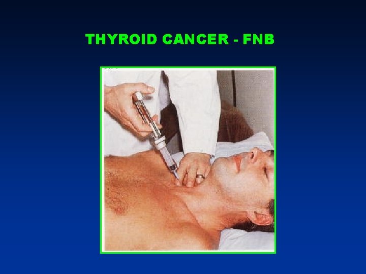
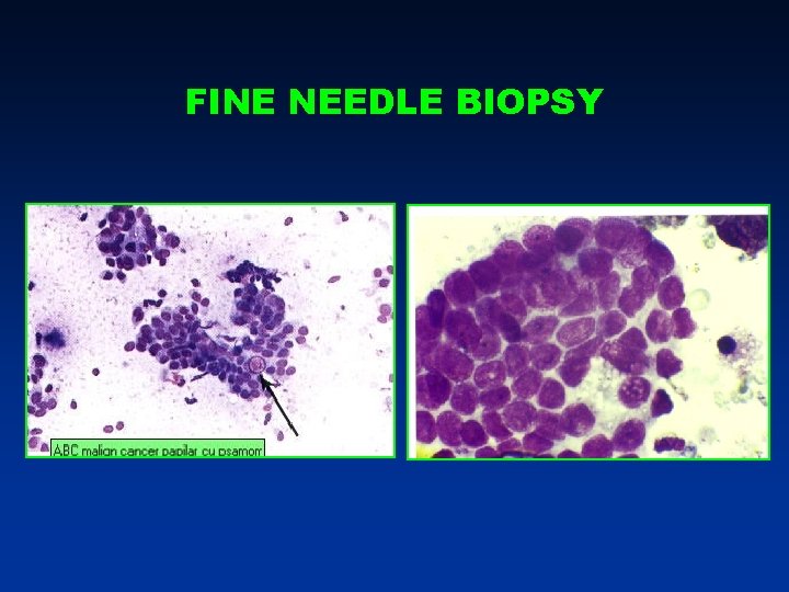
THYROID CANCER - FNB
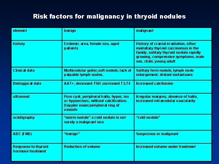
Risk factors for malignancy in thryoid nodules element benign malignant history Endemic area, female sex, aged patients History of cranial irradiation, other medullary thyroid carcinomas in the family, solitary thyroid nodule rapidly growing, compressive symptoms, male sex, child, young adult Clinical data Multinodular goiter, soft nodule, lack of palpable lymph nodes, Solitary ferm nodule, lymph node enlargement, distant metastases Biologgical data AAT+, deceased TSH , increased T 3, T 4 Increased calcitonine ultrasoud Pure cyst, peripheral hallo, hyper, iso or hypoechoic, without calcification. Doppler exam: peripheral ring of vassels Irregular margins, absence of hallo, increased intranodular vascularity scintigraphy “worm nodule” a cold nodule is not surely a malignant one “cold nodule” ABC (FNB) “benign” Suspicious or malignant Response to thyroid hormone treatment Reduction of volume Increased volume under treatment
FINE NEEDLE BIOPSY
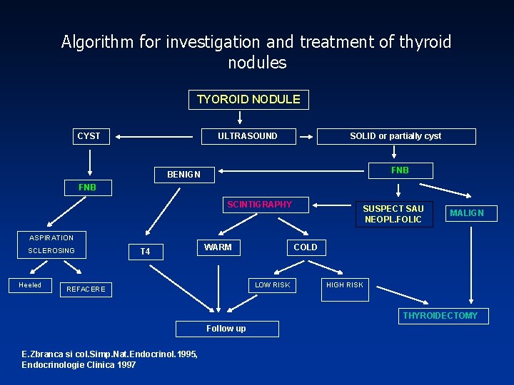
Algorithm for investigation and treatment of thyroid nodules TYOROID NODULE CYST ULTRASOUND SOLID or partially cyst FNB BENIGN FNB SCINTIGRAPHY SUSPECT SAU NEOPL. FOLIC MALIGN ASPIRATION SCLEROSING Heeled T 4 WARM COLD LOW RISK REFACERE HIGH RISK THYROIDECTOMY Follow up E. Zbranca si col. Simp. Nat. Endocrinol. 1995, Endocrinologie Clinica 1997
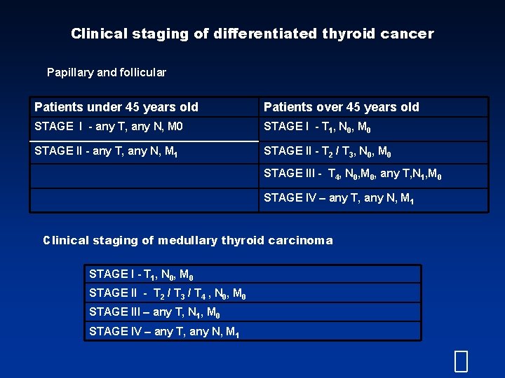
Clinical staging of differentiated thyroid cancer Papillary and follicular Patients under 45 years old Patients over 45 years old STAGE I - any T, any N, M 0 STAGE I - T 1, N 0, M 0 STAGE II - any T, any N, M 1 STAGE II - T 2 / T 3, N 0, M 0 STAGE III - T 4, N 0, M 0, any T, N 1, M 0 STAGE IV – any T, any N, M 1 Clinical staging of medullary thyroid carcinoma STAGE I - T 1, N 0, M 0 STAGE II - T 2 / T 3 / T 4 , N 0, M 0 STAGE III – any T, N 1, M 0 STAGE IV – any T, any N, M 1
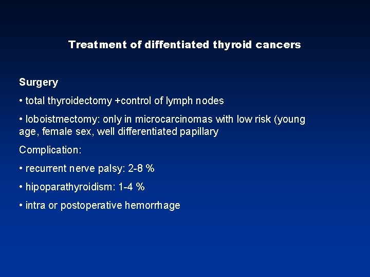
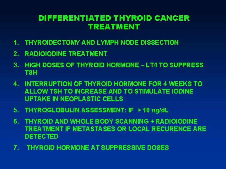
Treatment of diffentiated thyroid cancers Surgery • total thyroidectomy +control of lymph nodes • loboistmectomy: only in microcarcinomas with low risk (young age, female sex, well differentiated papillary Complication: • recurrent nerve palsy: 2 -8 % • hipoparathyroidism: 1 -4 % • intra or postoperative hemorrhage
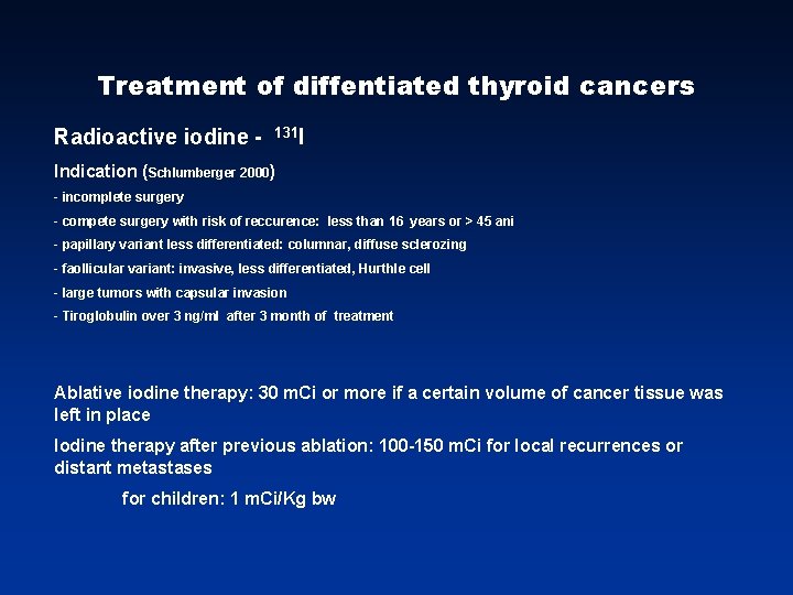
Treatment of diffentiated thyroid cancers Radioactive iodine - 131 I Indication (Schlumberger 2000) - incomplete surgery - compete surgery with risk of reccurence: less than 16 years or > 45 ani - papillary variant less differentiated: columnar, diffuse sclerozing - faollicular variant: invasive, less differentiated, Hurthle cell - large tumors with capsular invasion - Tiroglobulin over 3 ng/ml after 3 month of treatment Ablative iodine therapy: 30 m. Ci or more if a certain volume of cancer tissue was left in place Iodine therapy after previous ablation: 100 -150 m. Ci for local recurrences or distant metastases for children: 1 m. Ci/Kg bw
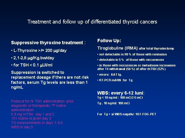
Treatment and follow up of differentiated thyroid cancers Suppressive thyroxine treatment : Follow Up: • L-Thyroxine >/= 200 g/day Tiroglobuline (IRMA) after total thyrodectomy • 2, 1 -2, 8 g/Kg. bw/day • for TSH < 0. 1 UI/ml Suppression is switched to replacement dosage if there are not risk factors, serum Tg levels are less than 1 ng/m. L • not detectable in 98 % of those with remission • detectable in 5 % of those with reccurences • in those with reccurences or metastases increaeses after T 4 withdrawal (56 %) of after rh. TSH (52%) • errors: AAT tg. • RT-PCR m. ARN for Tg WBS: every 6 -12 luni: Protocol for hr TSH adminstration prior diagnostic or therapeutic 131 Iodine administration 0. 9 mg hr. TSH day 1 and 2 131 Iodine id given day 3 TG measurements in days 1 -3 -5 WBS in day 5 Tg < 10 ng/ml : 100 m. Ci 2 -5 m. Ci Tg. 10 ng/ml: 100 m. Ci For Tg + si WBS negativ: 18 F-FDG-PET
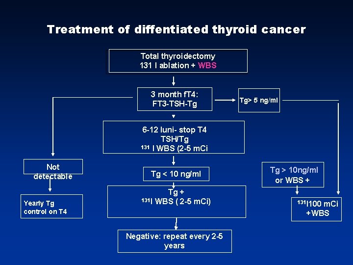
Treatment of diffentiated thyroid cancer Total thyroidectomy 131 I ablation + WBS 3 month f. T 4: FT 3 -TSH-Tg Tg> 5 ng/ml 6 -12 luni- stop T 4 TSH/Tg 131 I WBS (2 -5 m. Ci Not detectable Yearly Tg control on T 4 Tg < 10 ng/ml 131 I Tg + WBS ( 2 -5 m. Ci) Negative: repeat every 2 -5 years Tg > 10 ng/ml or WBS + 131 I 100 m. Ci +WBS
DIFFERENTIATED THYROID CANCER TREATMENT 1. THYROIDECTOMY AND LYMPH NODE DISSECTION 2. RADIOIODINE TREATMENT 3. HIGH DOSES OF THYROID HORMONE – LT 4 TO SUPPRESS TSH 4. INTERRUPTION OF THYROID HORMONE FOR 4 WEEKS TO ALLOW TSH TO INCREASE AND TO STIMULATE IODINE UPTAKE IN NEOPLASTIC CELLS 5. THYROGLOBULIN ASSESSMENT: IF > 10 ng/d. L 6. THYROID AND WHOLE BODY SCANNING + RADIOIODINE TREATMENT IF METASTASES OR LOCAL RECURENCE ARE DETECTED 7. THYROID HORMONE AT SUPPRESSIVE DOSES
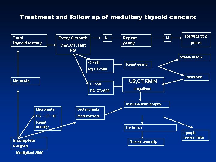
Treatment and follow up of medullary thyroid cancers Total thyroidecotmy Every 6 month N CEA, CT, Test PG Repeat yearly N Repeat at 2 years Stable. follow CT<50 Repat yearly Pg-CT<500 No meta CT>50 PG-CT>500 US, CT, RMIN increased negatives Immunoscintigraphy Micrometa Distant meta PG – CT =N Medical treat. Repat anually Incomplete surgery Modigliani 2000 No tumor Lymph nodes meta Repeat annually
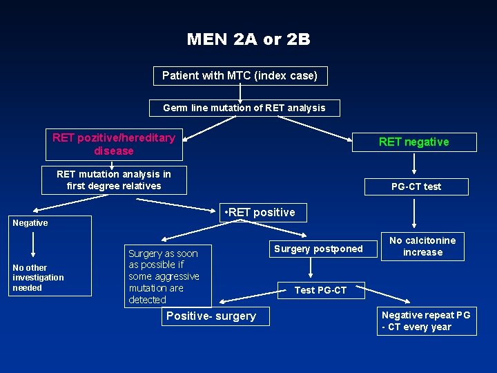
MEN 2 A or 2 B Patient with MTC (index case) Germ line mutation of RET analysis RET pozitive/hereditary disease RET negative RET mutation analysis in first degree relatives • RET positive Negative No other investigation needed PG-CT test Surgery as soon as possible if some aggressive mutation are detected Positive- surgery Surgery postponed No calcitonine increase Test PG-CT Negative repeat PG - CT every year
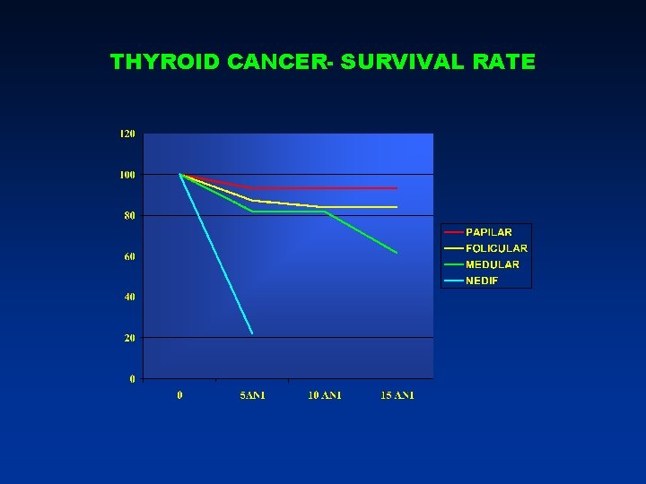
THYROID CANCER- SURVIVAL RATE