BREAST CANCER The Breast n n n A

BREAST CANCER

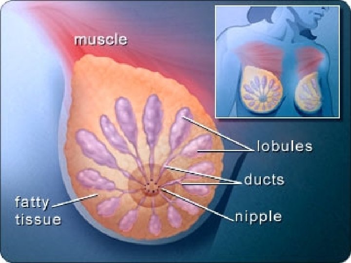
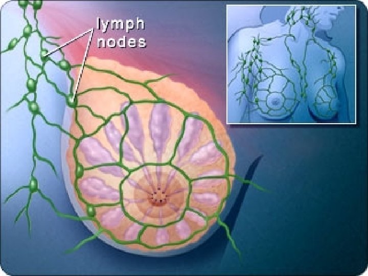
The Breast n n n A ducts B lobules C dilated section of duct to hold milk D nipple E fat F pectoralis major muscle G chest wall/rib cage Enlargement: A normal duct cells B basement membrane C lumen (center of duct)

Breast Carcinoma Incidence Ø Ø Ø Ø 20% of all cancers in women Commonest cause of death - 35 -55 y In UK 1 in 10 -12 chances 1 in 8 women in US Less incidence in Asia Majority of cancers arise in the ducts. Very rare before age 25

Risk Factors: Female sex. . !, Age, Obesity, high fat diet Ø Maternal relative with breast cancer. Ø Longer reproductive span. Ø Nulliparity, Oral contraceptives Ø Later age at first pregnancy. Ø Atypical epithelial hyperplasia. Ø Previous breast cancer/Endometrial Ca. Ø Geographic factors - country Ø BRCA 1 and BRCA 2 genes Ø

Breast Cancer Risk Factors that cannot be changed Age Family/Personal History Race Treatment with DES GENDER - All women are at risk Reproductive History Menstrual History Radiation Genetic Factors

Breast Cancer Risk Factors that can be controlled Obesity All women are at risk Exercise Breastfeeding Alcohol Hormone Replacement Therapy Not having children Birth Control Pills
 n n n Epithelial (mammary tissue) n Non invasive n")
Pathology ( WHO classification) n n n Epithelial (mammary tissue) n Non invasive n DCIS n LCIS n Invasive n Ductal 85 % n Lobular 9 % n Mucinous 5 % n Papillary < 5 % n Medullary < 5 % Mixed Ct & epithelial Miscellaneous n Paget’s disease n IBC
 n Neoplasm of mammary tissue proper n n Neoplasm of")
Pathology (Foot& Stewart classification) n Neoplasm of mammary tissue proper n n Neoplasm of lobular epithelium 9 - 10 % n LCIS 50 % n Lobular carcinoma invasive 50 % Neoplasm of ductal epithelium 85 % n DCIS n Ductal carcinoma Invasive ( IDC) n NOS ( simple type) n Special types ( scirrhous, medullary, mucinous, papillary, cribriform, comedo, tubular, secretory with metaplasia) n Unusual presentations n n Paget’s disease IBC
 n n Malignant mesenchymal neoplasm n Sarcoma n Lymphomas n")
Pathology (Foot& Stewart classification) n n Malignant mesenchymal neoplasm n Sarcoma n Lymphomas n Myeloid leukemia Miscellaneous malignancies n Skin n SCC n BCC n Skin adenxa ( carcinoma of sweat glands or sebaceous glands) Undifferentiated carcinoma Metastatic n Female ( other breast, lung, MM) n Male (prostate)

Carcinoma in situ It is a spectrum of pre invasive neoplastic changes in the breast includes; n DCIS 4 % symptomatic 25 % screen detected n LCIS <1 % symptomatic 1% screen detected n Hyper plastic appearance ( ductal or lobular)

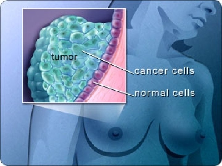
Ductal Carcinoma in Situ n n n It is the group of neoplasm arising from ductal epithelium & confined by basement membrane Ducts expanded by large irregular cells with lage irregular nuclei Malignant cells are confined by basement membrane
 n Comedo DCIS n n n High grade cytology")
Ductal Carcinoma in Situ (classification) n Comedo DCIS n n n High grade cytology Extensive necrosis Branched calcification Non Comedo DCIS • Low grade cytology • Lack necrosis • Lack calcification • Cribribriform • Solid • micropapillary Intermediate histology

Ductal Carcinoma in Situ n Clinical presentation Asymptomatic > 50 % in screening programs as abnormal mamographic finding n Nipple discharge n Paget’s disease n n n Risk of invasive BC is 40 % over 30 y Multicentricity in 50 %
 n n n Sterotactic CNB U/S guided CNB Wire")
Ductal Carcinoma in Situ (Diagnosis) n n n Sterotactic CNB U/S guided CNB Wire or ink guided excisional biopsy which is a must if; n n n Atypical ductal hyperplasia Radial scar Non specific diagnosis Lack correlation with mammogram Wedge biopsy if paget’s
 Depend on Van Nuys Prognostic Index which classify patients")
Ductal Carcinoma in Situ (Treatment) Depend on Van Nuys Prognostic Index which classify patients into 3 groups Depending on 3 factors 1 - Tumor size 2 - Histological grade 3 - Surgical free margin Low risk Wide local excision (BCS) Intermediate risk BCS & irradiation High risk Mastectomy SSM

Lobular Carcinoma In Situ n n n It constitute 25 % of CIS The risk of invasive cancer is 20 – 30 % life time and bilateral It is multicentric in 80 % Never palpable mass Treatment n Follow up by n n C/E every 4 months Mammography yearly Chemoprevention by Tamoxafen or raloxifene Mastectomy which is rarely used
")
Non Invasive (Carcinoma in Situ)

Invasive Breast Cancer n Epithelial Invasive BC Ductal 85 % n Lobular 9 % n Mucinous 5 % n Papillary < 5 % n Medullary < 5 % Mixed Ct & epithelial Miscellaneous n Paget’s disease n IBC n n n

n n n n Duct Carcinoma: small 5 Infiltrating % post menopausal(Atrophic with scirrhous ) shriveled breast NEA n Small size n Irregular in shape n Very hard in consistency MP n ++++ FT n + islads of malignant spheroidal cells n Infrequant mititic figures Very slowly progress 10 Y Very late metastases Best prognosis hard
 n n n n 75 % Middle aged 40")
Infiltrating Duct Carcinoma: Fibrosis (Scirrhous) n n n n 75 % Middle aged 40 – 60 Y NEA n Small size n Irregular in shape n hard in consistency MP n +++ FT n ++ scanty as finger like processes slowly progress late metastases Good prognosis

n n n Well developed breast of young woman NEA n Largr fleshy in size n Brain like cut section in shape with hge & necrosis n Soft in consistency MP n ++ delicate FT n ++ + highly malignant cells Rapidly progress Moderate metastases Good prognosis Medullary Carcinoma: Large soft n n n Rapid increase lead to early presentation Fungate more than infilttrate LN affection dt large cell size

Mucoid or Colloid Carcinoma n n n It form a bulky mass with mucoid degeneration & necrosis It grow slowly & disseminate late & may reach huge sizes so have good prognosis after surgery Signet ring shaped cells dt mucoid materials

Lobular Carcinoma n n n It constitute 9 % Arise in the terminal lobules It could take different presentation as ductal carcinoma

Paget’s Disease n n n It is a chronic eczematoid malignant eruption of the nipple 1 % in middle aged and old woman Etiology n Old theory ( skin tumor with secondary breast mass n New theory ( tumor in terminal ducts as in situ cancer then spread n Outward to nipple and skin n Inward breast mass

Paget’s Disease n n n Hyper plastic changes in all layers of epidermis (epidermal hypertrophy) Characteristic paget’s cells n Large vaculated cells n Deeply stained eccentric nucleus Subdermal round cell infiltration
 n n Persistent eczema like condition that affect old")
Paget’s Disease ( Clinical picture) n n Persistent eczema like condition that affect old female 50 Y which does not respond to topical treatment Unilateral erosion of the nipple which is red, thick, scaly & crusted without vesicles or itching Serosangious discharge Mass in the breast in 2 Years
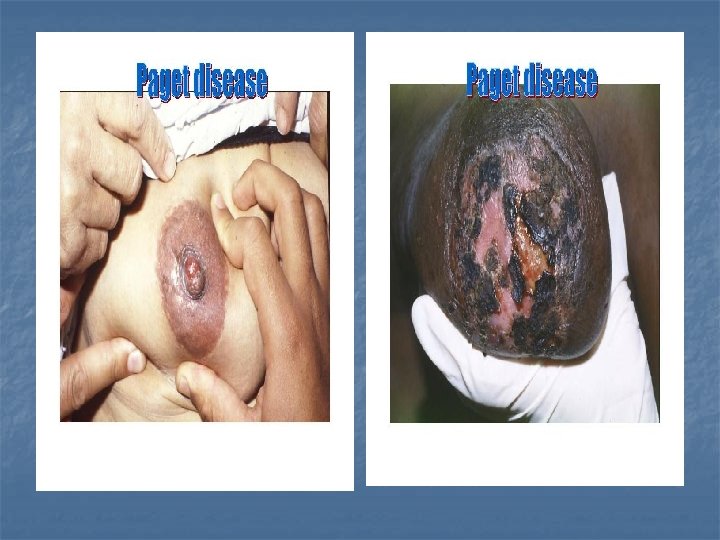
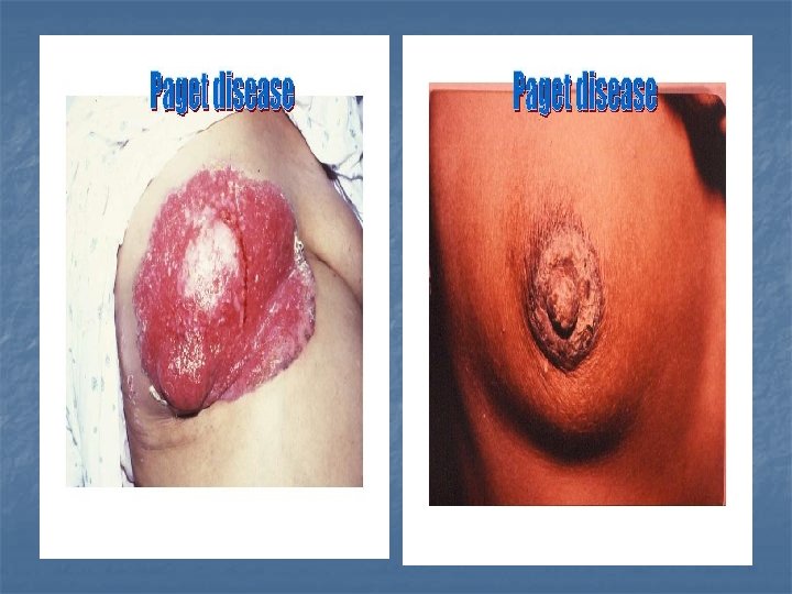

Paget’s n n n Menopause Unilateral No vesicles or itching Sub areolar mass after 2 years Not respond to topical treatment Biopsy paget cells Eczema n n n Lactation Bilateral Vesicles and itching No mass Respond to topical treatment No paget cells

Paget’s Disease Diagnosis n Mammography is a must n n Detect sub clinical mass Detect micro calcification Detect multi centricity Biopsy ( full thickness nipple biopsy) is diagnostic where there are 3 different types n n n Paget’s disease with DCIS ( high grade comedo) Paget’s disease with invasive cancer ( commonest) Paget’s disease confined to epidermis of nipple & areola ( rarest)
 n n The standard treatment is mastectomy Recently BCS is")
Paget’s Disease ( Treatment) n n The standard treatment is mastectomy Recently BCS is used with segmentectomy of nipple & areola & radiotherapy Paget’s disease with mass or with no mass invasive cancer Or with DCIS Segmentectomy Of N & A -Ve margins -No multicentric Radiotherapy Segmentectomy Of N & Axillary dissection + Ve margins multicentric Mastectomy
 Use of chemotherapy based on 5 prognostic indication of chemotherapy")
Paget’s Disease ( Treatment) Use of chemotherapy based on 5 prognostic indication of chemotherapy 1. 2. 3. 4. 5. Age < 35 year Tumor > 1 cm Tumor high grade + ve LN - ve ER

n n n pregnancy and lactation should be DD of abscess NEA n Diffuse swollen, hot on palpation , with dilated vein n Soft in consistency MP n + very little FT n ++ + + highly malignant anaplastic cells Rapidly progress Very early metastases Bad prognosis IBC( Inflammatory breast cancer

IBC( Inflammatory breast cancer n It is very similar to acute breast abscess with the following differences It is a diffuse lesion n No pyrexia n LN not tender n Progressive in nature n No lecucytosis n No respond to antibiotic n

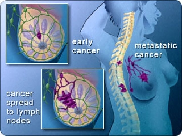
Spread of Breast Carcinoma: n n Methods of spread n Direct n Lymphatic n Blood n Trans- celomic Theories of spread n Loco-regional theory n Systemic theory

TNM Staging Tumor n Tx primary tumor can not be assessed n n n Tis In situ carcinoma & paget’s disease T 0 no palpable mass T 1 tumor < or = 2 cm n T 1 a < or = 0. 5 cm no deep fixation n T 2 b 0. 5 – 1 cm + deep fixation n T 3 c 1 – 2 cm + deep fixation T 2 tumor 2 – 5 cm n T 2 a no deep fixation n T 2 b deep fixation T 3 tumor 5 – 10 cm n T 3 a no deep fixation n T 3 b deep fixation T 4 tumor of any size n T 4 a direct chest extension n T 4 b skin ( Peau d’orange, skin nodule & ulceration) n T 4 c T 4 a + T 4 b n T 4 d inflammatory breast cnacer

Nodes n n n TNM Staging N x can not be assessed N 0 not palpable LN N 1 palpable homo-lateral axillary LN and mobile N 2 palpable homo-lateral axillary LN and fixed N 3 ipsilateral internal mammary LN Metastases n n n M X can not be assessed M 0 no known metastases M 1 distant metastases including supra-clavicular LN

TNM staging T 0 N 0 T 1 T 2 Stage I T 1 N 0 M 0 N 1 N 2 N 3 Stage II a T 1 N 1, T 2 N 0, T 0 N 1 Stage II b T 2 N 1, T 3 N 0 T 3 T 4

TNM staging T 0 T 1 T 2 T 3 N 0 N 1 N 2 N 3 Stage III a any N 2 any T 3 except T 3 N 0 Stage III b any N 3 any T 4
 7 -year Surv (%) I Tumor 2")
St ag Definition 5 -year Surv (%) 7 -year Surv (%) I Tumor 2 cm or less without spread 96 92 II Tumor 2 -5 cm with regional lymph node involvement but without distant metastases, OR > 5 cm in diameter without spread 81 71 52 39 18 11 Any size with skin/chest wall fixation, & axillary or internal mammary III nodal involvement, without distant metastases Tumor of any size with or without IV regional spread but with evidence of distant metastases
 n Mobile tumor n Free axilla")
Manchester classification n n Stage I ( 85%) n Mobile tumor n Free axilla n Paget’s Stage II ( 66 %) n Mobile tumor n Mobile axillary LN Stage III ( 41 %) n Tumor fixed n LN fixed Stage IV ( 10%) n Wide dissemination n suprac; lavicular LN

Prognosis n Clinical factors n n n n Pathological factors n n Age Sex Site Stage Grade Pregnancy Tumor type Grade Axillary LN Biological factors n n Receptors ER, Pg R Tumor markers DNA ploidy S phase fraction
 n Axillary LN involvement n n n 1 no node")
Nottingham Prognostic Index (NPI) n Axillary LN involvement n n n 1 no node 2 1 -3 node 3 4 or more node Grade (1, 2, 3) Tumor size in cm x 0. 2 Prognostic group NPI 10 Y survival Excellent < or = 2. 4 94 Good < or = 3. 4 83 Moderate I < or = 4. 4 70 Moderate II < or = 5. 4 31 Poor > 5. 4 20

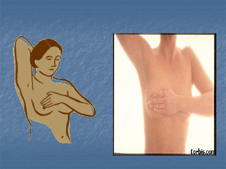
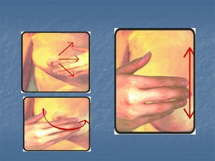
Breast self examination for early detection

 n Main symptoms n n Lump Discharge ( blood stained) Pain")
Clinical Features: (symptoms) n Main symptoms n n Lump Discharge ( blood stained) Pain ( late) Symptoms of spread n n n Direct ( skin, nipple, Areola) Lymphatic LN Blood n n Lung ( respiratory distress & hemoptsis) Bone ( aches & patholgical fracture) Malignant ascites Met static nodules any where
 1. Breast a whole n n n Examination while sitting (")
Clinical Features: (signs) 1. Breast a whole n n n Examination while sitting ( puckered or displaced Raising the arms above the head (pulled upward) Patient leaning forward ( not protrude freely)
 2. Nipple changes n Recent retraction n n dt neoplastic fibrosis")
Clinical Features: (signs) 2. Nipple changes n Recent retraction n n dt neoplastic fibrosis & lactiferous ducts invasion Should be DD from n n Congenital Chronic inflammation Nipple erosion (should be DD of eczema) Discharge which could be serous or bloody
 3. Skin manifestations 1. 2. 3. Peau d’ orange dt obstruction")
Clinical Features: (signs) 3. Skin manifestations 1. 2. 3. Peau d’ orange dt obstruction of skin lymphatic Cancerous nodule or satellites Ulceration or fungation dt skin invasion
")
Clinical Features: (signs)
")
Clinical Features: (signs)
")
Clinical Features: (signs)
 4. 5. 6. 7. 8. 9. Dimpling and puckering dt pull")
Clinical Features: (signs) 4. 5. 6. 7. 8. 9. Dimpling and puckering dt pull on cooper ligaments Dilated veins Skin lymphoedema Tumor fixation to the skin Inflammatory signs as in IBC Nipple and areola changes
 10. Cancer en cuirasse 1. 2. 3. 4. 5. Atrophic breast")
Clinical Features: (signs) 10. Cancer en cuirasse 1. 2. 3. 4. 5. Atrophic breast Hard Pigmented Fixed to chest wall Studded with nodules
 4. Breast lump n n n 5. Mostly in UOQ in")
Clinical Features: (signs) 4. Breast lump n n n 5. Mostly in UOQ in 60 % Irregular in shape Hard in consistancy Ill deined borders Fixed within the breast my be fixed to skin or chest wall Opposite breast examined first before the diseased one to exclude metastases
 6 - lymph nodes should be examined Central and apical groups")
Clinical Features: (signs) 6 - lymph nodes should be examined Central and apical groups Pectoral or anterior group Lateral or brachial groups
 Posterior or subscapular group Supraclavicular group")
Clinical Features: (signs) Posterior or subscapular group Supraclavicular group
 7 - general examination Chest effusion, deposites , mediastinal LN n")
Clinical Features: (signs) 7 - general examination Chest effusion, deposites , mediastinal LN n Abdomen ascites, hepatomegally n Pelvis by PR and PV n Krukenberg n Plummer shelf n n Bones tenderness , weakness, deformity and fractures

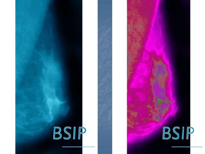
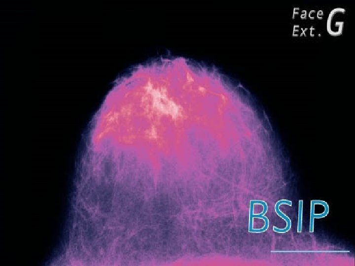
Diagnosis: n Laboratory n n n General Liver function Kidney function Cytological examination of nipple discharge Tumor markers Radiological n n Plain x ray Breast imaging n n n n Mammography Thermo graphy Galactography Ultrasound CT MRI Light spectroscopy Radioactive isotope scanning of LN

Diagnosis: n Biopsy Fine Needle Aspiration Biopsy n Core Biopsy n Excision Biopsy n Frozen section n Drill biopsy n Sentinal node biopsy n n n Immunoperoxidase, Molecular techniques – Gene detection.

History of Mammography n n Used in clinical practice since 1927 in diagnosis of breast abnormalities. In the 50’s and 60’s it was developed to the point that benign and malignant tumors could be differentiated. 1963 -1967 screening program for the detection of breast cancer conducted by the Health Insurance Plan of New York (60, 000 women screened). 1973 Breast Cancer Detection Demonstration Project (B. C. D. D. P. ) – 15 annual screenings of 270, 000 women.

Low Dose X-rays n n n Electrons originating at the cathode are accelerated towards the rotating anode. Upon contact the kinetic energy of the electron is converted into x-rays and heat (0. 5% x-rays) Collimator system, composed of lead for complete absorption, focuses the x-ray beam

X-ray/ Breast Interaction n n As with most x-ray images greater contrast occurs when there is a large difference in attenuation between tissues. The breast is compressed and the x-ray beam is applied. Contrast is best seen between fatty tissue and functional glandular tissue, but contrast is poor between glandular tissue and cancerous tissues. Thus, in older women, post-menopause, the reduction in functional glandular tissue provides for a distinct contrast between cancerous masses and fatty tissues.

Two Types of Mammograms n n n A screening mammogram is an x-ray examination of the breast in a woman who has no breast complaints (asymptomatic). The goal of screening mammography is to find cancer when it is still too small to be felt by her doctor or the woman. A screening mammogram usually takes 2 x-ray pictures (views) of each breast. A diagnostic mammogram is an x-ray examination of the breast in a woman who either has a breast complaint (for example, a breast mass, nipple discharge, etc. ) or has had an abnormality found during a screening mammogram. During a diagnostic mammogram, more pictures will be taken to carefully study the breast condition.

Two Methods of Mammograms n n Ordinary film Xero or zeno mammography n over selinium plates gave different colors blue and white

Mammogram Equipment n n A mammography unit is a rectangular box that houses a tube in which xrays are produced. Attached to the unit is a device that holds and compresses the breast and positions it so images can be obtained at different angles. Modern technique uses a special machine exclusively for breast x-rays to produce studies that are high quality but have a low radiation dose (usually about 0. 1 to 0. 2 rad dose per picture).

Mammogram Equipment Cont. n n A mammogram device has special accessories that allow only the breast to be exposed to the x-rays do not penetrate tissue as easily as the xray used for routine chest films or x-rays of the arms or legs.
 2) 3) 4) 5) The breast is first placed")
Mammogram Procedure n n 1) 2) 3) 4) 5) The breast is first placed on a platform and squeezed between 2 plates Breast compression is necessary to: even out the breast thickness so all tissue can be visualized spread out tissue so small abnormalities won't be obscured by overlying breast tissue allow the use of a lower x-ray dose since a thinner amount of breast tissue is being imaged hold the breast still to eliminate blurring of image caused by motion reduce x-ray scatter to increase sharpness of picture.

Indications of Mammography 4 - Evaluation of contralateral breast 1 - Breast with mass 2 - Breast with discharge 3 - Follow up of breast lesions 5 - Screening of BC Follow up is needed in the following 6 - breast that is difficult to be examined Premalignant lesions, papillomatoso cystic lesions , atypia, lobular neoplasia 7 – work up of met static Aden carcinoma Patient at high risk of cancer breast Patients with previous BC

n n n Reading the Mammogram Best if read by radiologist specializing in mammography Important to recognize even the smallest abnormalities Multiple films and angles are often necessary Sometimes two physicians will read the same film for the most thorough assessment Computer based digital mammography is used to get maximum information from each mammogram taken Comparison with older films is also extremely useful
 Average-size lump found")
Mammography Average-size lump found by woman practicing occasional breast self-exam (BSE) Average-size lump found by woman practicing regular breast self-exam (BSE) Average-size lump found by first mammogram Average-size lump found by getting regular mammograms

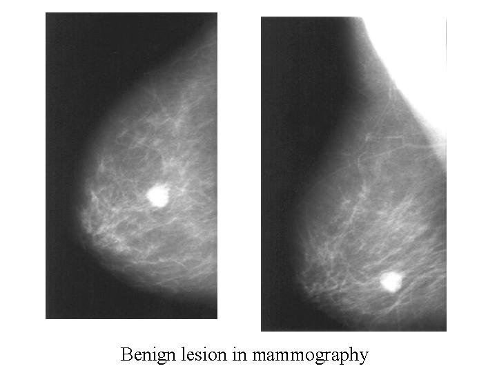
Abnormal Mammographic findings Micro calcifications Circumscribed lesion Speculated lesion Satellite lesion Rounded Linear branching punctuate Mammographic signs of malignancy 1. Breast lump 2. Linear or branching micrcalcification 3. Skin or nipple thickening 4. Mammary duct distortion or asymmetry


Ultrasound n n n It is the intial investigation in a woman < 35 yeaers DD solid and cystic lesions Positive predictive value is 92 % with palpable mass

Sentinel Node Biopsy n n An evolving technique to identify node status without formal axillary dissection A radioactive tracer and/or blue dye is identified in the first draining node Potentially gives accurate staging with decreased morbidity Sensitivity exceeds 90% and accuracy exceeds 95% for experienced surgeons
 Treatment of advanced")
Breast Cancer Treatment of early BC ( stage I& II a) Treatment of advanced BC • (stage II b, III& IV) • Metastatic disease • Local recuurence Neoadjuvant chemotherapy Surgery& observation Surgery& Adjuvant therapy Surgery either Mastectomy or BCS + or - Radiotherapy + or - Chemotherapy

Treatment of early BC n Surgery & Observation n Indication T 1 N 0 n ER + ve n Patient under willing close observation n n Surgery MRM n MRM + breast reconstruction n n Observation Monthly C/ E n Chest x ray, U/S abdomen every 6 months n

Treatment of early BC n Surgery & Adjuvant therapy n Why use of adjuvant therapy n n Decrease local recurrence ( Radiotherapy) Decrease distant metastases as Radiotherapy) micro metastases are present in 50 % of cases at diagnosis (chemotherapy) Good response to adjuvant therapy Types of adjuvant therapy n n n Radiotherapy Chemotherapy Hormonal treatment
 n Old operation that lost popularity (Radical Mastectomy) n Remove")
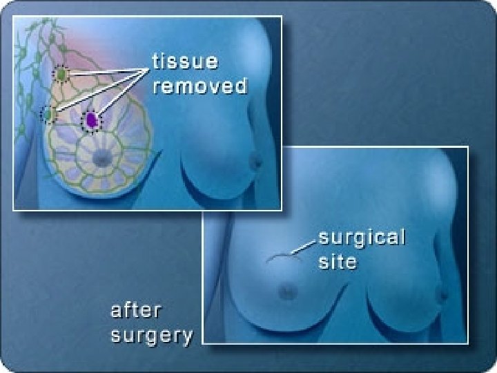
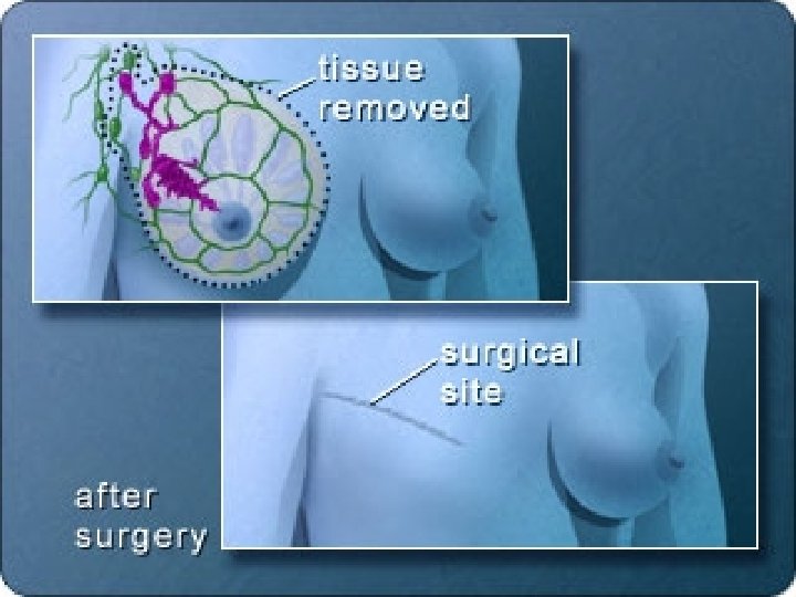
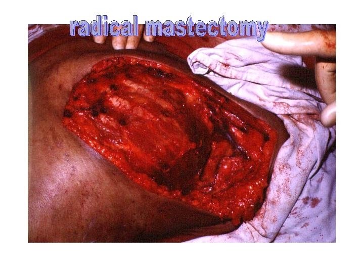
Breast Cancer Treatment (Surgery) n Old operation that lost popularity (Radical Mastectomy) n Remove the whole breast, P Major & minor, axillary LN and wide margin of skin & soft tissue n Its rationale is loco regional theory of spread n Obsolete operations n Extended Radical Mastectomy ( RM + internal mammary LN removal) Used with medial lesions, +ve Axillary Ln & M 0 n Supra Radical Mastectomy ( RM + clavicle excision and supaclavicular LN removal) Operations that recently gained popularity n Modified Radical Mastectomy 70 % in USA n Simple mastectomy (Total Mastectomy) 70 % in UK Breast Conservative procedures n Lumpectomy n Partial Mastectomy (Quadrantectomy) n Segmental mastectomy n Tylectomy n QUART (Quadrantectomy +Axillary clearance + RT) n n
 1. 2. 3. 4. 5. Indications for Use: Tumor size 1.")
Conservation Therapy (BCT) 1. 2. 3. 4. 5. Indications for Use: Tumor size 1. 2 cm in small breast 2. 4 cm in large breast Tumor location favorable for good aesthetic result (peripheral location) Unifocal single tumor with negative margins Patient’s preference for breast conservation Patient’s inability to tolerate general anesthesia Advantages of BCS • Better cosmetics • Not affect survival • Not affect local recuurence which if occur not in the chest wall and MRM could be done

Contraindications to Conservation 1. 2. 3. 4. 5. 6. Tumor size > 5 cm Tumor multi centric (Two or more primary tumors in separate quadrants) Diffuse tumors ( Diffuse malignant appearing micro calcifications) High grade tumors Distant metastases Any contraindication to irradiation n Previous breast irradiation n Pregnancy (unless radiation is provided after delivery) n Collagen vascular disease (relative contraindication) n Large breast size

dissection Aim of axillary surgery n Provides staging information n Provides local control if node positive n Provide prognostic information n No reliable imaging technique Complications n Wound infection n Arm lymphoedema n Arm morbidity Standard Axillary Dissection
 Surgical Treatment Options n n n Procedure is still")
Sentinel Lymph Node Biopsy (SLNB) Surgical Treatment Options n n n Procedure is still under investigation to determine if patients’ survival will not be affected if lymph nodes that may have cancer in them are left behind and untreated Not the standard of care for breast cancer at this point Success rate of about 92 %

Indications of MRM n n n Tumor size > 5 cm Tumor multi centric (Two or more primary tumors in separate quadrants) Diffuse tumors ( Diffuse malignant appearing micro calcifications) High grade tumors Distant metastases Any contraindication to irradiation n Previous breast irradiation n Pregnancy (unless radiation is provided after delivery) n Collagen vascular disease (relative contraindication) n Large breast size
 Depend on Van Nuys Prognostic Index which classify patients")
Ductal Carcinoma in Situ (Treatment) Depend on Van Nuys Prognostic Index which classify patients into 3 groups Depending on 3 factors 1 - Tumor size 2 - Histological grade 3 - Surgical free margin Low risk Wide local excision (BCS) Intermediate risk BCS & irradiation High risk Mastectomy SSM

Lobular Carcinoma In Situ n n n It constitute 25 % of CIS The risk of invasive cancer is 20 – 30 % life time and bilateral It is multicentric in 80 % Never palpable mass Treatment n Follow up by n n C/E every 4 months Mammography yearly Chemoprevention by Tamoxafen or raloxifene Mastectomy which is rarely used
 n n The standard treatment is mastectomy Recently BCS is")
Paget’s Disease ( Treatment) n n The standard treatment is mastectomy Recently BCS is used with segmentectomy of nipple & areola & radiotherapy Paget’s disease with mass or with no mass invasive cancer Or with DCIS Segmentectomy Of N & A -Ve margins -No multicentric Radiotherapy Segmentectomy Of N & Axillary dissection + Ve margins multicentric Mastectomy
 Use of chemotherapy based on 5 prognostic indication of chemotherapy")
Paget’s Disease ( Treatment) Use of chemotherapy based on 5 prognostic indication of chemotherapy 1. 2. 3. 4. 5. Age < 35 year Tumor > 1 cm Tumor high grade + ve LN - ve ER
 Breast Cancer")
Post-Treatment Follow-up of the Patient with Early Stage (I and II) Breast Cancer
 and Axillary Lymph Node")
Infiltrating Cancer Surgical treatment Options Breast Conservation (followed by RT) and Axillary Lymph Node Dissection n n Modified Radical Mastectomy (with/without reconstruction)

Long Term Side Effects of Surgery for Breast Cancer n n n Loss of part or of the whole the breastchange of self image and sexuality Nerve Function Deficits/Neuropathy Lymphedema Motor (Muscle) Function Deficits Pain

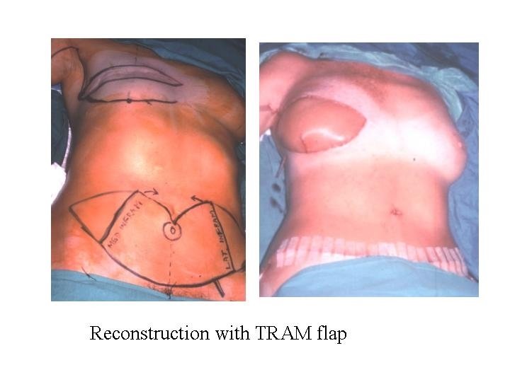
Breast Reconstruction n n Indicated in women undergoing mastectomy who desire reconstruction Radiation after reconstruction may produce less desirable results Autogenous tissue vs. prosthetic vs. combination Immediate vs. delayed- no survival difference

Prosthetic Silicon implants

Latissmus Dorsi Mycutaneus flap

TRAM Flap

TRAM Flap

n n n Most women with breast cancer may be treated with breast conservation if they so desire Most women requiring/choosing mastectomy may undergo immediate breast reconstruction Optimal treatment involves multimodality therapy provided by multidisciplinary teams

Radiotherapy n n Aim to destruction of local micro metastases to decrease local recurrence Indications Radiotherapy to breast area After all BCS Radiotherapy to Axilla After mastectomy 1. 4 or more + ve LN 2. Extracapsular invasion 3. + ve or close margin T 3 , T 4 & pectoral fascia affection All ABC Used only if 1. 4 or more + ve Axillary LN 2. Extra capsular invasion

Radiotherapy n When ? n n Dose n n 1. 2. 3. 4. 2 - 3 weeks after mastectomy 40 – 50 Gy delivered at 15 – 25 fraction Complications T 1 N 0 it decrease 5 y survival Lymphatic destruction Increase cancer in contralateral breast Local complications n n Skin burn Arm lymph-oedema Interfere with breast reconstruction Increase interstitial pulmonary fibrosis
 First line n Mechanism n n n Advantages n")
Hormonal therapy n Anti-estrogen (Tamoxifen) First line n Mechanism n n n Advantages n n n Decrease annual recurrence by 25 % Decrease annual mortality by 17% Decrease risk of CB in contra-lateral breast by 40 % Benefits observed in pre & post menopausal Great benefit in ER + ve but also in ER – ve Dose n n Decrease estrogen uptake by tissue Increase TGF inhibitor 20 mg/ day for 2 - 5 years Side effects n n Hyper-calcemia Bone pains Hot flashs phlebitis

Hormonal therapy n Aromatase Inhibitor Second line n n Progestin Third line n n It block conversion of androgen to estrogen Megestrol acetate 40 mg 4 times daily LHRH agonists n Reversible ovarian suppression in premenopausal female

Chemotherapy n Aim to n n Indications 5 major n n n killing of malignant micro-metastases any where in the body Age < 35 years Tumor > 1 cm Tumor high grade ER + ve LN + ve of metastases Methods n given 6 cycles post operative in early CB

Chemotherapy Classic CMF CA Cyclophosphamide 100 600 Methotrexate 40 40 5 FU 600 A ( Doxorubicin) Cyclic frequency 4 weeks 3 w 600 FAC 400 -500 (day 1) 400 -500 60 40 -50 3 w 4 weeks
 Treatment of advanced")
Breast Cancer Treatment of early BC ( stage I& II a) Treatment of advanced BC • (stage II b, III& IV) • Metastatic disease • Local recuurence Neoadjuvant chemotherapy Surgery& observation Surgery& Adjuvant therapy Surgery either Mastectomy or BCS + or - Radiotherapy + or - Chemotherapy

Neo-adjuvant Chemotherapy n Advantages 1. 2. Assessment of tumor response 70 % of tumors show clinical response n n n 3. 4. n 20 - 30 % complete response 80% still have histological evidence of the tumor Surgery is required even with complete response Increase incidence of BCS Improve cosmetic results Disadvantages 1. 2. 3. Delayed local treatment Loss of prognostic information of LN and tumor size Induction of drug resistance

Neo-adjuvant Chemotherapy n What to give n n When to give n n n CMF VAP CHOP SE n n n 3 months pre-operative 9 months post-operative BM suppression Alopecia Cystitis Cardio-toxic Neuro-toxic GIT disturbance

Treatment of ABC Neo-adjuvant chemotherapy No response Change regime n Partial response Stop treat RT until the tumor Is operable MRM +/- RT + Chemo Complete response Radio alone then Chemo for a year BCS with PALND Then Radio Then Chemo for a year

Treatment of ABC n Hormonal treatment used in all patients regardless age n Given continuously until relapse occur n n Postoperative chemotherapy n Life threatening disease Rapidly growing tumor n Liver metastases n Lung metastases n ER – ve n Failure of hormonal treatment n

Treatment of ABC Radiotherapy If No response RT until the tumor Is operable Partial response MRM +/- RT + Chemo Complete response Radio alone then Chemo for a year BCS with PALND Then Radio Then Chemo for a year

Treatment of ABC n Palliative Radiotherapy Single brain metastases n Chest wall recurrence n Multiple metastases n Bone n Spinal cord n Liver n Brachial plexus n

Male BC n n n 4 quadrant from the start Absent pad of fat Lymphatic spread in 4 directions Rapid blood spread Radical surgery is difficult due to lack of soft tissue Recently male and females are equal except male with + ve LN
- Slides: 124