The Cell Division Cycle Cell cycle Cell Division


 – like ropes with hooks will")
 - The glue holding the")


")

 Chromosomes (duplicated,")


- Slides: 50
The Cell Division Cycle Cell cycle Cell Division Mitosis
Science Starter Mitosis is going on as we speak in your body. Do you know which cells are dividing as we speak?
Cell Theory 3. �All Cells arise only from pre-existing cells How is this accomplished? Cell division
Cell division is needed for - Reproduction (mostly unicellular organisms, asexual reproduction in multi-cellular organisms) - Growth and development of multi-cellular organisms - Repair, regeneration of tissues in multi-cellular organisms
§ A multi-cellular organism needs to coordinate cell division across different tissues & organs – lack of coordination= cancer u critical for normal growth, development & maintenance § coordinate timing of cell division § coordinate rates of cell division § not all cells can have the same cell cycle
§ Frequency of cell division varies by cell type u embryo § cell cycle < 20 minute u skin cells § divide frequently throughout life § 12 -24 hours cycle u liver cells § retain ability to divide, but keep it in reserve § divide once every year or two u mature nerve cells & muscle cells § do not divide at all after maturity § permanently in G 0
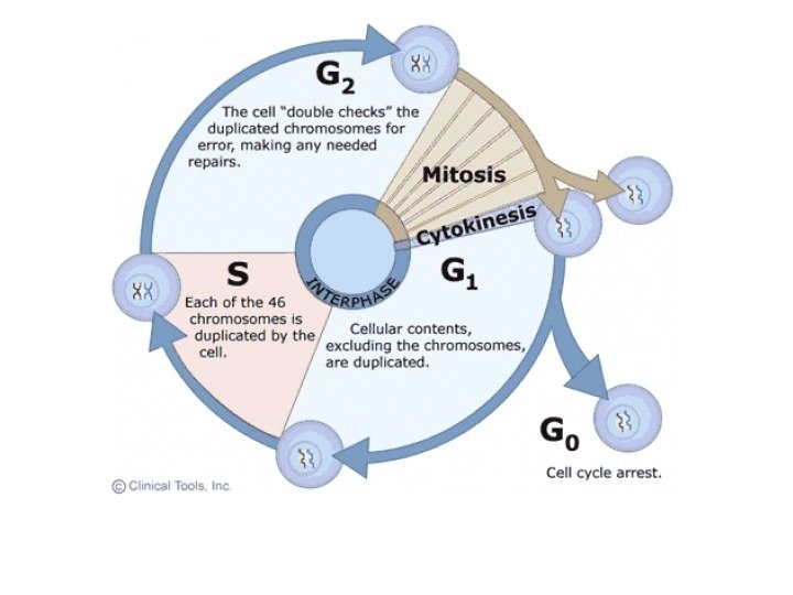
The Cell Cycle Phases S - Synthesis - DNA replication occurs M - Mitosis - nuclear division and cytokinesis Gap phases: G 1 prior to S G 2 prior to M M G 2 G 1 R S G 1, S and G 2 collectively called Interphase
The Logic of the Mitotic Cell Division Cycle - Goal - Faithful Duplication of the Genetic Material - that is mother and daughter cells are identical genetically - Goal - achieved through highly ordered sequences of events and feed-forward and feed-back links and loops: 1) an event is dependent on the correct completion of a preceding event; 2) an event determines the timing and nature of following events; 3) an event can negatively influence a preceding event ensuring a forward movement and occurrence of an event only once per cycle (e. g. DNA replication)
M G 2 G 1 S S - Synthesis phase - Cell will duplicate, make an exact second copy of all it's chromosomes (1 chromosome = 1 molecule DNA) - This is when DNA Replication takes place G 2 – Gap phase 2 - Cell gets ready to divide - Checks if all DNA is completely replicated - Check if there are no errors during Replication
Gap phase 1 - G 1 - Occurs after Mitosis, prior to the next S-phase; - Couples growth to cell division; - Major control/decision point in a cell’s life cycle - Is absent/mostly absent in early embryonic cycles M G 2 G 1 S
A Cells life can be viewed as a shifting balance between the Mitogenic and Anti-mitogenic signals it receives Signals: Cell-to-cell communication, secreted factors (in the case of tissue cultured cells also from serum) and internal signals. Mitogenic signals - usually through the MAP kinase signaling pathway - grow and divide Anti-mitogenic - varry: apoptotic signals; starvation; contact inhibition; differentiation signals…….
The Restriction Point M A Apoptosis M Differentiation M G 2 G 1 S R Senescence Division M A A M - mitogenic – generates mitosis – signal to divide A - anti-mitogenic = opposite of above – don't divide!
SPF and MPF Synthesis promoting factor Maturation promoting factor
Figure 9. 14 Experiment 1 S G 1 Experiment 2 M G 1 Results S S G 1 nucleus immediately entered S phase and DNA was synthesized. M M G 1 nucleus began mitosis without chromosome duplication. Conclusion Molecules present in the cytoplasm control the progression to S and M phases.
The Cell Cycle Machinery Cyclin-dependent Kinases D cdk 4, 6 B M cdk 1 A cdk 1 G 2 G 1 R E cdk 2 S A cdk 2
Cyclins - Name – first discovered due to highly periodic appearance and disappearance of protein in sea urchins - Fluctuations in protein levels linked to cell cycle stage - Genetic evidence for cell cycle regulation (S. cerevisiae and S. pombe), followed by biochemical evidence – yeast and mammals periodicity is achieved by control of expression - transcription - m. RNA stability - translation -protein degradation
Cyclin-CDK = 2 protein complex
Cycling of CDK activity - The cell cycle is ‘driven’ by fluctuations in Cyclin-CDK activity - Cyclin-CDK activity is regulated in several ways: 1. Cyclin protein abundance (transcription, translation, protein degradation) 2. CDK phosphorylation (inhibitory and activating) 3. Cyclin-CDK complex assembly 4. CDK inhibitors
Distinct CDKs drive different transitions
CDK-regulated events are. . . Mitotic CDK disappears at the end of Mitosis until the next G 2 phase = You can't do Mitosis until you have done G 1 and S phase – why?
MPF activity ensures One-way progression
Stages of Mitosis • 1. Interphase/Early Prophase – chromosomes look like tangled rope, no individual chromosomes are visible – not condensed • 2. Prophase – chromosomes condense – DNA gets packaged tightly with the help of proteins, we clearly see the chromosomes – they have a shape; nuclear membrane falls apart, breaks down – centrioles move to opposite ends of cell
Prophase
Metaphase - Spindle fibers (come out of centrioles) – like ropes with hooks will attach to the chromosomes and pull them to the middle of the cell – the metaphase plate - The two identical pairs of chromosomes are sticking together – they are called sister chromatids
Anaphase - The spindle fibers contract (similar to muscles) - The glue holding the two identical chromosomes (sister chromatids) breaks down - The two sister chromatids are pulled by the spindle fibers to opposite ends of the cell
Telophase - Chromosomes have moved to opposite ends -Nuclear membrane re-forms around "new" chromosomes - Cell begins Cytokinesis – it pinches in in preparation for splitting
Cytokinesis - The Cytoplasm and all its content divides in two - Cell division is complete: 2 new, daughter cells are formed - The daughter cells have identical chromosomes/DNA
10 m Figure 9. 7 a G 2 of Interphase Centrosomes (with centriole pairs) Nucleolus Prophase Chromosomes Early mitotic Centromere (duplicated, spindle Aster uncondensed) Nuclear envelope Plasma membrane Two sister chromatids of one chromosome Prometaphase Fragments of nuclear envelope Kinetochore Nonkinetochore microtubules Kinetochore microtubule
10 m Figure 9. 7 b Metaphase Anaphase Metaphase plate Spindle Centrosome at one spindle pole Telophase and Cytokinesis Cleavage furrow Daughter chromosomes Nuclear envelope forming Nucleolus forming
Figure 9. 7 c G 2 of Interphase Centrosomes (with centriole pairs) Chromosomes (duplicated, uncondensed) Nucleolus Nuclear envelope Plasma membrane Prophase Early mitotic Centromere spindle Aster Two sister chromatids of one chromosome
Figure 9. 7 d Prometaphase Fragments of nuclear envelope Kinetochore Metaphase Nonkinetochore microtubules Kinetochore microtubule Metaphase plate Spindle Centrosome at one spindle pole
Figure 9. 7 e Telophase and Cytokinesis Anaphase Cleavage furrow Daughter chromosomes Nuclear envelope forming Nucleolus forming
10 m Figure 9. 7 f G 2 of Interphase
10 m Figure 9. 7 g Prophase
10 m Figure 9. 7 h Prometaphase
10 m Figure 9. 7 i Metaphase
10 m Figure 9. 7 j Anaphase
10 m Figure 9. 7 k Telophase and Cytokinesis
Remember the Direction
Mitotic Cyclin is degraded AFTER Chromatids split and move apart
The Cell Cycle - Sequence of the phases in the life of a cell - Has 4 phases: G 1 S G 2 these 3 are called Interphase M - Mitosis
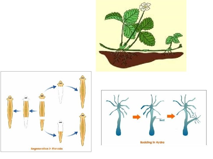
Asexual Reproduction • Reproduction with one parent • Offspring are genetically identical to the parent – Binary fission: type of asexual reproduction • Organism splits in half to become two new identical organisms – Cloning: production of identical genetic copies of a parent • Plants reproduce this way using mitosis
Asexual Reproduction – Regeneration: type of asexual reproduction • Star fish can loose an arm and regenerate a whole starfish; planaria – a type of worm – Budding: type of asexual reproduction • Yeast, sponges and hydra can reproduce this way • A new small individual begins to grow out of the side of the parent organism • This is called a bud • The bud breaks free when it is large enough to live on its own
Budding Yeast Fission Yeast
Checkpoints • Checkpoint – checking that an even is completed BEFORE proceeding to the next event OR checking for damage/defects • Checkpoint – triggered only when something is wrong • Checkpoints – several – at different places in the cell cycle
What happens when a Checkpoint is triggered? • Cell division stops – cell cycle arrest (temporary) and cell attempts to correct problem OR • Cell undergoes apoptosis (programmed cell death) – damage is too extensive • Arrested cells will eventually proceed with cell cycle
Mitotic Checkpoints
Unusual Cell cycles 1. Endoreduplication cycles - alteranting S-phases without intervening mitoses - poliploid cells 2. Embryonic cell division - rapid cycles without/with short Gap phases - S-M-S-M……- non-cell cycle regulated CDK activity (constant) - the appearance of G 1 probably signals the beginning of cellular differentiation