PERIODONTIUM PERIDONTIUM n 1 2 3 4 THE


- Slides: 72

PERIODONTIUM

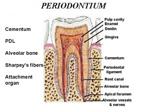
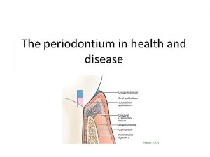
PERIDONTIUM n 1. 2. 3. 4. THE PERIDONTIUM IS DEFINED AS THOSE TISSUES SUPPORTING AND INVESTING THE TOOTH THAT CONSISTS OF CEMENTUM PERIODONTAL LIGAMENT BONE LINING THE ALVEOLUS PART OF THE GINGIVA FACING THE TOOTH

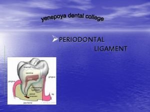
PERIODONTAL LIGAMENT n THE PDL IS THAT SOFT SPECIALIZED CONNECTIVE TISSUE SITUATED BETWEEN THE CEMENTUM COVERING THE ROOT OF THE TOOTH AND THE BONE FORMING THE SOCKET WALL n THE PDL RANGES IN WIDTH FROM 0. 15 TO 0. 38 MM WITH ITS THINNEST PORTION IN THE MIDDLE OF THE ROOT, THE AVERAGE WIDTH IS 0. 21 MM AT 11 TO 16 YEARS, 0. 18 MM AT 32 TO 52 YEARS 0. 15 MM AT 51 TO 67 YEARS n n n

FUNCTIONS OF PDL. SUPPORT: PDL supports teeth in their socket. It prevents loosening of teeth. n MASTICATORY LOAD: PDL permits teeth to withstand the considerable forces of mastication. n SENSORY: PDL is supplied by abundant receptors and nerves that sense the movement when teeth are in function. Helps in the proper positioning of the jaws during normal function. n

NUTRITIVE: Blood vessels of ligament provide essential nutrients for the ligament’s vitality and hard tissue of cementum and alveolar bone. n Fibroblasts, osteoblasts, cementoblasts, and even resorptive osteoclasts and macrophages require nutrition. n

CLINICAL CORRELATION: Bone, PDL, and the cementum together form a functional unit of special importance when the orthodontic tooth movement is undertaken. Orthodontic forces causes compression and constriction of blood vessels, soft tissue changes occur. n Hence loss of alveolar bone occurs, now blood flow occurs in the spaces. And the mesenchymal cells of PDL repair the tissues. n

MAINTAINENCE: Tissues are maintained under the influence of heavy masticatory forces. n ADAPTIVE ROLE n n SHOCK ABSORBER : It absorbs the shock of chewing.

PDL FORMATION PDL is formed within the developing dental follicle. Ligament space consists of unorganized connective tissue with short fiber bundles extending into it from bone and cemental surfaces. Ligament mesenchymal cells begin to secrete collagen The developing periodontal ligament (mainly collagen type I). fiber bundles extend into the unorganized ligament space from both the cementum surface and the surface of the alveolar bone.

n n n Collagen bundles extend from the bone and cementum surfaces, establishing an attachment of the tooth to the bone. In addition several non collagenous proteins are secreted. They play role in maintenance of PDL space. Eruptive tooth movement and the establishment of occlusion then modify this attachment. When the tooth erupts the crest of the alveolar bone is above the CEJ junction and the developing fibers are directed obliquely.

n n Then when the tooth moves during eruption, the level of the alveolar crest coincides with the CEJ junction and the oblique fibers become horizontally aligned. When the tooth finally comes in function, the alveolar crest fibers is positioned nearer the apex.

n n The horizontal fibers termed as alveolar crest fibers have become oblique once again but with the difference that now the CEJ has reversed its relation to the alveolar attachment and is positioned in a coronal direction. Only after the teeth come into function do the fiber bundles of the PDL thicken appreciably.

COMPONENTS OF THE PDL n n n The PDL consists of cells, an extracellular compartment of collagenous fibers and noncollagenous extracellular matrix. The cells include osteoblasts, osteoclasts, fibroblasts, epithelial rest cells of Malassez, macrophages, undifferentiated mesenchymal cells and cementoblasts. The extracellular compartment consists of well defined collagen fibers bundles embedded in the ground substance comprising of glycosinaminoglycans, glycoprotiens, and glycolipids.


PERIODONTAL FIBERS The predominant collagens of PDL are types I, III and XII. n They are arranged in distinct and definite fiber bundles. n They are able to adapt to the continual stresses placed on them. n

PRINCIPAL FIBER BUNDLES OF PDL 1. THE ALVEOLAR CREST GROUP These are attached to the cementum just below the cementoenamel junction and running downward and outward to insert into the rim of the alveolus.

2. THE HORIZONTAL GROUP n These are just apical to the alveolar crest fibers and running at right angles to the long axis of the tooth from the cementum to the bone below the alveolar crest.

3. n THE OBLIQUE GROUP They are the most numerous in the PDL and running from the cementum in an oblique direction to insert into the bone coronally

4. THE APICAL GROUP: n These are radiating from the cementum around the apex of the root to the bone forming the base of the socket.

5. THE INTERADICULAR GROUP n Found only in the multi -rooted teeth and running from the cementum into the bone forming the crest of the Interradicular septum.

SHARPEY’S FIBERS n At each end all the principal fibers of the PDL are embedded in the cementum or the bone and this embedded portion is referred to as the SHARPEY’S FIBERS therefore providing anchorage.

n SHARPEY’S FIBERS in primary acellular cementum are mineralized fully. n Those in cellular cementum and bone generally are mineralized only partially at their periphery. n Sharpey’s fibers pass uninterruptedly through the bone of alveolar process to continue as principal fibers of PDL.


CEMENTUM n DEFINITION: n CEMENTUM IS A HARD, AVASCULAR CONNECTIVE TISSUE THAT COVERS THE ROOTS OF THE TEETH.

CEMENTUM n n n CEMENTUM is excreted by cells called CEMENTOBLASTS within the root of the tooth. CEMENTOBLASTS are the formative cells of cementum. It is thickest at the root apex.

DEVELOPMENT OF CEMENTUM The development of cementum has been subdivided into n PREFUNCTIONAL STAGE, which occurs through out root formation. n FUNCTIONAL STAGE, which starts when the tooth is in occlusion and continues through out life. n

TYPES OF CEMENTUM n n n CEMENTUM is classified according to the presence or absence of cells within its matrix. CELLULAR CEMENTUM, which has an adaptive role in response to tooth wear and movement and is associated with repair of periodontal disease. ACELLULAR CEMENTUM, which provides attachment for the tooth. A- CELLULAR CEMENTUM B-ACELLULAR CEMENTUM

PHYSICAL CHARACTERSTICS AND COMPOSITION COLOR: YELLOW. n HARDNESS: LESS THAN DENTIN. n COMPOSITION: ORGANIC MATRIX (50%) AND INORGANIC ELEMENT (45 -50%) n ORGANIC MATRIX: COLLAGENOUS AND NON-COLLAGENOUS PROTEINS. n

TYPES OF COLLAGEN: Type I Collagen is predominant in cementum, constitutes 90% of organic component of cellular cementum. n Type III, Type XII, are also present. n Type XII is found in high concentration in PDL. n Traces of Type V, VI and XIV are also found in cementum. n

NON-COLLAGENOUS PROTEINS IN CEMENTUM n n n n n These proteins are also associated with bone: ALKALINE PHOSPHATASE. BONE SIALOPROTEIN. DENTIN SIALOPROTEIN. OSTEOCALCIN OSTEOPONTIN. OSTEONECTIN. FIBRONECTIN. DENTIN MATRIX PROTEIN. PROTEOGLYCANS. ETC.

INITIATION OF CEMENTUM FORMATION n n n CEMENTUM formation takes place along the entire root. At the advancing root edge, HERTWIG’S EPITHELIAL ROOT SHEATH (HERS), which is derived from the extension of inner and outer epithelium releases enamel proteins. Possibly sends inductive message to the ectomesenchymal cells of pulp.

n n These ectomesenchymal cells of pulp now differentiate into odontoblasts and produce a layer of predentin. HERS now become disintegrated. (HERS separates the dentin from the surrounding tissues of dental follicle). Therefore the inner layer of dental follicle comes in contact with predentin. Cells of the dental follicle now differentiate into CEMENTOBLASTS.

n The next series of event results in formation of cementum on the root surface. n The trigger factors and specific cells responsible for its formation could be: n DENTAL FOLLICLE CELLS receiving signals from HERS or dentin, and differentiate into cementoblasts. n HERS cells transform into cementoblasts.

EPITHELIAL REST CELLS OF MALASSEZ n During these two processes, some cells within the HERS undergo APOPTOSIS. n Some cells within the root sheath form discrete masses surrounded by BASAL LAMINA, known as EPITHELIAL REST CELLS OF MALASSEZ.

n n n n These rest cells persists in mature PDL. These cells are not only residual cells but they also participate in maintenance and regeneration of periodontal tissues. CIFC: cellular intrinsic fibers of cementum. ERM: epithelial rest cells of malassez. Cb: Cementoblasts. Cc: Cementocytes. AEFC: Acellular extrinsic fibers of cementum.


ENAMEL PEARLS n If some HERS cells remain attached to forming root surface they can produce focal deposits of enamel like structures called ENAMEL PEARLS.

ORIGIN OF PDL CELLS AND DIFFERENTIATION OF CEMENTOBLASTS. n n n PRECURSOR CELLS FOR CEMENTOBLASTS AND PDL: DENTAL FOLLICLE. REGENERATION OF PDL TISSUE: STEM CELLS & LOCAL PROGENITOR CELLS. CEMENTOBLASTS FORMATION: EPITHELIAL CELLS FROM HERS MAY UNDERGO EPITHELIAL-MESENCHYMAL TRANSFORMATION INTO CEMENTOBLASTS.

MOLECULAR FACTORS REGULATING CEMENTOGENESIS These molecules and factors: n CONTROL DEVELOPMENT. (CEMENTOGENESIS). n MAINTAIN PDL. n REGENERATE PDL. n

MATRIX PROTEINS BONE SIALOPROTEIN & OSTEOPONTIN. n FUNCTIONS: n CEMENTOGENESIS. n REPAIR & REGENERATION OF PDL. n OSTEOPONTIN: Regulate Mineral Growth. n BONE SIALOPROTEIN: Promotes mineral formation on root surface. n

COLLAGEN TYPE I COLLAGEN: Abundant in cementum as well as in PDL. n TYPE III: Abundant during early stages of Cementogenesis. And during development and Repair. n TYPE XII: Abundant in PDL and lower levels in Cementum. n

OTHER FACTORS: TRANSCRIPTION FACTORS n ALKALINE PHOSPHATASE. (promote cementogenesis). n GROWTH FACTORS. ( Insulin-like growth factor, platelet-derived growth factor). n METALLOPROTEINASE. n PROTEOGLYCANS. ( Accumulate at DCJ). n

CEMENTUM VARIETIES n n ACELLULAR (PRIMARY). EXTRINSIC FIBERS: From cervical margin to apical third. FUNCTION: Anchorage. CELLULAR (SECONDARY). INTRINSIC FIBERS: Middle to Apical third and Furcations. FUNCTION: Adaptation and Repair. A. CELLULAR CEMENTUM. B. ACELLULAR CEMENTUM.


MIXED ( ALTERNATING LAYERS OF ACELLULAR AND CELLULAR CEMENTUM). INTRINSIC AND EXTRINSIC: Present in Apical portion and Furcations. n FUNCTION: Adaptation. n ACELLULAR AFIBRILLAR: Spurs and patches over enamel and dentin along the CEJ. n

ACELLULAR EXTRINSIC FIBER CEMENTUM (PRIMARY CEMENTUM). n During root development, first cementoblasts align along with Predentin after disintegration of HERS. n Cementoblasts exhibit fibroblastic nature i. e. they extend cell processes in Unmineralized Predentin.

n Cementoblasts deposit collagen within Predentin so that the Dentin and Cementum fibrils INTERMINGLE. n Thereby CEMENTODENTINAL JUNCTION is established. SERIES OF EVENTS IN ACELLULAR EXTRINSIC FIBERS: n Initial fibers consist of mineralized layer with a short fringe of collagen fibers perpendicular to the root surface. n

n n n Once the fiber fringes are incorporated, cells from root surface migrate away and continue to deposit collagen. Hence thickening and lengthening of fiber bundles occur. Cells on the root surface also secrete non-collagenous matrix proteins that fill in spaces between collagen fibers.

Once 15 -20 micrometer cementum has been deposited then: n Cementoblasts secrete only NONCOLLAGENOUS PROTEINS. n And the PDL fibroblasts secrete the collagen fibrils embedded in cementum. n Acellular cementum develops slowly as the tooth is erupting. n It is considered to be ACELLULAR because the cells that form it remain on its surface. n

STRUCTURE OF ACELLULAR CEMENTUM n n These fibers are relatively Structure less. These fibers cover the root from CEJ to apical third. Parallel striations to the root surface indicate incremental deposition. Short striations perpendicular to the root surface indicates insertion of PDL fibers.

n n Acellular cementum appear as homogenous layer in ground section. Overall degree of mineralization of this cementum is about 45 -60%. Innermost layers are less mineralized The outer layers are characterized by alternating bands of more and less mineral content that run PARALLEL to root surface.

CELLULAR INTRINSIC FIBER CEMENTUM (SECONDARY CEMENTUM). n n n After at least half the root is formed, Cellular cementum starts depositing. It forms more rapidly. It is less mineralized than Acellular cementum. It is deposited on unmineralized dentin. It contains cells called CEMENTOCYTES.

Cementoblasts extend cell processes in the unmineralized dentin. n And deposit collagen fibers so that the collagen fibers of dentin and cementum intermingle. n Cementoblasts also secrete various noncollagenous proteins. n A layer of unmineralized matrix is present at the surface of mineralized cementum called CEMENTOID. n


n n n Unlike OSTEOID or PREDENTIN, cementoid is not as regular and distinct. Cells become entrapped in extra cellular matrix with REDUCED SECRETORY ACTIVITY, called CEMENTOCYTES. They reside in LACUNAE. OSTEOCYTES reside in bone lacuna. Similarly CEMENTOCYTES resides in cementum lacuna. Cementocytes have processes that lodge in Canaliculi.

CONSTITUENTS OF CELLULAR CEMENTUM. n n n Cementocytes within lacunae, with processes in canaliculi directed towards tooth surface. Cementoid. ( Secondary cementum is deposited first as an unmineralized layer, called Cementoid, which gradually calcifies). 1. CEMENTOCYTE LACUNA. 2. CEMENTOCYTE CANALICULI.

ACELLULAR AFIBRILLAR CEMENTUM. This type of cementum lacks COLLAGEN, hence called AFIBRILLAR. n It consists of Acellular and Afibrillar mineralized matrix. n It plays no role in tooth attachment. n It is in proximity to the CEJ. n

CEMENTOENAMEL JUNCTION n n n Relation of Cementum to Enamel at the Cementoenamel Junction (CEJ) "OMG rule" In 60% of the teeth cementum OVERLAPS enamel. In 30% of the teeth cementum just MEETS enamel forming a butt joint. In 10% of the teeth there is a small GAP between cementum and enamel.


ALVEOLAR PROCESS. n n n Alveolar process is that bone of jaws containing the sockets (alveoli) for the teeth. The alveolar process consists of OUTER (BUCCAL & LINGUAL) CORTICAL PLATE. Central bone is SPONGIOSA.


n n n Bone lining the alveolus is called ALVEOLAR BONE. Cortical plates and alveolar bone meets at the alveolar crest (1. 5 -2 m. m. ) below the level of CEJ. Alveolar bone is perforated by foramina because it is vascular, . Nerves and vessels pass through it. Sometimes referred to as cribriform plate.

n n Alveolar bone is referred as LAMINA DURA, radiographically, because of its increased RADIOPACITY. The bone directly lining the socket i. e. the inner aspect of alveolar bone is known as BUNDLE BONE.

GINGIVAL GROUP. Other groups of collagen fibers found in the lamina propria of the gingiva collectively form the gingival ligament. 1. CIRCULAR GROUP: n These fibers form a band around the neck of the tooth and help to bind the free gingiva to the tooth.

2. DENTOGINGIVAL GROUP: n These are the most numerous fibers extending from the cervical cementum to the lamina propria of the free and attached gingiva.

3. DENTOPERIOSTEAL GROUP: n Running apically from the cementum over the periosteum of the alveolar bone , these fibers insert into the alveolar process or the vestibular muscle and floor of the mouth.

4. ALVEOLOGINGIVAL GROUP n These fibers radiate from the bone of the alveolar crest and insert into the free and attached gingiva.

5. TRANSEPTAL FIBER SYSTEM n These fibers run interdentally from the cementum just apical to the base of the junctional epithelium of one tooth over the alveolar crest and insert into the cementum of the adjacent tooth. n Thus they form a interdental ligament connecting all the teeth in the arch.


THANK YOU !!
 Dentoperiosteal group
 Definition of periodontal ligament
 Non collagenous proteins of periodontium
 Periodontium
 Theories of pdl
 độ dài liên kết
 Môn thể thao bắt đầu bằng từ chạy
 Thiếu nhi thế giới liên hoan
 Hình ảnh bộ gõ cơ thể búng tay
 điện thế nghỉ
 Một số thể thơ truyền thống
 Trời xanh đây là của chúng ta thể thơ
 Hệ hô hấp
 Số nguyên tố là số gì
 Voi kéo gỗ như thế nào
 Tia chieu sa te
 đặc điểm cơ thể của người tối cổ
 Các châu lục và đại dương trên thế giới
 Thế nào là hệ số cao nhất
 ưu thế lai là gì
 Slidetodoc
 Tư thế ngồi viết
 Bàn tay mà dây bẩn
 Mật thư tọa độ 5x5
 Tư thế ngồi viết
 Chụp phim tư thế worms-breton
 Thẻ vin
 Ví dụ về giọng cùng tên
 Thể thơ truyền thống
 Từ ngữ thể hiện lòng nhân hậu
 Các châu lục và đại dương trên thế giới
 Sự nuôi và dạy con của hươu
 Bổ thể
 Diễn thế sinh thái là
 Vẽ hình chiếu vuông góc của vật thể sau
 Làm thế nào để 102-1=99
 Alleluia hat len nguoi oi
 Lời thề hippocrates
 đại từ thay thế
 Vẽ hình chiếu đứng bằng cạnh của vật thể
 Quá trình desamine hóa có thể tạo ra
 Công thức tiính động năng
 Thế nào là mạng điện lắp đặt kiểu nổi
 Khi nào hổ mẹ dạy hổ con săn mồi
 Tỉ lệ cơ thể trẻ em
 Các loại đột biến cấu trúc nhiễm sắc thể
 Thế nào là sự mỏi cơ
