
INTRODUCTION 1 2 3 4 The periodontium pert




- Slides: 50
INTRODUCTION. . � 1. 2. 3. 4. The periodontium (pert= around , odontos= tooth) comprises the following tissues The gingiva (G) The periodontal ligament (PL) The root cementum (RC) The alveolar bone (AP)
ALVEOLAR BONE IN HEALTH
DEFINITION. . . The alveolar process is defined as the parts of the maxilla and the mandible that form and support the sockets of the teeth.
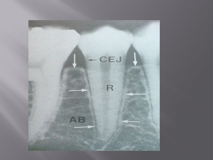
The alveolar bone consists of 2 components, the alveolar bone proper (ABP) and the alveolar process. The alveolar bone proper, also called “bundle bone” is continuous with the alveolar process and forms the thin bone plate that lines the alveolus of the tooth.
DEVELOPMENT OF MAXILLA AND MANDIBLE MAXILLA: The human maxilla is homologous to two bones The maxilla proper &pre maxilla The incisors form the anterior part of hard palate The composition of human maxilla from premaxilla &maxilla is indicated by incisor fissures
MANDIBLE: The mandible makes its appearence as a bilateral structure in the sixth week of fetal life as thin plate of bone lateral to meckel’s cartilage. The mandible is a paired bone Right and left mandible are joined in the midline by fibrocartilage in the mandibular symphisis
NORMAL ANATOMY OF ALVEOLAR BONE � The alveolar process develops in conjunction with the development and eruption of the teeth. The alveolar process consists of bone which is formed both by cells from the dental follicle (alveolar bone proper) and cells which are independent of tooth development. Together with the root cementum and the periodontal membrane, the alveolar bone constitutes the attachment apparatus of the teeth, the main function of which is to distribute and resorb forces generated by , for example, mastication and other tooth contacts.
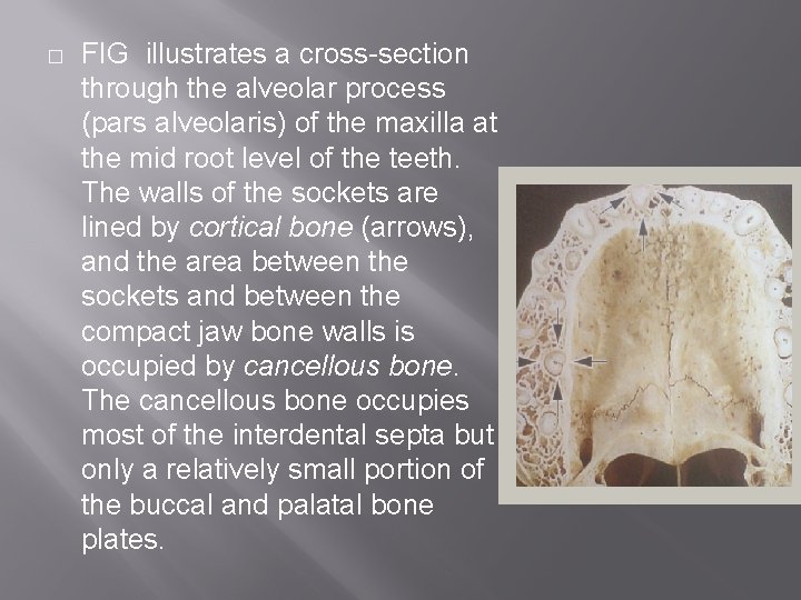
� FIG illustrates a cross-section through the alveolar process (pars alveolaris) of the maxilla at the mid root level of the teeth. The walls of the sockets are lined by cortical bone (arrows), and the area between the sockets and between the compact jaw bone walls is occupied by cancellous bone. The cancellous bone occupies most of the interdental septa but only a relatively small portion of the buccal and palatal bone plates.
The cancellous bone contains bone trabeculae, the architecture and size of which are partly genetically determined and partly the result of the forces to which the teeth are exposed during function.
The alveolar process starts to form early in foetal life, with mineral deposition at small foci in the mesenchymal matrix surrounding the tooth buds. Increase in size, fuse, and become resorbed and remodelled until a continuous mass of bone has formed around the fully erupted teeth. The mineral content of bone, which is mainly hydroxyapetite, is about 60% on a weight basis.
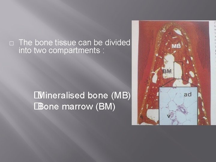
� The bone tissue can be divided into two compartments : � Mineralised bone (MB) � Bone marrow (BM)
COMPOSITION OF ALVEOLAR BONE � a. b. It has two basic constituents : The cells consists of osteoblasts, osteoclasts, and osteocytes Extracellular matrix consists of 65% inorganic and 35% organic matter.
� � � The inorganic component is composed of minerals such as calcium, phosphate along with hydroxyl, carbonate, citrate and trace amounts of other ions, such as sodium, magnesium and flourine. The minerals are in the form of hydroxyapetite crystals. The organic matrix consists of 90% of type I collagen, with small amounts of noncollagenous proteins such as osteocalcin, osteonectin, bone morphogenetic proteins, proteoglycans and glycoproteins
� a. b. c. Cellular components Osteoblasts : they are cuboidal cells with well developed rough endoplasmic reticulum, a large golgi apparatus and secretory vesicles. Osteoclasts : they are cells responsible for resorption of bone. Osteocytes: a bone cell: an osteoblast that has ceased activity and has become embedded in the bone matrix
CELLS AND INTERCELLULAR MATRIX � � Alveolar bone is formed during foetal growth by intramembraneous ossification and consist of calcified matrix with osteocytes enclosed within space called lacunae. the osteocytes extend processes into canaliculi that radiate from the lacunae. The bone consist of two thirds inorganic mater and one third organic matrix. . the inorganic mater is composed of minerals such as calcium and phophate along with hydroxyl carbonates citrate and trace amount of other ions.
The organic matrix consist mainly(90%) of collagen type 1 with small amount of noncollagenous protiens which are osteocalcin, osteonectin, bone morphogenic protien, phospho protien and proteoglycans. Bone deposition by osteoblast is balanced by resorption by osteoclast during tissue removal and renewal.
SOCKET WALL Socket wall consist of dense, lamellated bone, some of which is arranged in haversian systems and bundle bone is the term given to bone adjacent to the PDL that contains a greater number of sharpey’s fibers. Bundle bones is not unique to the jaws; it occurs through out the skeletal system where ever the ligaments and muscle attached
Cancellous bone: it is found predominantly in the inter radicular and inter dental spaces and in limited amounts facially or lingually except in the palate. In the adult, there is more cancellous bone in the maxilla than in the mandible.
BONE MARROW In the embryo and new born, the cavities of all bones are occupied by red hematopoetic marrow. in jaws common locations are the maxillary tuberosity and the maxillary and mandibular molar and premolar areas, which may be visible radiographically as zone of radiolucency
PERIOSTEUM & ENDOSTEUM All bone surfaces are covered by layers of differentiated osteogenic connective tissue. The tissue covering the outer surface of bone is termed periosteum. Tissue lining the internal bone cavities is called endosteum.
INTERDENTAL SEPTUM The interdental septum consists of cancellous bone bordered by socket walls of approximating teeth and the facial and lingual cortical plates. If the interdental space is narrow, the septum may consist of only lamina dura.
REMODELLING OF ALVEOLAR BONE In contrast to its apparent rigidity, alveolar bone is the least stable bone of the periodontal tissues, because its structure is in a constant state of flux. Remodelling of alveolar bone affects its height,
Contour and density and is manifested in three areas → adjacent to the periodontal ligament. → in relation to the periosteum of the facial and lingual plates. → along the endosteal surface of the bone marrow.
ALVEOLAR BONE IN DISEASE
OSSEOUS DEFECTOsseous defects are those defects, which are formed as a result of destruction of alveolar bone due to periodontal disease.
FENESTRATION AND DEHIESENCEFenestration are isolated areas in which the root surface is covered only by the periosteum and gingiva, the marginal bone remains intact.
DEHISCENCESWhen the marginal bone is denuded the defect is called dehiscence.
RADIUS OF ACTION On the basis of Waerhaug’s measurements, it was postulated that there is a range of effectiveness of about 1. 5 to 2. 5 mm within which bacterical plaque can inducde bone loss, beyond 2. 5 mm there is no effect. Interproximal defects can appear only in spaces wider than 2. 5 m. Larger defects far exceeding 2. 5 mm can be seen in certain conditions like localised juvenile periodontitis and Papillon-Lefevre Syndrome.
BONE DESTRUCTION CAUSED BY EXTENSION OF GINGIVAL INFLAMATION
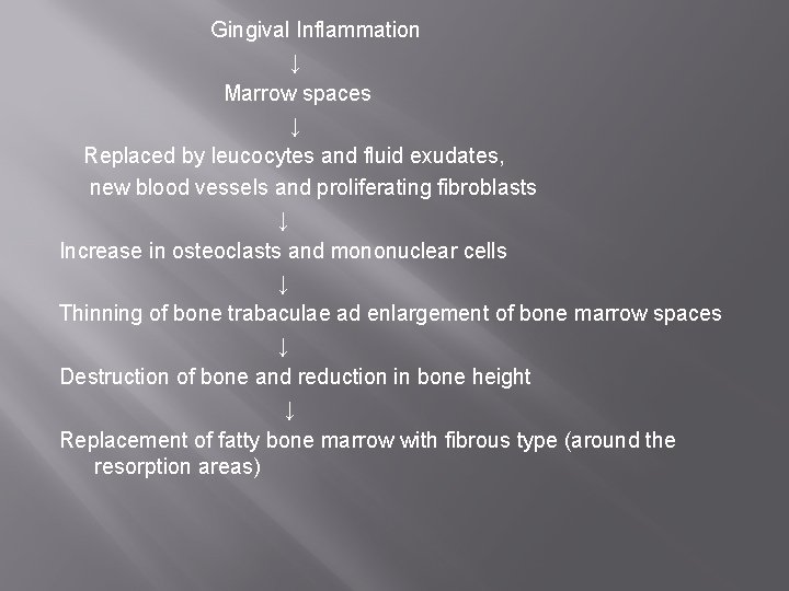
Gingival Inflammation ↓ Marrow spaces ↓ Replaced by leucocytes and fluid exudates, new blood vessels and proliferating fibroblasts ↓ Increase in osteoclasts and mononuclear cells ↓ Thinning of bone trabaculae ad enlargement of bone marrow spaces ↓ Destruction of bone and reduction in bone height ↓ Replacement of fatty bone marrow with fibrous type (around the resorption areas)
DESTRUCTION CAUSED BY TRAUMA FROM OCCLUSION
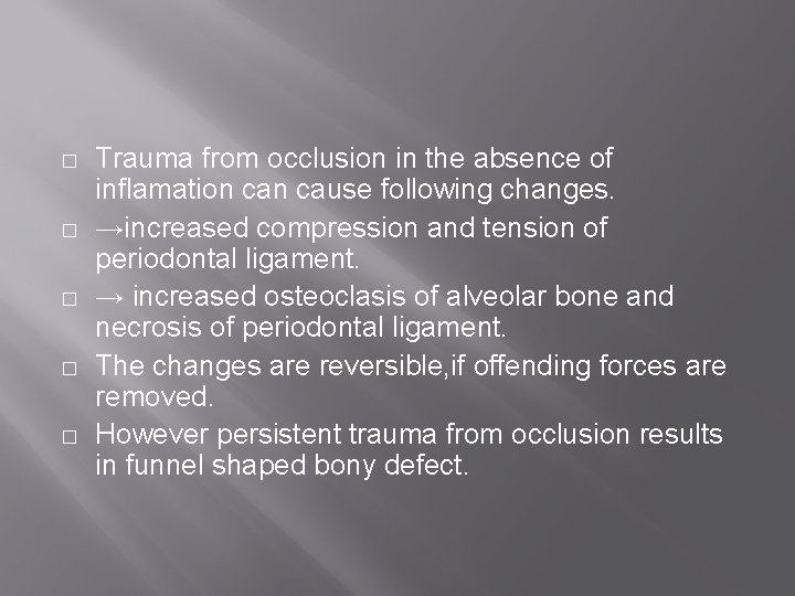
� � � Trauma from occlusion in the absence of inflamation cause following changes. →increased compression and tension of periodontal ligament. → increased osteoclasis of alveolar bone and necrosis of periodontal ligament. The changes are reversible, if offending forces are removed. However persistent trauma from occlusion results in funnel shaped bony defect.
Destruction Caused by Systemic Disorder
Osteoporosis is a physiologic conditio of post menapausal women, resulting in loss of bone mineral content and micro structural bone changes. Periodontal bone loss may occur in generalised bone disturbances (eg. Hyperparathirodism, leukemia etc. ) by mechanism that may not be related to the usual periodontal bone destruction.
NORMAL VARIATION IN ALVEOLAR BONE Thickness, width and crestal angulation of the interdental septa Thickness of facial and lingual alveolar plates The presence of fenestrations and dehiscences Increased thickness of alveolar bone margins to accommodate functional demands Allignment of the teeth, root trunk anatomy.
BONE DESTRUCTION PATTERN IN PERIODONTAL DISEASE
HORIZONTAL BONE LOSS VERTICAL OR ANGULAR DEFECT OSSEOUS CRATERS BULBOUS BONY CONTOURS REVERSED ARCHITECTURE LEDGES FURCATION INVOLVEMENT
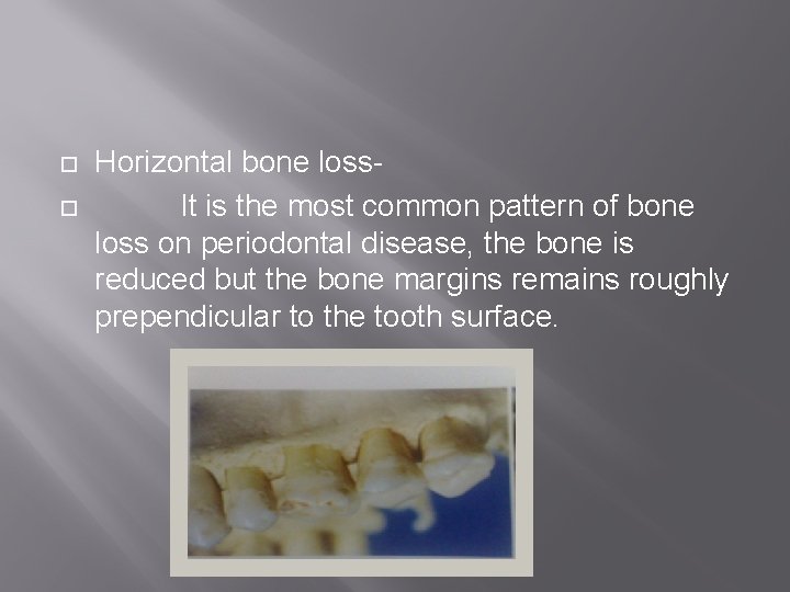
Horizontal bone loss. It is the most common pattern of bone loss on periodontal disease, the bone is reduced but the bone margins remains roughly prependicular to the tooth surface.
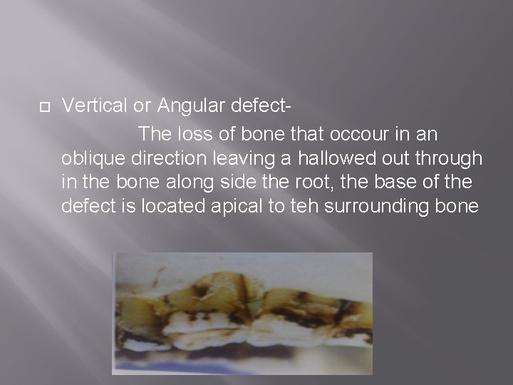
Vertical or Angular defect. The loss of bone that occour in an oblique direction leaving a hallowed out through in the bone along side the root, the base of the defect is located apical to teh surrounding bone
Angular defects are classified as↓ One walled or hemiseptal defect Two walled defect Three walled or infra bony pocket Combined osseous defect
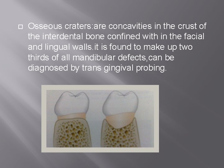
Osseous craters: are concavities in the crust of the interdental bone confined with in the facial and lingual walls. it is found to make up two thirds of all mandibular defects, can be diagnosed by trans gingival probing.
� � Bulbous Bony Contours: they are bony enlargement caused by exostoses, adaption to function or buttresing bone formation. They are found more frequently in maxilla than mandible. Reversed Architecture: these defects are produced by loss of interdental bone, including the facial and lingual plates without concomitant loss of radicular bone, there by reversing the normal architecture. it is more commonly seen in maxilla
Ledges: they are plateau like bone margins caused by resorption of thickened bony plates. Furcation involvement: It refers to commonly occuring conditions in which the bifurcation and trifurcation of multi rooted teeth or involved by the disease process. The mandibular first molar are most common sites and least common are maxillary premolars
ALVEOLAR BONE IN AGING
� � Changes in alveolar bone with aging are similar to those occuring in the skeletal system They are →osteoporosis →decreased vascularity →reduction in metabolic rate and healing capacity. →bone resorption may be increased or decreased →density of bone may be increased or decreased depending on its location in the body.
REFERENCES
� Clinical periodontology&Implants(4 th E) →JAN LINDHE � Clinical periodontology(8 th &10 th E) →CARRANZA’S � Essentials of clinical periodontology&periodontics(2 nd E) →SHANTIPRIYA REDDY
THANK YOU