GENERAL History and Physical Examination Dr Duaa Hiasat


 • Short/ specific in one clear sentence communicating major/present problem/issue •")


 Characteristics of pain (SOCRATES) • Site: Somatic pain often well localized, e.")

 Since onset Episodic or continuous? If episodic, duration")

 • Cough, Wheeze •")

 • Timing and regularity of periods")


 • Warm envirnoment. • Examination bed. .")


. • Pink tinge…CO poisining.")
…anemia • Pink flushing…vasodilation(perimenapausal women or carcinoid tumour)")


 • Low")


 Shivering")


. If the condition persists,")


- Slides: 132
GENERAL History and Physical Examination Dr. Duaa Hiasat
Objectives: At the end of this session you should be able to: - Know how to define History - Know the importance of History taking - Know how to approach to the patient - Know parts of History - Know how to do General physical examination
History taking • Is defined as planned professional conversation that enables the patient to communicate his/her symptoms, feelings, and fears to the clinician so as to obtain an insight into the nature of patients illness and his/her attitude towards them.
Importance of taking history • Obtaining an accurate history is the critical first step in determining the etiology of a patient’s illness • Most of times you will actually be able to make a diagnosis based on history alone
General approach • Introduce yourself Note-never forget patient’s names Create patient appropriatly in a friendly relaxed way Confidentiality and respect patient’s privacy • Try to see things from patient point of view, understand patient anxiety, irritation or depression. Listening is very important. • Questioning: simple, clear, avoid medical terms , open, leading, interrupting, direct questions and summerizing.
Taking history and recoding • Always record personal details: -name -marital status -sex -address -occupation -ethnicity -age -record date of exam
Complete history taking -chief complaint -present illness -past medical history -systemic inquiry -family history -drug history -Social history
Chief complaint • Is the main reason that push the patient to seek for visiting physician or for help • Usually single symptoms, occasionally more than one eg; chest pain, palpitation, SOB, ankle swelling etc. • The patient describes the problem in their own words, & should be recorded in their own words. • What brings you here? How can I help you ? . .
Chief complaint( CC) • Short/ specific in one clear sentence communicating major/present problem/issue • Timing: fever for last two weeks or since Monday • Recurrent • Any major disease important eg: DM, HTN, asthma, IHD, pregnancy • Note: should be put in patient’s language
History of present illness/Tips • Elaborate in to the chief complaint in details • Ask relevant associated symptoms • Have differential dignosis in mind that Lead the conversation and thoughts • Decide and weight the importance of minor complaints • In details of present problem with time of onset/mode of evolution/any investigation/treatment and outcome/and associated +’ve or –’ve symptoms
• Sequential presentaion: -always relay story in days before visit -narrate in details
• In details of symptomatic presentation: if patient has more than one symptom like chest pain, swollen leg and vomiting, take each symptom individually and follow it through fully mentioning important negatives as well. Eg: the pain was central crushing, radiating to left jaw, lasted less than 5 minutes relieved by rest, no associated symptoms with pain/never had this pain before/no relation to food/
History of present illness/Tips • Avoid medical terminology and make use of descriptive language that is familiar to them • Describe each symptom in a choronological order
Pain (SOCRATES) Characteristics of pain (SOCRATES) • Site: Somatic pain often well localized, e. g. sprained ankle Visceral pain more diffuse, e. g. angina pectoris • Onset: Speed of onset and any associated circumstances • Character: Described by adjectives, e. g. sharp/dull, burning/tingling, boring/stabbing, crushing/tugging, preferably using the patient's own description rather than offering suggestions
• Severity : Difficult to assess as so subjective. Sometimes helpful to compare with other common pains, e. g. toothache • Radiation : Through local extension, or referred by a shared neuronal pathway to a distant, unaffected site, • Associated symptoms : Visual aura accompanying classical migraine , Numbness in the leg with back pain suggesting nerve root irritation
• Timing: (duration, course, pattern) Since onset Episodic or continuous? If episodic, duration and frequency of attacks. if continuous, any changes in the severity Variation. Exacerbating and relieving factors : Circumstances in which pain is provoked or exacerbated e. g. food Specific activities or postures, and any avoidance measures that have been taken to prevent the onset.
Systematic inquiry - cardinal symptoms • General health Well-being Energy Appetite Sleep Weight change Mood • Cardiovascular System • Chest pain on exertion (angina) • Breathlessness: lying flat (orthopnoea) at night (paroxysmal nocturnal dyspnoea) on minimal exertion • Palpitation, Pain in legs on walking (claudication) • Ankle swelling
• Respiratory system • Shortness of breath (exercise tolerance) • Cough, Wheeze • Sputum production (colour, amount)Bloody? (haemoptysis) • Chest pain (due to inspiration or coughing) • Gastrointestinal system • Mouth (oral ulcers, dental problems) • Difficulty swallowing (dysphagia ) or pain (odynophagia) • Nausea and vomiting
• Abdominal pain • Change in bowel habit • Change in colour of stools (pale, dark, tarryblack, blood) • Genitourinary system • Pain passing urine (dysuria) • Frequency passing urine (at night, nocturia) • Blood in the urine (haematuria) • Men If appropriate: difficulty starting - hesitancy • poor stream or flow, incontinence, urethral discharge
• Women Last menstrual period (consider pregnancy) • Timing and regularity of periods • Length of periods, Abnormal bleeding, Vaginal discharge • Contraception • Nervous system • Headaches, Weakness • Dizziness (vertigo or light-headed) • Faints, Fits • Altered sensation (numbness or tingling (paraesthesiae))
• Musculoskeletal • Joint pain, stiffness or swelling • Mobility • Falls • Endocrine • Heat or cold intolerance • Change in sweating • Excessive thirst (polydipsia) • Other • Bleeding or bruising • Skin rash
Past medical illness • Start by asking the patient if they have any medical problems. • IHD, Heart attack, DM, TB etc. , if DM – mention the time of diagnosis/current medication/clinic check up There is often a relationship between the past medical history and the presenting problem. Past surgical/operation history: -time/place/and what type of operation. note any blood transfusion and blood grouping
Drug History • Ask about prescribed drugs and any other medications your patient is taking. Include over-the-counter remedies and alternative medicine treatments, • Always use generic name or put trade names in brackets with dosage, timing and how long Compliance/concordance the traditional model of medical care where patients take their medication as prescribed by the doctor. Half of all patients do not take medicines as directed.
• Drug allergies/reactions Always ask if your patient has ever had an allergic reaction to medication. Ask about other allergies, e. g. foodstuffs, animal hair, pollen or metal. Record true allergies prominently in the patient's case records and drug chart.
Family history Obtain the family history by asking open-ended questions, e. g. 'Are there any illnesses that run in your family? ' It is possible that the presenting complaint directs you to a particular line of inquiry • Any familial disease/running in families • Eg: breast ca, IHD, DM, developmental delay, asthma etc.
Social History • Upbringing • Birth injury or complications • Schooling, academic achievements or difficulties • Further or higher education and training • Behaviour problems Home life • Emotional, physical or sexual abuse • Experiences of death and illness • Interest and attitude of parents
Occupation • Current and previous • Exposure to hazards, e. g. chemicals, asbestos, • Unemployment, reason and duration • Attitude to job Finance • Circumstances including debts
Relationships and domestic circumstances • Married or long-term partner • Quality of relationship • Problems • Partner's health, occupation and attitude to patient's illness • Who else is at home, any problems, e. g. health, violence?
House • Type of home, size, owned or rented • Details of home including stairs, toilets, heating, etc. • Community support • Social services involvement, e. g. home help, • Attitude to needing help Leisure activities • Exercise, hobbies and pastimes, pets Substance misuse
Social history The social history places a disease in the context of the patient's life and reveals factors relevant to the presenting illness. A social history can be expansive, incorporating everything from childhood experiences to coffee intake.
• Occupational history Some occupations known to be associated w/ certain illnesses. in particular, any chemical or dust exposure, protective devices and other workers have become ill. • Tobacco Ask if your patients have ever smoked; for how long and how much, when started and stopped. If they smoke ask what type, quantity(No. of cigarettes/day), duration
Key points: • Encourage patients to tell the story in their own words. • Facilitate this by using open questions. • Ask closed questions later to clarify symptoms. • Clarify any medical or other terms the patient uses so that you both understand what is meant. • Ask sensitive questions in an objective and non- judgemental way. • Check drug history and other facts with the patient's GP.
Key points • Quantify the use of tobacco, alcohol and illicit drugs. • Summarize your understanding of the problems and reflect it back to the patient. • Patients' previous experience and attitude influence their symptoms and presentation. • You should have a good idea of the diagnosis after obtaining the history. If not, you are unlikely to make one during the examination
The setting for P/E : • Privacy…(curtains) • Warm envirnoment. • Examination bed. . (adjustable hieght, backrest) • Blanket. • Chaperone. • Equipment. • VS: Blood pressure, Temp, Respiratory rate, Heart rate.
Sequence of P/E: • Inspection. • Palpation. • Percussion. • Auscultation.
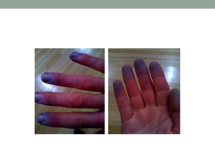
The handshake: • Cold, sweaty hands. . Anxiety. • Cold, dry hands…Raynaud Phenomenon. • Hot, sweaty hands…Hyperthyroidism. • Large, fleshy, sweaty hands. . Acromegaly. • Delayed relaxation of grip. . myotonic dystrophy • Dry, coarse skin. . hypothyroidism, regular water exposure, manual occupation. • Deformed hands. . dupuytrens contracture, rhumatoid arthritis
Facial expressions: • Poverty of expression…parkinsonism. • Startled expression. . hyper thyroidism. • Apathy w poor eye contact. . depression. • Apathy w pale puffy skin. . hypothyroidism. • Agitated expression. . anxiety, hyperthyroidism, hypomania.
Clothing • Young, dirty clothes. . poor state • Elderly, dirty clothes. . unable to look after themselves.
Complexion: • Yelloish_brownish tinge. . uremia • Bluish tinge…methaemoglobinemia(oxidized Hb). • Pink tinge…CO poisining. • bluish_grey…. metabolite of drugs e. g amiodarone.
Hb • Pallor…vasocostriction(pt who faint or frightend)…anemia • Pink flushing…vasodilation(perimenapausal women or carcinoid tumour) • Plethoric …polycythemia, cushing syndrome)
Cyanosis • Abnormal bluish discoloration of the skin and mm & require an absolute con of deoxygenated Hb of>50 g/l. ØCentral cyanosis. (hypoxemia, methemoglobinemia. . etc) ØPeriphral cyanosis. (cold, CHF, Pneumonia. . )
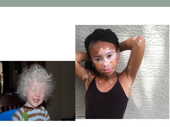
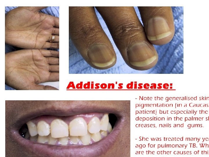
Melanin • Underproduction… ØVitiligo ØAlbinism ØHypopituitrism § Overproduction: ØAdrenal insufficiency ØCushing syndrome ØPregnancy & OCPs Øhemochromatosis
Carotene • Hypercarotenemia: v. Excessive intake of raw carotene & tomato vhypothyroidism
Iron • Bronzed diabetes…haemochromatosis • Erythema ab igne…. low level of heat chronic exposure
Sounds: • Normal speech depend upon: • Tongue • Lips • Palate • Nose • larynx
Abnormality of speech: • Hoarseness. . (infective laryngitis, heavy smoking, neurological causes) • Low pitch, slow speech. . (hypothyroidism) • Musical wheezing. . (breathlessness)
Odours • 1. 2. 3. 4. Excessive body odour occur in: Very elderly. Physical disability preventing normal hygiene. Alcohol or drug misuse. Severe learning difficulties.
Diagnostic odour: • Fetor hepaticus: liver failure • Sweet smell: DKA, starvation • Fishy smell: uraemia • Fetid smell: lung abcess
Halitosis: • Decomposing food bw teeth. • Gingivitis. • Stomatitis. • Atrophic rhinitis • Tumours of nasal passage. • Bronchiectesis.
Tremor: • Anxiety. • Hyperthyroidism. • B agonist.
Flaping tremor: • Hepatic failure • CO 2 retention
The hands Common abnormalities
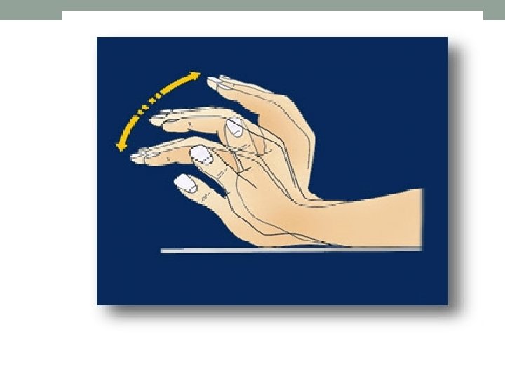
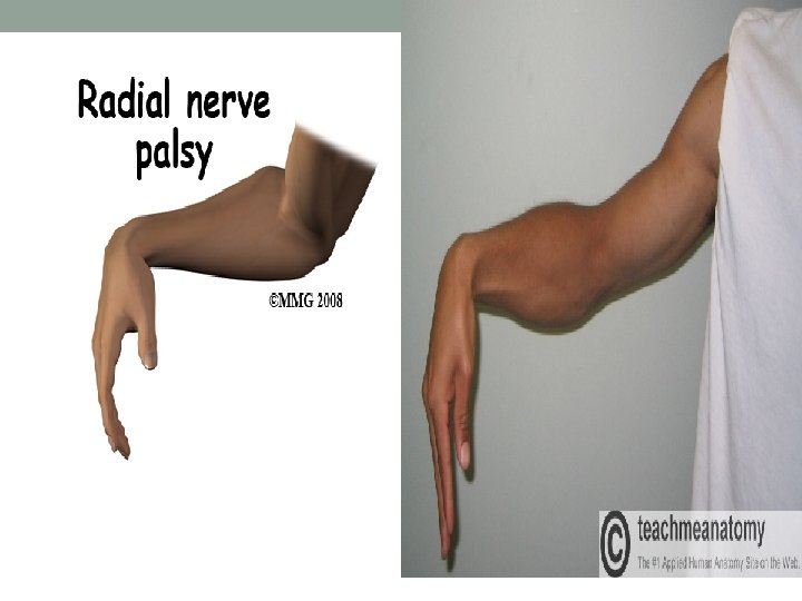
Posture: • Flexed hand arm in hemiplagia, radial palsy • Ulnar deviation in RA.
Shape: • Archnodactyly. . marfan syndrome. • Small metacarpal…pseudohypoparathyroidism.
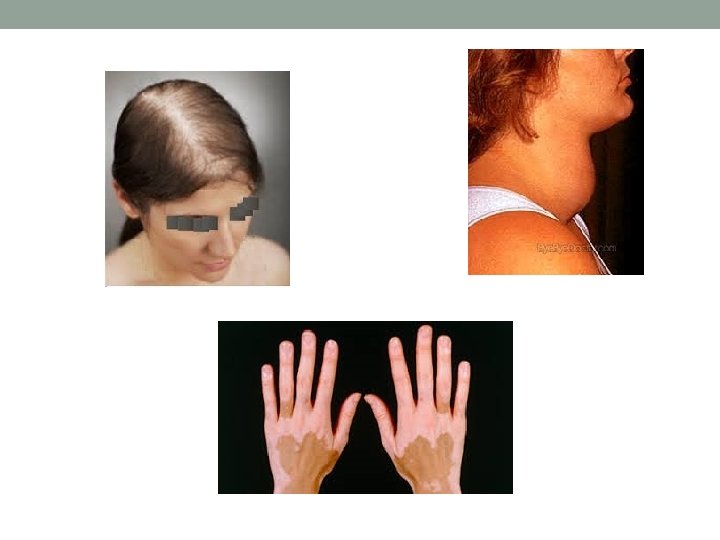
Size • Large broad fleshy hands…acromegally. • Thick soft tissue…myxodema. • Localized edema of the hand…venous or lymphatic obstruction.
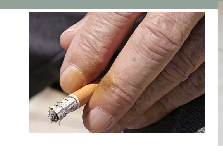
Colour • Tobacco staining of the fingers. • Pigmentation of creases
Tempreture: • Cold hands in HF • Warm in COPD, Hyperthyroidism
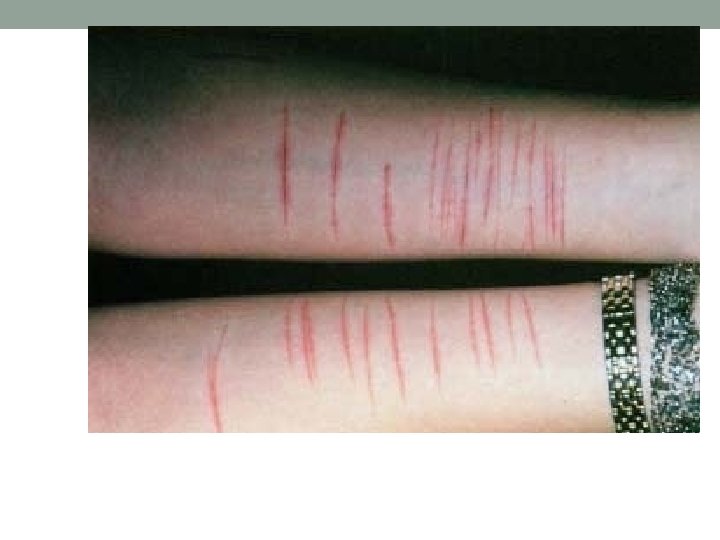
Skin • Hairless skin in male…hypogonadism • Multiple linear scars. . self cutting
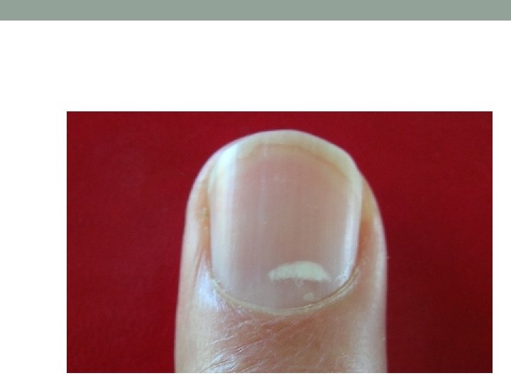
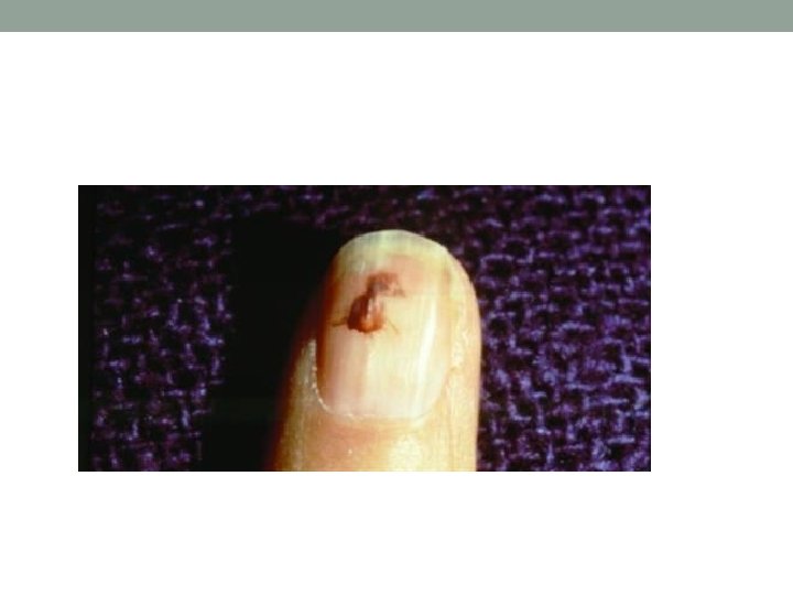
Nails • Koilonychia…IDA • Leukonychia…Hypoalbuminaemia • Beuas lines. . severe illness(malnutr, MI, liver dis. . etc) • Splinter Haemorrhage…infective endocarditis
Spoon nails - koilonychia
Beus line
Subcutaneus tissues • Thyroid acropachy
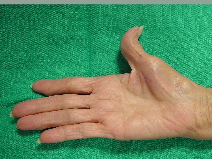
Dupuytren’s Contractures • Palmar or digital fibromatosis • Flexion contracture • Painless nodules near palmar crease • Male> Female • Epilepsy, diabetes, alcoholism
Joints: • Arthritis fequently involve small joints of the hand
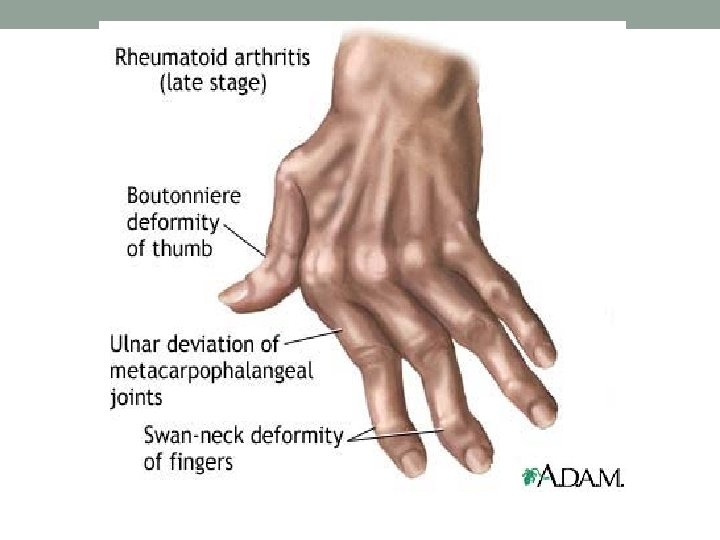
Rheumatoid Arthritis • MCP swelling • Swan neck deformities • Ulnar deviation at MCP joints • Nodules along tendon sheaths
Osteoarthritis • Heberden’s nodes: DIP • Bouchard’s nodes: PIP
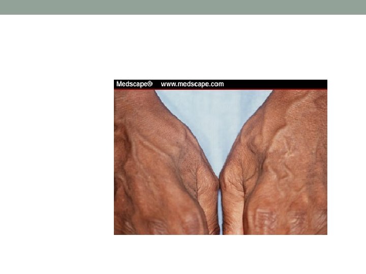
Muscles • Muscle wasting w dorsal guttering. . RA • Thenar muscle wasting…carpal tunnel syndrome
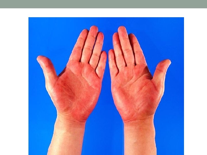
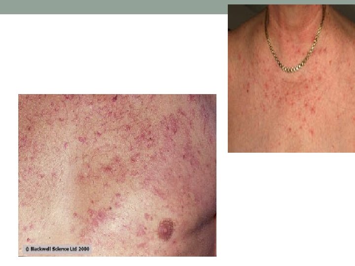
Blood vessels • Palmer erythema…hyperthyroidism, chronic liver disease. • Spider naevi. . chronic liver disease, thyrotoxicosis, HRT…
Lymph glands: • 1. 2. 3. 4. 5. Normally lymph glands are: Palpaple. size<=. 5 cm Soft rubbery consistacy. Not tender, Not fixed to underlying structure.
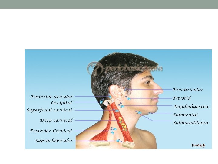
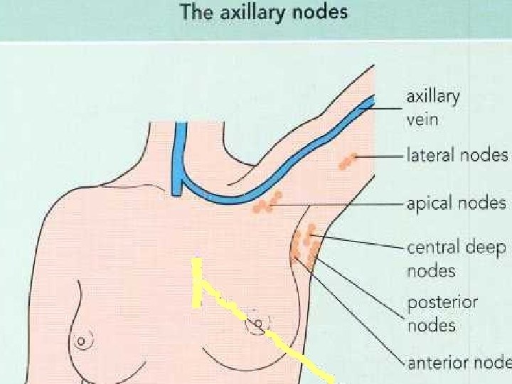
The Lymphatic System: Palpable superficial lymph nodes Occipital Posterior auricular Preauricular Tonsillar Submaxillary Submental Superficial cervial Deep cervical Supraclavicular Axillary Lateral Subscapular Pectoral Epitrochlear Inguinal
Examination sequence General principles : • Inspect for any visible lymphadenopathy. • Palpate one side at a time using the fingers of one hand. • Compare with the glands on the contralateral side. • Assess: Site, Size, consistency. • Record the measurements of the main glands. • Note any tenderness. • Determine if the gland is fixed to: surrounding and deep structures , skin.
Cervical glands • From behind, examine the submental, submandibular, preauricular, tonsillar, supraclavicular and deep cervical glands in the anterior triangle of the neck. • From the front of the patient, examine the posterior triangles, up the back of the neck and the posterior auricular and occipital nodes.
Axillary glands • From in front of the patient, support the arm on the side under examination. • Palpate the right axilla with your left hand vice versa. • place your finger tips into the vault of the axilla and then draw them downwards • feeling the medial, anterior and posterior axillary walls in turn.
Epitrochlear glands • Support the patient's right wrist with your left hand, grasp the patient's partially flexed elbow with your right hand use your thumb to feel for the epitrochlear gland. • Examine the left epitrochlear gland with your left thumb.
Inguinal glands • Palpate over the horizontal chain, which lies just below the inguinal ligament, • then over the vertical chain along the line of the saphenous vein.
Popliteal glands • Use both hands to examine the popliteal fossa with the knee flexed and limb muscles relaxed.
Causes of generalized lymphadenopathy: • Viral. . CMV, EBV, HIV • Bacterial…brucellosis, syphilis • Malignancy. . lymphoma, leukemia. • Inflammatory. . RA, SLE, sarcoidosis
localized • Infection • Malignancy. . mets, lymphoma
THE THYROID GLAND
THE THYROID GLAND • Examination sequence • Inspect the neck from the front. • Look for a thyroid swelling while the patient swallows a sip of water. • Ask the patient to sit with the neck muscles relaxed and stand behind the patient. • Place your hands gently on the front of the patient's neck with your index fingers just touching. Ask the patient to swallow a sip of water while feel over the gland. • Note the size, shape and consistency of any goitre, and the presence or absence of a thrill. • Measure any discrete nodules with calipers. • Listen by stethoscope for a thyroid bruit.
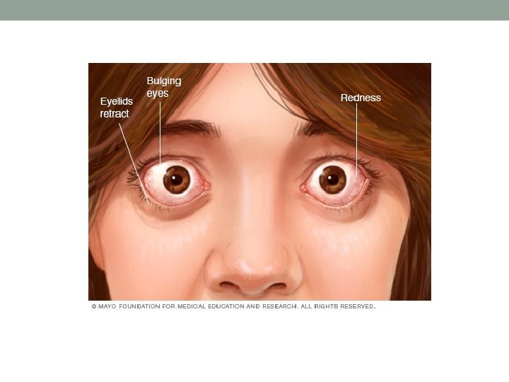
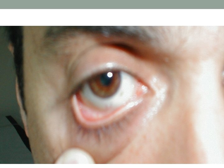
Abnormal findings • Shape and surface. • Simple goitres are relatively symmetrical in their earlier stages but often become nodular with time. • The surface of the thyroid gland is usually smooth and diffuse in Graves' disease. • Mobility. • Most goitres move upwards with swallowing. However, invasive thyroid cancer may fix the gland to surrounding structures. • Very large goitres may also be immobile. • Consistency. • A very hard consistency suggests malignant change in the gland. • Large, firm lymph nodes near a goitre also suggest thyroid cancer. • Tenderness. • Diffuse tenderness is typical of viral thyroiditis. • localized tenderness may follow bleeding into a cyst. • Thyroid bruit. • hyperthyroidism.
SWELLINGS • • • Size Position Attachments Surface Edge Consistency Transillumination Inflammation: redness tenderness heat Thrills, bruits and noises
MEASUREMENT OF HEIGHT AND WEIGHT • Body mass index • Underweight < 18. 5 • Normal 18. 5 -24. 9 • Overweight 25 -29. 9 • Obese 30 -39. 9 • Morbid obesity =40
Cot… • Body mass index does not describe the distribution of body fat. • To assess this measure waist circumference, which correlates with visceral fat and indirectly measures central adiposity. • There is an increased risk to health when waist circumference exceeds 94 cm (37 inches) for men and 80 cm (32 inches) for women.
Tall stature: • Abnormal causes of increased height are: • Marfan's syndrome • hypogonadism • pituitary gigantism.
Short stature: • usually familial. • any significant childhood illness will reduce the rate of growth and may , limit final height. • renal tubular acidosis • intestinal malabsorption • hypothyroidism. • compression fractures of the spine due to osteoporosis.
Obesity • It is associated with: • sleep apnoea • hypertension, • Hyperlipidaemia • type 2 diabetes mellitus • certain cancers • shortens life expectancy.
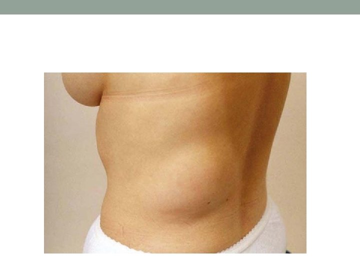
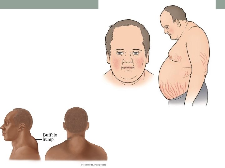
Specific abnormalities of fat distribution: • Lipomas are commonly found around the trunk and are benign soft, fluctuant, circumscribed, lobulated swellings. • Localized atrophy of subcutaneous fat may develop in diabetic patients at the sites of insulin injection. • In Cushing's syndrome the distribution of fat is abnormal. Fat deposition tends to be centrally distributed.
MEASUREMENT OF TEMPERATURE : • The 'normal' oral or ear temperature is 37°C but may range between 35. 8 and 37. 2°C. • Rectal temperature is usually about 0. 5°C higher than in the mouth which, in turn, is 0. 5°C higher than the axilla, but the axilla is not recommended as a site for reliable measurement. • Body temperature may be taken beneath the tongue, or in the rectum or the external auditory meatus. • Skin warmth to the touch usually provides an indication of fever, but the skin of a patient with a normal temperature may feel cold, and an apparently normal skin temperature does not exclude hypothermia.
Increase in body temperature • heat illness. • Fever is an increase in body temperature caused by a cellular response to infection, immunological disturbance or malignancy. • A rigor is an episode of shivering or shaking followed by excessive sweating that follows a rapid increase in body temperature.
Hypothermia: • Hypothermia is a core temperature < 35°C. It is easily missed and may mimic clinical death. • rectal temperature the best approximation to core temperature.
Causes of hypothermia: • elderly immobile patients living alone, particularly during the winter • water immersion and near-drowning • prolonged unconsciousness in low ambient temperatures, esp with alcohol intoxication • drug overdosage. • stroke or head injury • severe hypothyroidism
Clinical features of hypothermia: • 36°C Increased metabolic rate, vasoconstriction • 35°C (hypothermia) Shivering maximal, impaired judgement • 34°C Uncooperative • 33°C Depressed conscious level • 28_32°C (severe hypothermia) Progressive depression of conscious level, muscle stiffness, Failure of vasoconstrictor response and shivering, Bradycardia, hypotension, J waves present on ECG, risk of arrhythmias • 28°C< Coma, patient may appear dead, absent pupillary and tendon reflexes Spontaneous ventricular fibrillation • 20°C Asystole/profound bradyarrhythmias
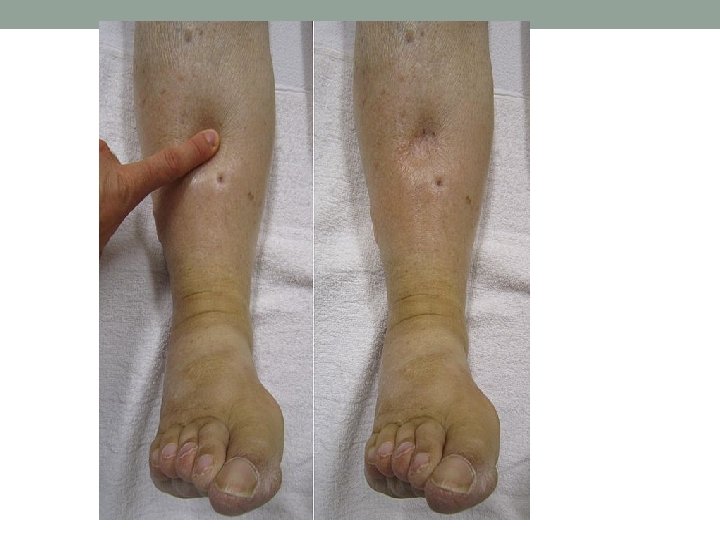
ASSESSMENT OF HYDRATION • Assess the state of hydration by testing skin turgor. Gently pinch a fold of skin on the neck or anterior chest wall, hold it for a few seconds, then release it. • Record the pulse rate and supine/erect blood pressures. Look for postural hypotension (a fall > 15 mm. Hg) in systolic pressure. • Check for oedema in the ankles and legs. In patients in bed check for sacral oedema. • Examine the jugular venous pulse (JVP).
Oedema • Oedema means swelling of tissues due to an increase in interstitial fluid. • Generalized oedema may be due to a disorder of the heart, kidneys, liver or gut or may be nutritional in origin. • Localized oedema may arise from venous or lymphatic obstruction, allergy or inflammation. • Postural oedema is relatively common in the lower limbs of inactive patients.
Clinical manifestations of oedema • When oedema is due to generalized fluid retention, its distribution is determined by gravity. It is usually found in the legs, backs of the thighs and the lumbosacral area in the semirecumbent patient. If the patient lies flat, it may involve the face and hands, as in children with nephrotic syndrome. • The cardinal sign of subcutaneous oedema is pitting of the skin, made • • by applying firm pressure with your fingers or thumb for a few seconds. The pitting may persist for several minutes until it is obliterated by the slow return of the displaced fluid. Pitting on pressure may not be demonstrable until body weight has increased by as much as 10 -15%. Day-to-day alterations in body weight are usually the most reliable index of changes in body water. Hypothyroidism is characterized by mucinous infiltration of the tissues (myxoedema). In contrast to oedema. myxoedema and chronic lymphoedema do not pit on pressure.
Generalized oedema • There are two principal causes of generalized oedema: 1. 2. hypoproteinaemia fluid overload. • These may be distinguished by the jugular venous pulse (JVP), which is usually elevated in fluid overload but not in hypoproteinaemia.
Hypoproteinaemia • Inadequate protein intake or absorption • Kwashiorkor develops when there is inadequate intake of protein. • Failure of digestion of dietary protein results from impairment of pancreatic exocrine function, as in chronic pancreatitis. • Malabsorption of the products of digestion may occur in conditions such as Crohn's disease or coeliac disease, or after extensive small bowel resection.
Hypoproteinaemia • Inadequate protein production • Reduced synthesis of albumin is found in chronic liver disease including cirrhosis. • When the portal venous pressure is high, ascites may be more prominent than dependent oedema.
Hypoproteinaemia • Excessive loss of protein • Heavy protein loss in the urine (> 5 g/day) results in the nephrotic syndrome. • Repeated removal of body fluids, especially ascites, may cause protein depletion. • protein-losing enteropathy, e. g. coeliac disease. • In addition, a low intravascular volume may cause secondary hyperaldosteronism via the reninangiotensin system, promoting sodium retention and causing a further increase in oedema.
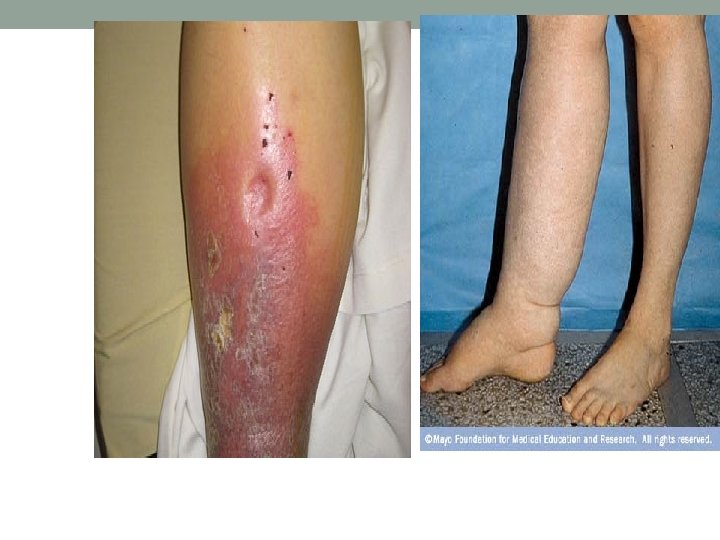
Causes of Localized oedema • Venous causes • increased venous pressure. This may be a problem with the vein itself, e. g. external pressure from a tumour, thrombosis, or valvular incompetence from previous thrombosis or surgery. • Conditions which impair the normal pumping action of muscles, e. g. hemiparesis, forced immobility. • Oedema may accordingly occur in immobile, bedridden patients, in a paralysed limb, or even in a healthy person sitting for long periods, e. g. during air travel.
Causes of Localized oedema • Lymphatic causes. • oedema (lymphoedema). If the condition persists, fibrous tissues proliferate in the interstitial space and the affected area becomes hard and no longer pits on pressure. Lymphoedema is common in some tropical countries because of lymphatic obstruction by filarial worms. The legs, external genitalia and the female breast are most frequently involved. Eventually the skin in affected areas becomes very thick and rough (elephantiasis). • in. UK, lymphoedema is most often due to congenital hypoplasia of the lymph vessels of the legs (Milroy's disease), • recurrent lymphangitis (resulting in lymphatic fibrosis), or may affect an arm after radical mastectomy and/or irradiation for breast cancer.
Causes of Localized oedema • Inflammatory causes. • Any cause of tissue inflammation including infection, injury or ischaemia, liberates mediators which cause vasodilatation and increase capillary permeability. • Allergic causes.
THANK YOU!
sources • Macleod’s Clinical Examination • www. medescape. com • www. healthline. com • www. nailsmag. com