Clinical examination in orthopaedics Z Rozkydal L Pazourek


 height 2 m 2 • •")


 - 0 - flexe (plantiflexe) 20 - 50")


 BMD-")


- Slides: 74
Clinical examination in orthopaedics Z. Rozkydal L. Pazourek
Clinical examination The aim- establish the diagnosis 1. History 2. Objective examination - general 3. Objective examination - local 4. Laboratory tests 5. Imaging methods
History Family Personal Pharmacological Social Occupation Epidemiological Current problems Analysis of pain
Family • Congenital abnormalities • Important diseases in family (heart, DM, haemophilia, oncological diseases, neurological diseases, TB • Birth, miscarriage
Personal • Important general diseases (hypertension, DM, heart, tumors, lung problems • Coagulopathies • Infections • Injuries: consequences, complications • In children- pregnancy psychomotor development
Current symptoms Local Pain, motor function, limping, deformity, ROM, swelling, loss of sensation General: fever, shivering, cachexia Cause of the problem – – injury overloading Infection systemic diseases (endocrine, metabolic, inflammation, neurological, haemotological. )
• Development of symptoms – Onset, duration – Intensity – Aleviation, increasing factors • Present management – Examination in the past time – Conservative therapy – Operative therapy • Mobility, occupation • Emotions, psychological condition • Simulation, dissimulation, aggravation
Analysis of pain Intensity, frequence, duration Acute, chronic Local, irradiating Visceral Type- sharp, blunt, burning, stubbing Neuralgia Nerve root pain Phantom pain Neurogenic claudication
Analysis of pain Localised, diffuse Psychological background Durig activity or in rest VAS – visual analogue scale Scale of ten degrees 0 - no pain 10 - the most sever pain not bearable Pain 5 or more- change of management
Pharmacology • Medicines used currently • Important medecines: warfarin, heparin, other anticoagulants, antiepileptics, cytostatics, immunosuppresives, NSA, corticoids, biological treatment, • Alcohol, smoking, drugs • Alergy (antibiotics, metal, dissinfections)
Occupation and social • • Occupation, type of work, manual labor Rent Social situation (living, marriage) Subsequent management
Gynecological history • Cycles, gravidity, menopause, current gynecological problems • Epidemiologiocal history influenza, viral infections, herpes simplex, focal infections (UTI, stomatological infections, ulcers, erysipel)
Objective examination General orthopaedic examination Local orthopaedic examination Posture and gait
Somatotype asthenic pycnic normosthenic
Gigantisms Fröhlich syndrom Nanisms Achondroplasia Marfan syndrom
Nutrition • Body mass index: weight kg (BMI) height 2 m 2 • • • Below 20 - cachexia 20 -25 - normal weight 25 -30 - overweight 30 -35 - obesity Over 35 - severe obesity
Skin • Colour
• pigmentation, naevus • Trophicity, turgor
• Fistulas, ulcers • Subcutaneus nodes • nails • Lymfadenopathy, soft tumors, inflammations
Swelling • Local • General • Anasarca • Decollement Local signs of inflammations: readness, swelling, pain, warm, limited function, soft mass, effusion, discharge
Soft mass
• Haematoma • Lymphonodes • Tumor
Effusion
Congenital deformity • 1. Shape, size • 2. Differential • 3. Duplicity • 4. Gigantisms • 5. Hypoplasia
Malalignment • varus x valgus • antecurvation x recurvation • rotation deformity odchylka
Deformity of spine • Scoliosis • Hyperkyphosis, hyperlordosis
Hand deformities RA Boutonniere deformity Swan neck deformity OA
Foot deformities
Length of extremity Lower extremity – Spinomaleolar distance – Umbilicomaleolar distance – Support during standing – X- ray of the hip, knee, ankle joint Upper extremity: acromion- 3. finger • Circumferential measurement
ROM • Active and passive movements • • Sagital Frontal Transversal = horizontal Rotation
Shoulder S: extenze - 0 - flexe 50 - 180 F: abdukce - 0 addukce 180 - 25 T: abdukce - 0 addukce 110 - 30 R: ZR - 0 - VR 90 - 90
Elbow S: extenze - 0 - flexe 10 - 150 R: supinace - 0 - pronace 90 - 90
Wrist F: rad. dukce - 0 - uln. dukce 20 - 40 S: extenze (dorz. flexe) - 0 – flexe (palm. flexe) 80 - 80
Hip S: extenze - 0 - flexe 15 - 0 - 140 F: abdukce - 0 addukce 60 - 40 T: abdukce - 0 addukce 80 - 30 R: ZR - 0 - VR 50 - 40
Knee S: extenze - 0 - flexe 0 - 140
Ankle S: extenze (dorziflexe) - 0 - flexe (plantiflexe) 20 - 50
Ancylosis • Extrarticular • Intraarticular
Stability of joints • Stable joint • Unstable joint • Instability – acute – chronic – habitual
Shoulder Apperhension test Drawer sign
Knee
Laxity • test
Maneuvers • Maneuvers
Sound phenomenons • Crepitus
Contracture • Lumbago, torticollis • Cerebral palsy
Muscles • Trophicity • Tonus • Cramps • Power
Muscle test 0 1 2 3 4 5 - no activity - trace - motion without gravity - motion against gravity and slight resistance - normal activity 0% 10 % 25 % 50 % 75 % 100 %
Posture Correct Wrong
Gait • 1. heel strike 2. standing 3. toe off 4. swing phase
Limping • • Antalgic gait Shortening of a lower extremity Ancylosis Trendeleburg sign and gait Hemiparetic gait Spastic gait Drop foot gait Parkinson gait
Imaging methods • • X-ray, artrography Angiography Ultrasonography CT, MRI Scintigraphy DEXA Biopsy
X-ray In two planes - bone hypertrophy - bone atrophy - osteolysis - osteonecrosis
Kellgren- Lawrence clasification of O. A. I. III. IV.
Fistulography Artrography
Angiography Clasical CT angiography MR angiography Digital subtraction angiography
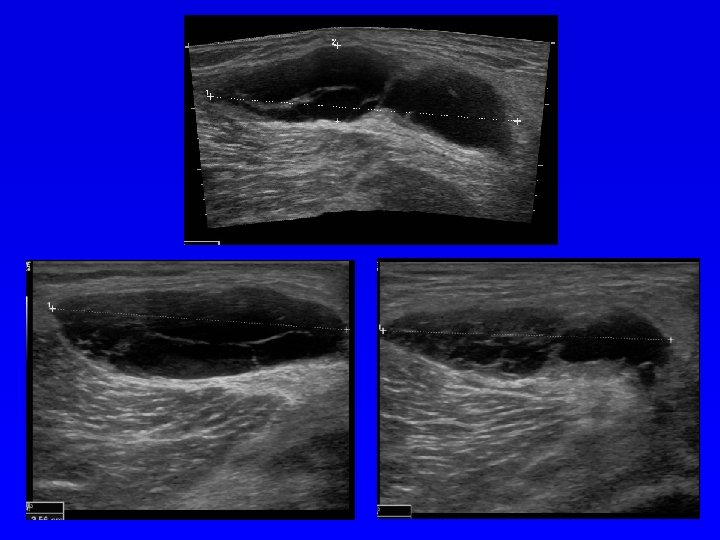
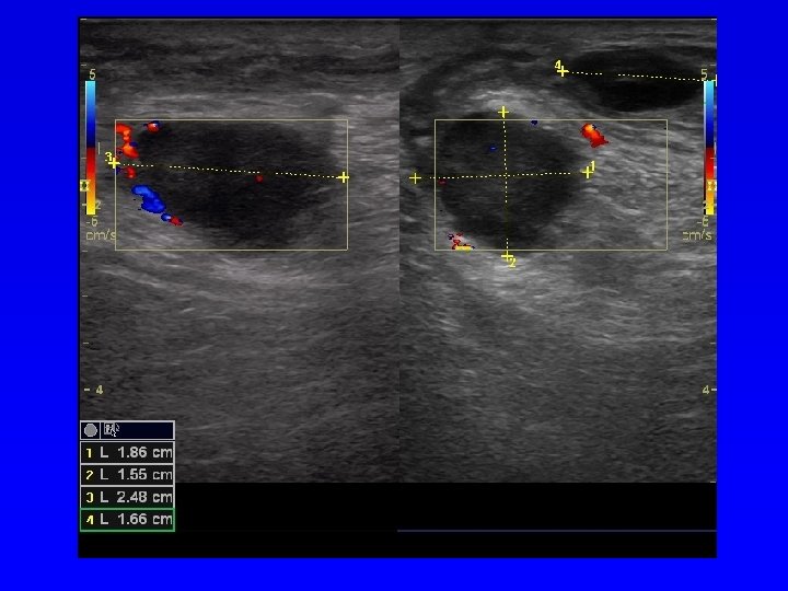
Ultrasonography Echogenity of tissues Bone, fibrous tissue, muscles, adipous tissue, cartilage, fluid Anechogenic structure- black Hypoechogenic structure- grey Hyperechogenic structure- white Soft tissues Tumors DDH Effusion in joints
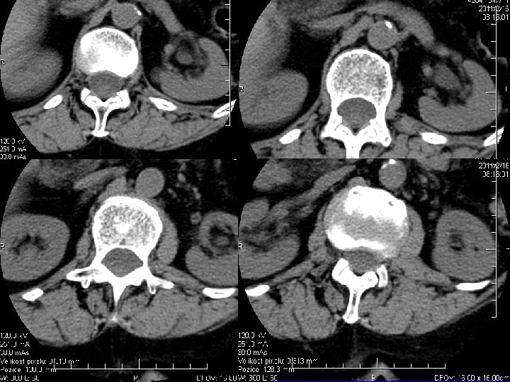
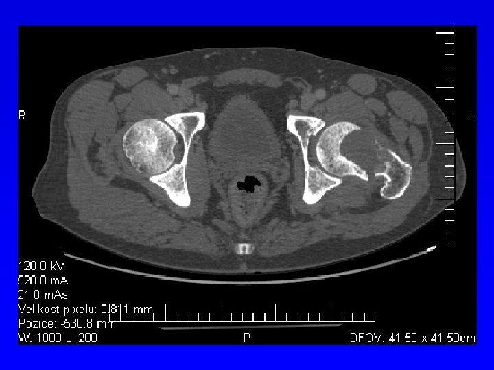
CT scann Absorption of X-ray beams Air – 1000 H. U. Water 0 H. U. Bone + 1000 H. U. Enhancement with a dye Bone lesions Bone tumors
CT
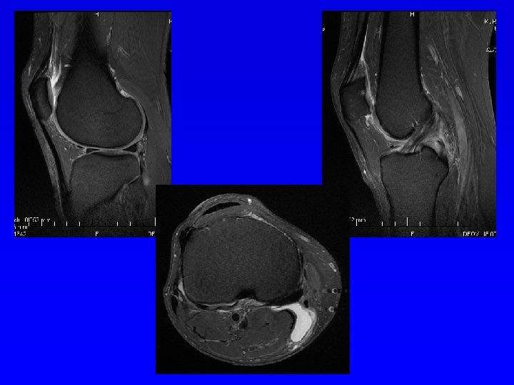
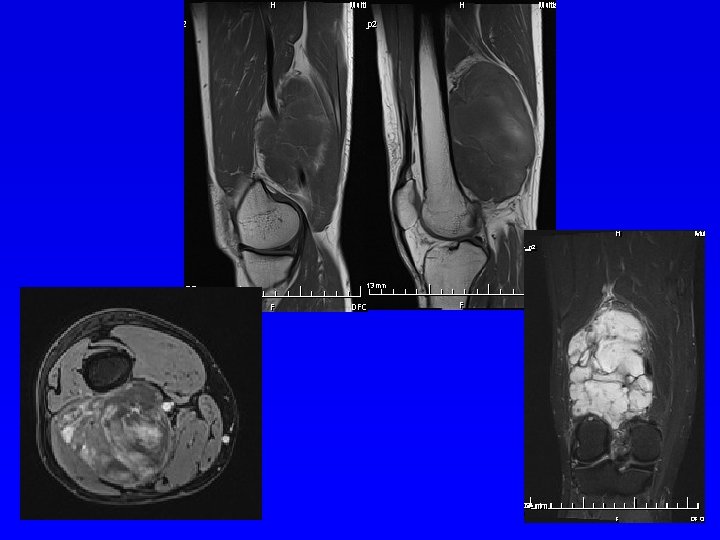
MRI Magnetic field Hyposignal- dark Hypersignal - white Soft tissue tumors Soft tissue mass Spine
MRI
Perimyelography Myelography Radiculography
Scintigraphy
Densitometry DEXA Absorption of X-ray of two energies (70 and 140 k. V) BMD- bone mineral density in g/cm 2 T- score - difference from peak bone mass Z- score - difference in the same age Change - difference from previous examination 1 SD = 10% of bone mass
DEXA
WHO definition of osteoporosis
Laboratory tests • Inlammation: ESR, leu, CRP, differencial, ELFO • Osteopathy: Ca, P, ALP, bone isoenzyme of ALP osteokalcin, osteonectin, PTH, vitamin D • Bone markers- PSA
Biochemistry • • Proteins Glucose Lactate Uric acid
Joint effusion • • • Microscopic Biochemic Bacteriologic Immunologic Cytologic
Biopsy Histological examination Biopsy – CT, ultrasonograpy