Semen analysis Introduction n A semen analysis measures


 are very small mucus secreting")


, sterile, nontoxic,")


. Using either a graduated cylinder with a")


 Motility")


 Eosin")
 and dead (stained) spermatozoa")
, increase")


- Slides: 45
Semen analysis
Introduction n A semen analysis measures the amount of semen a man produces and determines the number and quality of sperm in the semen sample. A semen analysis is usually one of the first tests done to help determine whether a man has a problem fathering a child (infertility). A problem with the semen or sperm affects more than one-third of the couples who are unable to have children (infertile).
Purpose of seminal fluid analysis n There are basically four indications for the examination of seminal fluid: 1. 2. 3. 4. The investigation of fertility: male infertility is primarily responsible in 30%-50% of infertile marriages. To determine the effectiveness of vasectomy. To determine the suitability of semen for artificial insemination. Medicolegal: testes to detect semen are frequently requested in alleged rape or in association with other sexual crimes of violence.
Fluid Fractions 1. Bulbourethral & Urethral glands (2 -5%) are very small mucus secreting glands, add alkaline mucus to neutralize prostatic acid and vaginal acidity 2. Prostate: (produce about 13 -33 % of the fluid volume of semen) Prostate glands secretion is a milky, alkaline fluid that plays a role in activating sperm, the secretion contains acid phosphatase and proteolytic enzymes that act on the fluid from the seminal vesicles, resulting in the coagulation and liquefaction of the semen.
3. 4. Seminal vesicles (produce about 46 -80 % of the fluid volume of semen) Viscous, yellowish secretion is rich in fructose, vitamin C, prostaglandin, and other substances, which nourish and activate the sperm passing through the tract. This component has high flavin content, which is largely responsible for the fluorescence of semen. Testis & Epididymis: (5%) Spermatozoa are produced in the testis under the influence of testosterone, and then the epididymis (is the first part of the duct system) provides a temporary storage site for the immature sperm that enter it from testis. This fraction still in the inactive form until ejaculation due to the high content of carnitine, glyceryle-phosphorylcholine and diminished oxygen supply.
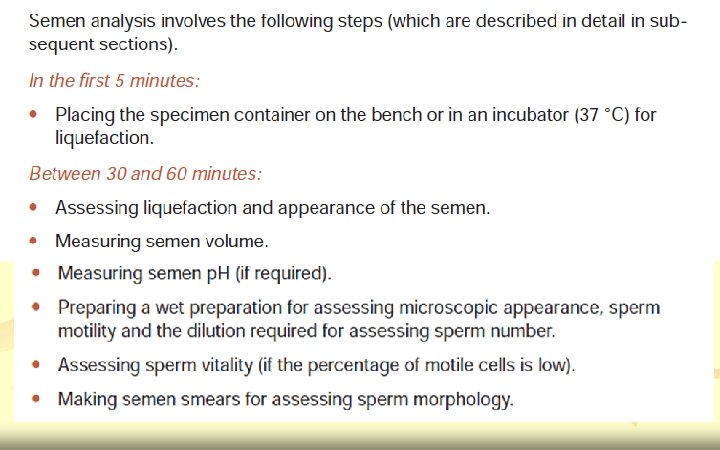
Coagulation and liquefaction q Coagulation and subsequent liquefaction are believed to be three stage processes: § § § Coagulation results from the actions of a prostatic clotting enzyme on a fibrinogen-like precursor formed by the seminal vesicles. Liquefaction is initiated by enzymes of prostatic origin. The protein fragments are degraded further to free amino acids and ammonia by the action of several poorly characterized proteolytic enzymes, including an amino peptidase and pepsin. Clearly, a semen analysis should not be performed immediately following sample production. The sample should be mixed well in the original container by swirling for several seconds prior to removing the lid. Do not invert the container.
Specimen collection n Specimen should be collected into prewarmed (21 o. C), sterile, nontoxic, wide-mouth container, after a couple has abstained from sexual activity for 2 -3 days. Verbal and written instructions should be given to the patient to ensure appropriate collection & delivery of semen sample to the laboratory. Ideally the sample should be collected in a room set aside for this purpose at the clinic laboratory in order to reduce ejaculation-analysis interval but this is not always possible. The patient should be advised to urinate and then wash and dry his hands and genitals thoroughly prior to ejaculation to avoid bacterial contamination. It is important to note that contamination of the semen sample with either soap or water may adversely affect sperm quality.
Methods of collection 1. 2. 3. 4. Masturbation (the method of choice for all seminal fluid tests) The use of condom: it is not recommended for fertility testing because the condoms may contain spermicidal agents (used to determine the effectiveness of vasectomy). By coitus interrupts (withdrawal method): the sample may be mistimed and part of the ejaculate may thus be lost. TESA: Testicular sperm extraction (TESE)
Labeling n The sample should be clearly labeled with: 1. 2. 3. the patient's name ID or clinic number (if available) Date and time of sample collection.
• • The following should be recorded on the laboratory analysis form: 1. The period of abstinence (in days). 2. If sample collection was complete or incomplete. 3. The time interval from collection to analysis. The sample should be transported upright, at body temperature if possible, and should be delivered to laboratory as soon as possible after collection and certainly within one hour of ejaculation. If the sample is cold on receipt, this should be noted in laboratory records. Patients should be advised not to expose the sample to extremes of temperature.
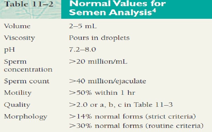
Examination of seminal fluid n When evaluating semen specimens in cases of infertility, the following parameters are routinely measured: ü ü ü volume viscosity p. H sperm count (concentration) motility morphology.
Macroscopic examination n n After ejaculation, the seminal secretions form a coagulum, which gradually liquefies 10 -20 min. In most cases, the semen sample should become fully liquefied within 60 minutes of production. Once liquefaction is complete then the physical appearance of the sample should be recorded in the laboratory records. If liquefaction does not occur then this abnormality should be noted.
Viscosity of the ejaculate n n n Estimate the viscosity of the semen by aspirating the semen into the measuring pipette and allowing the semen to drop by gravity and will not appear clumped. Observe the length of the thread. With excessively viscous samples, thorough mixing can be difficult and accurate estimation of sperm concentration and Normal droplets form a thin thread when released from the pipette. Droplets with threads longer that 2 centimeters are considered highly viscous. Ratings of 0 (watery) to 4 (gel-like) can be assigned to the viscosity report. Viscosity can also be reported as low, normal, and high. Increased viscosity and incomplete liquefaction impede sperm motility
Volume n Normal is (2 -5 milliliters). Using either a graduated cylinder with a conical base or a disposable wide- mouthed pipette (accurate to 0. 1 ml) measure the ejaculate volume to the nearest 0. 1 ml. n Excessively small or large volumes are important in the transport of semen within the female reproductive tract and should be noted. n The volume may be low if a man is anxious when producing a specimen, if all of the specimen is not caught in the collection container, or if there are hormonal abnormalities or ductal blockages.
Color of seminal fluid n Semen is normally a gray-yellow opalescent fluid. Its opacity is due to the most part, to its high protein content but is of course also produced by the many millions of spermatozoa as well as the cellular debris that is normally suspended within it.
PH n n n The normal p. H of semen is slightly alkaline (7. 2 - 8. 0) but increases with time. Increased p. H is indicative of infection within the reproductive tract. A decreased p. H is associated with increased prostatic fluid.
Microscopic examination
Microscopic examination 1. 2. 3. 4. Concentration (sometimes referred to as the "count") Motility (sometimes referred to as the "mobility") Agglutination Morphology
Concentration "count" n n This is a measurement of how many million sperm there are in each milliliter of fluid. There are various techniques for obtaining this number - some prove to be more accurate than others are. Average sperm concentration is more than 60 million per milliliter (60 -150 million/ml). Counts of less than 20 million per milliliter (<20 million/ml) are considered subfertile.
n n n Several terms are used to describe both sperm concentration and sperm count: Azoospermia describe a total absence of spermatozoa in semen. (After centrifuge sperm count is zero/HPF). Oligozoospermia refers to a reduced number of spermatozoa in semen and is usually used to describe a sperm concentration of less than 20 million/ml. Sperm count 5 -10 sperm/HPF. Severe oligospermia, sperm count 1 -2 sperm/HPF. Polyzoospermia denotes an increased number of spermatozoa in semen and is usually refers to a sperm concentration in excess of 350 million/ml.
Methods of measuring sperm concentration A. By using hemacytometer 1. 2. 3. 4. The sperm count is performed in the same manner as blood and CSF counts; that is by diluting the specimen and counting the spermatozoa in a neubauer chamber. Sperm can be counted by make dilution 1: 20 in WBC pipette or by automatic pipette (which is more accurate) with a solution containing sodium bicarbonate (5 g) and formalin (1 ml) (immobilize & preserve the spermatozoa), tap water (100 ml) will suffice as a diluent. The sperm should then be counted - do not count headless or "pinheads" sperm and do not count tailless heads. Traditionally, the sperm concentration is expressed in millions per milliliter (x 106/ml) of semen and the total sperm/ejaculate is reported in millions (x 106) per ejaculate.
Calculations 1. 2. Using a 1: 20 dilution and two large WBC’s squares counted The sperm concentration/ml = No of sperms counted x 100, 000 Using a 1: 20 dilution and five small RBC’s squares counted The sperm concentration /ml = No of sperms counted x 1, 000
n Using a 1: 20 dilution, an average of 60 sperm are counted in the five RBC counting squares on both sides of the hemocytometer. Calculate the sperm concentration per milliliter and the total sperm count in a specimen with a volume of 4 m. L. n 60 sperm counted x 1, 000 = 60, 000 sperm/m. L n 60, 000 sperm/m. L x 4 m. L = 240, 000 sperm/ejaculate
B. n Direct smear The application of a drop of well-mixed semen to a clean glass slide under a lightly applied glass coverslip will allow visualization of the sperm in a specimen of semen.
Motility "mobility" n This describes the percentage of sperm, which are moving. 50% or more of the sperm should be moving. In order to achieve fertilization, a sperm must not only be able to move but be capable of movement that results in forward progression is often also known as progressive activity.
n There are four classifications of motility 1. 2. 3. 4. Rapid progressive motility - the sperm are moving swiftly across the field usually in a straight line Slow or sluggish progressive motility - the sperm may be less linear in their progression Non-progressive motility - sperm are also described as twitching or shaking Immotility - sperm do not move at all.
Vitality Assessment 1. 2. 3. 4. n n Eosin-nigrosin (dead sperm stain pink/red) Eosin (1%) (dead sperm stain pink/red) Trypan (0. 4%) blue (dead sperm stain blue) Hypo-osmotic swelling test (HOS) (live sperm shows tail curling Test 1, 2 and 3 for diagnostic uses. Usually 1: 1 ratio of semen to dye mixture, mix well and smear onto a slide. Read immediately at x 40 objective, count 200 sperms Test 4 is use to choose live (immotile) sperm for ICSI. Dead sperms will not react in HOS while live sperm will take up fluids causing their tails to curl within 5 min and stabilize at 30 min. Therefore viable sperms may be selected for ICSI
n Eosin stain is used to differentiate live (unstained) and dead (stained) spermatozoa
Other cells in semen 1. 2. 3. 4. 5. leukocytes normally (1 -4/HPF), increase number (leukocytospermia) indicates reproductive tract infection Epithelial cells normally (1 -2/HPF) Spermatocytes (Immature germ cells) 1 -2/HPF. Erythrocytes (1 -2/HPF). Increased number may indicate a reproductive tract infection or damage to a small capillary during sample production. Bacteria and protozoan such as Trichomonas vaginalis are uncommon in human semen but their presence is indicative of possible male reproductive tract infection and should be reported to the referring doctor further evaluation.
Agglutination n n The presence of agglutination should be recorded as this may indicate immunological infertility. Assess the spermatozoa in 10 random fields - estimate the average percentage of spermatozoa clumped together to the nearest 5%. Only count motile sperm attached to other motile sperm - do not assess immotile sperm stuck together or motile sperm adhering to mucus threads, other cells or debris , this is non-specific aggregation.
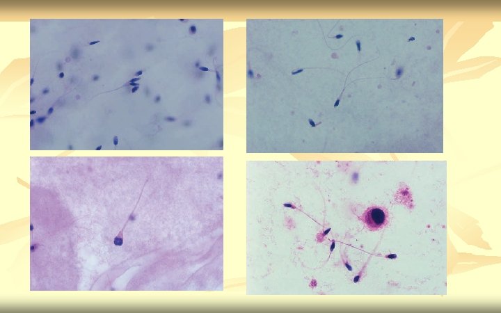
Morphology n n This describes the shape of the sperm. 70% of the sperm should be normal by these criteria. Generally accepted that a high incidence of morphologically abnormal spermatozoa in a semen sample is associated with reduced fertility. Human sperm can be visualized using bright field microscopy on fixed stained specimens.
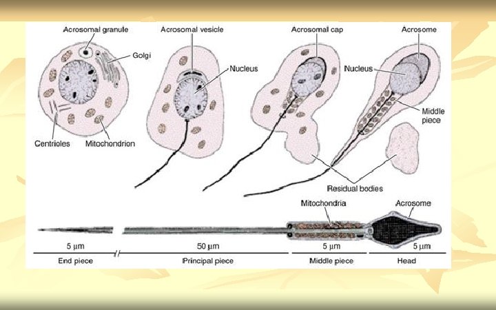
n n n Examples of fixed stained preparations (Papanicolaou stain, Vital staining with eosin/nigrosin, giemsa stain). Normal spermatozoa should have an oval shaped head (4 -5. 5µm long and 2. 5 -3. 5µm wide). The midpiece should be cylindrical (3 -5µm long and 1. 0µm wide). The tail should also be cylindrical (45 -50µm long and 0. 5µm wide) with a narrower terminal segment (4 -6µm long). There should be no head, midpiece or tail defects, and no cytoplasmic droplet more than one-third the size of a normal sperm head.
n Normal spermatozoa structure
Defects to be scored n n Head shape/size defects - such as large, small, tapering, pinhead form, amorphous, vacuolated, multiple heads or any combination of these. Neck and midpiece defects - such as non-inserted or bent tail, distended, irregular / bent midpiece, thin midpiece (no mitochondrial sheath), absent tail (free or loose heads) or any combination of these. Tail defects - such as short, multiple, hairpin, broken, irregular width, coiled tails, tails with terminal droplets or any combination of these. Cytoplasmic droplets - greater than one-third the size of a normal sperm head.
n n Each spermatozoa is scored as either normal or abnormal with each of the defects being tallied separately. If a majority of the cells have a particular morphological defect this should also be noted. In stained preparations 100 -200 sperm should be scored using a x 100 oil-immersion bright field objective
Abnormalities of sperm heads and tails are illustrated
Hematoxylin-Eosin Staining o o o Hematoxylin-Eosin Fix slide in Et. OH/Me. OH 95% Wash in running tap water Dry on absorbent paper Hematoxylin (Sigma, HHS-128) Wash in running tap water Acid alcohol (99 ml 70% Et. OH + 1 ml H 2 SO 4) Eosin (Sigma, HT 1102128) Et. OH 70% Et. OH 90% Absolute Et. OH (99. 9%) Xylene n 20 min 5 min Dip (2) 5 min 2 min (2) n n Fairly good differentiation The acrosomal area and cytoplasmic fragments is stained pink and the post -acrosomal area is stained dark purple. Abnormally stained sperms (nuclear/ chromatin material) may be differentiated. Takes longer and need experience to produce good staining
Abnormal Sperms
Abnormal Sperms Triple head. 1 sperm Acrosome. 2 reacted sperm Sperm with no. 3 acrosome Sperm with a. 4 tapering head and swollen midpiece