
Neurological Examination Sherif Elwatidy MSc FRCSSN MD Professor
- Slides: 41
Neurological Examination Sherif Elwatidy MSc, FRCS(SN), MD Professor of Neurosurgery, College of Medicine - KSU
Neurologic History • Like history in Medicine & Surgery – Personal history – History of the present complaints – Social History – Past medical History
• From the history we should be able to answer 2 important questions: 1. Where is the problem ? (brain, spine –Cx. , Thoracic, lumbar) 2. What is the nature of the problem ? (Congenital, inflammatory, neoplastic, degenerative, …. )
The objective of a neurological exam is threefold. 1. To identify an abnormality in the nervous system. 2. To differentiated peripheral from central nervous system lesions.
Neurologic examination includes: I- General Appearance, including posture, motor activity, vital signs and perhaps meningeal signs if indicated. II- Mini Mental Status Exam, including speech observation. III- Cranial Nerves, I through XII. IV - Motor System, including muscle atrophy, tone and power. V- Sensory System, including vibration, position, pin prick, temperature, light touch and higher sensory functions. VI- Reflexes, including deep tendon reflexes, clonus, Hoffman's response and plantar reflex. VII- Coordination, gait and Rhomberg's Test Examining the comatose patient
General appearance • • • Level of consciousness Personal hygiene and dress Posture and motor activity Height build and weight Vital signs
POSTURE • Chorea refers to sudden, ballistic movements, • Athetosis refers to writhing, repetitive movements. • Fasciculations are fine twitching of individual muscle bundles, most easily noted on the tongue. • Dystonia refers to sudden tonic contractions of the muscles of the tongue, neck (torticollis), back (opisthotonos), mouth, or eyes (oculogyric crisis). • Early signs of tardive dyskinesia are lip smacking, chewing, or teeth grinding. • Damage to the substantia nigra may produce a resting tremor. • This tremor is prominent at rest and characteristically abates during volitional movement and sleep. • Damage to the cerebellum may produce a volitional or action tremor that usually worsens with movement of the affected limb. • Spinal cord damage may also produce a tremor, but these tremors do not follow a typical pattern and are not useful in localizing lesions to the spinal cord.
Higher mental functions • Consciousness (GCS) • Intelligence – Nominate week days forward & backward – Nominate months Forward & backward – Digit span (6 forward & 4 backward) – Spelling short word forward & backward e. g W-O-R-L-D and D-L-R-O-W - • Memory – Short term – Long term • Language – Spoken – written
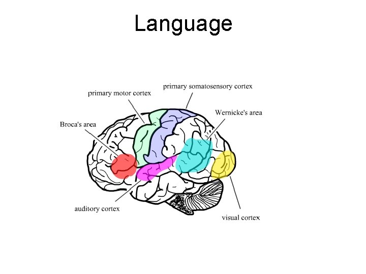
Language
Cranial nerve examination • • • I: Olfactory II: Optic III-IV-VI: extraoculars V: Trigeminal VII: Facial VIII: Vestibulocochlear IX-X: Glossopharyngeal, Vagus XI: Accessory XII: Hypoglossal
CN I: Olfactory • Usually not tested. • Observe for rash, deformity of nose or discharge (CSF). • Test each nostril with essence bottles of coffee, vanilla, peppermint.
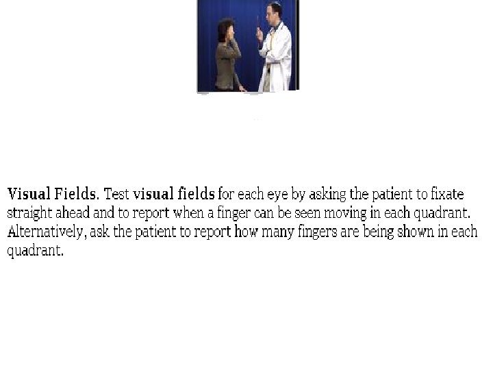
CN II: Optic • With patient wearing glasses. • Test each eye separately on eye chart/ card using an eye cover. • Examine visual fields by confrontation , keep examiner's head level with patient's head. • If poor visual acuity, map fields using fingers and a quadrant-covering card. • Look into fundi.
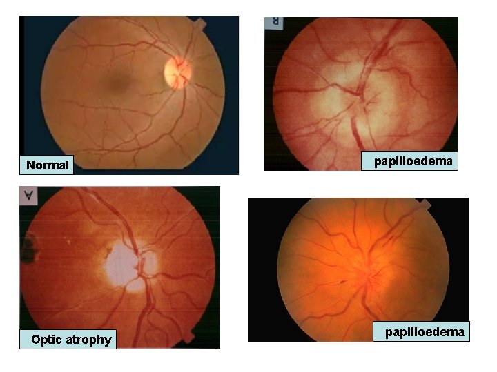
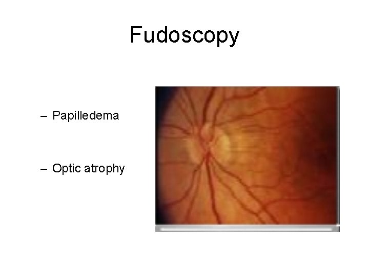
Normal Optic atrophy papilloedema
Light Reflex
Fudoscopy – Papilledema – Optic atrophy
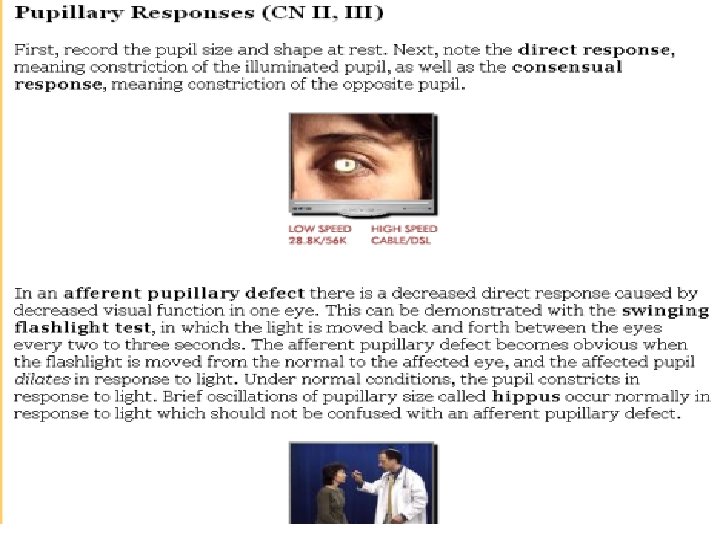
CN III, IV, VI: Oculomotor, Trochlear, Abducens • Look at pupils: shape, relative size, ptosis. • Shine light in from the side to gauge pupil's light reaction. • Assess both direct and consensual responses. • Assess afferent pupillary defect by moving light in arc from pupil to pupil. unne). Optionally: as do arc test, have pt place a flat hand extending vertically from his face, between his eyes, to act as a blinder so light can only go into one eye at a time. • "Follow finger with eyes without moving head": test the 6 cardinal points in an H pattern. • Look for failure of movement, nystagmus [pause to check it during upward/ lateral gaze]. • Convergence by moving finger towards bridge of pt's nose. • Test accommodation by pt looking into distance, then a hat pin 30 cm from nose. • If MG suspected: pt. gazes upward at Dr's finger to show worsening ptosis.
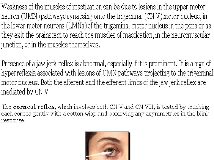
CN V: Trigeminal • Corneal reflex: patient looks up and away. • Touch cotton wool to other side. • Look for blink in both eyes, ask if can sense it. • Repeat other side [tests V sensory, VII motor]. • Facial sensation: sterile sharp item on forehead, cheek, jaw. • Repeat with dull object. Ask to report sharp or dull. • If abnormal, then temperature (heated/ water-cooled tuning fork), light touch (cotton). • Motor: pt opens mouth, clenches teeth (pterygoids). • Palpate temporal, masseter muscles as they clench. • Test jaw jerk (pseudobulbar palsy).
CN VII: Facial • Inspect facial droop or asymmetry. • Facial expression muscles: pt looks up and wrinkles forehead. • Examine wrinkling loss. • Feel muscle strength by pushing down on each side [UMNL preserved because of bilateral innervation]. • Pt shuts eyes tightly: compare each side. • Pt grins: compare nasolabial grooves. • Also: frown, show teeth, puff out cheeks. • Corneal reflex already done. See CN V.
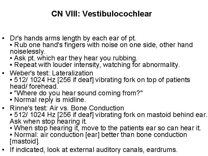
CN VIII: Vestibulocochlear • Dr's hands arms length by each ear of pt. • Rub one hand's fingers with noise on one side, other hand noiselessly. • Ask pt. which ear they hear you rubbing. • Repeat with louder intensity, watching for abnormality. • Weber's test: Lateralization • 512/ 1024 Hz [256 if deaf] vibrating fork on top of patients head/ forehead. • "Where do you hear sound coming from? " • Normal reply is midline. • Rinne's test: Air vs. Bone Conduction • 512/ 1024 Hz [256 if deaf] vibrating fork on mastoid behind ear. Ask when stop hearing it. • When stop hearing it, move to the patients ear so can hear it. • Normal: air conduction [ear] better than bone conduction [mastoid]. • If indicated, look at external auditory canals, eardrums.
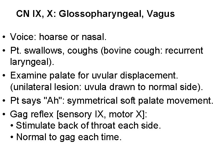
CN IX, X: Glossopharyngeal, Vagus • Voice: hoarse or nasal. • Pt. swallows, coughs (bovine cough: recurrent laryngeal). • Examine palate for uvular displacement. (unilateral lesion: uvula drawn to normal side). • Pt says "Ah": symmetrical soft palate movement. • Gag reflex [sensory IX, motor X]: • Stimulate back of throat each side. • Normal to gag each time.
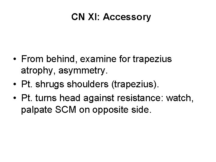
CN XI: Accessory • From behind, examine for trapezius atrophy, asymmetry. • Pt. shrugs shoulders (trapezius). • Pt. turns head against resistance: watch, palpate SCM on opposite side.
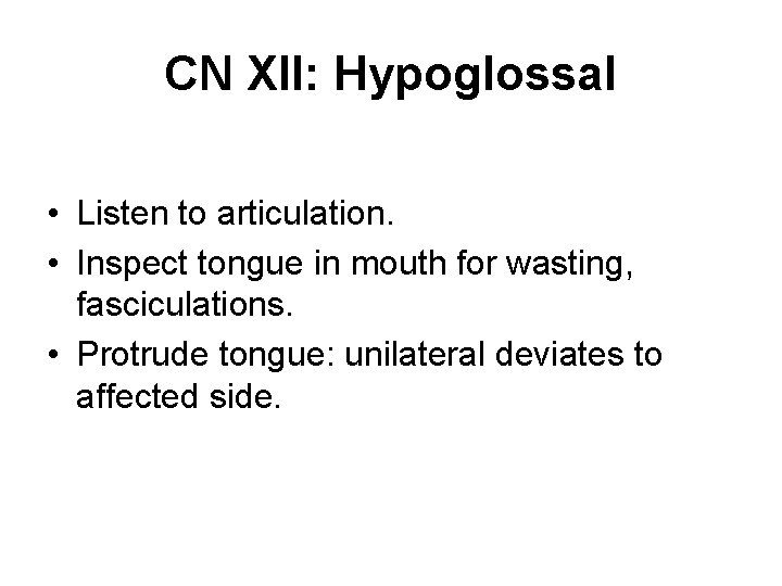
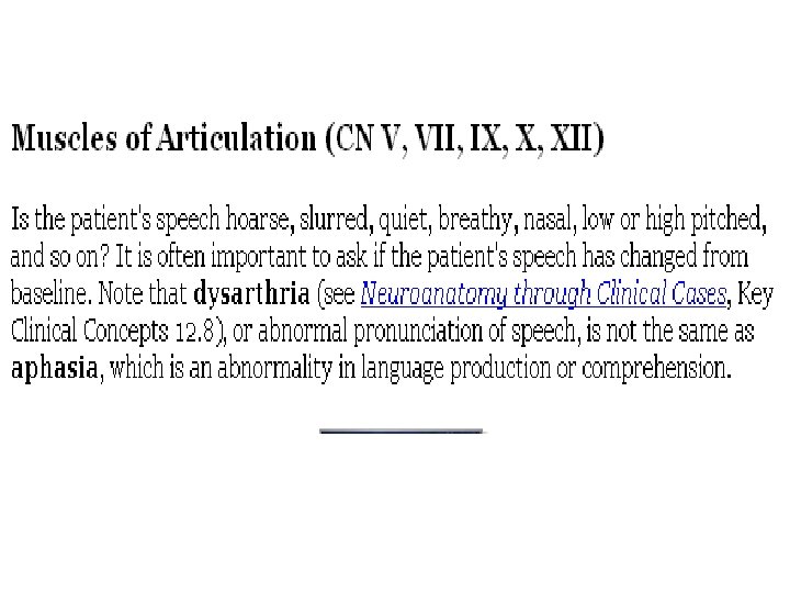
CN XII: Hypoglossal • Listen to articulation. • Inspect tongue in mouth for wasting, fasciculations. • Protrude tongue: unilateral deviates to affected side.
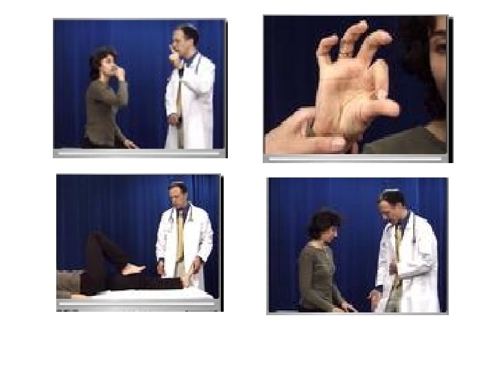
Coordination • Gait • Tandem walking • Limb coordination – Rapid alternating movement – Finger - nose – Finger – finger – Heel - shin
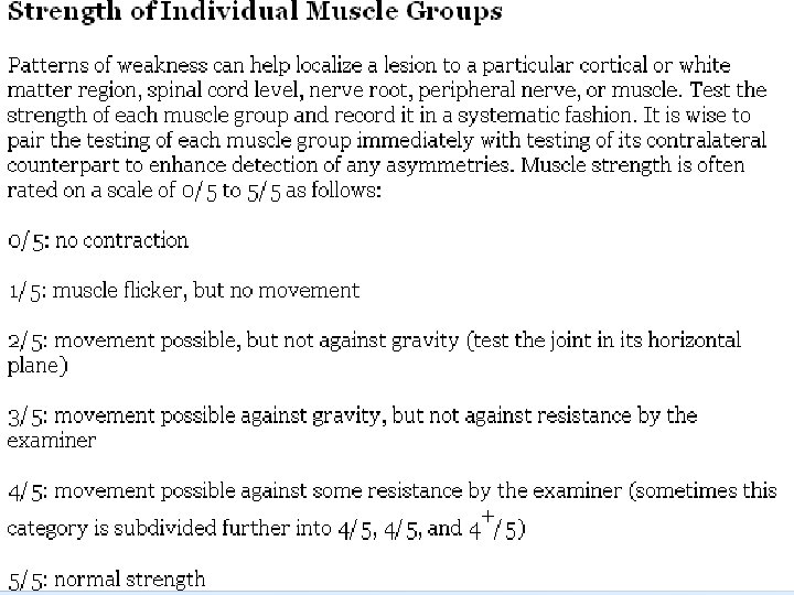
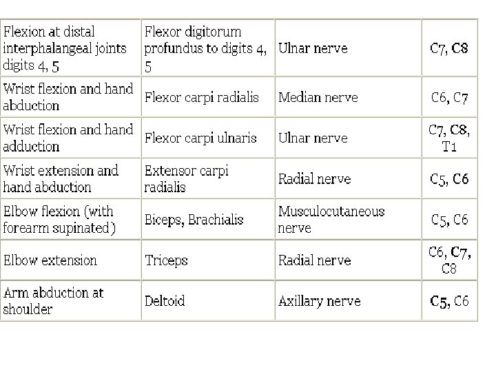
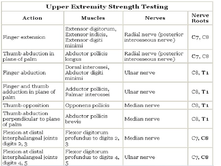
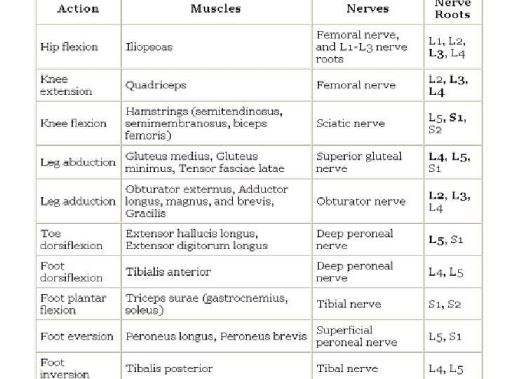
Motor examination • • • Muscle status Muscle tone Muscle power Tendon reflexes Gait & coordination
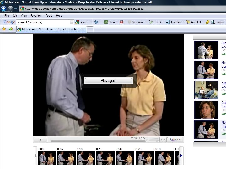
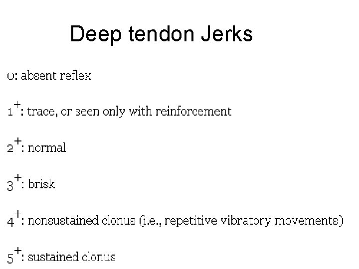
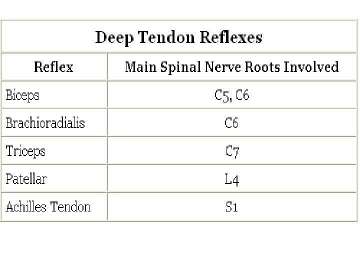
Deep tendon Jerks
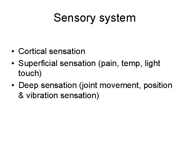
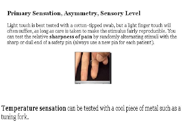
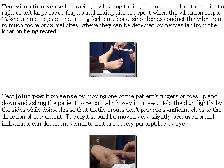
Sensory system • Cortical sensation • Superficial sensation (pain, temp, light touch) • Deep sensation (joint movement, position & vibration sensation)