Rectum Anatomy and Physiology Clinical features of Rectal


.")


 • Rectal carcinoma •")


 should be considered")


 Commonly referred to as the Thiersch procedure")
 Commonly referred to as the Delorme procedure")


- Slides: 36
Rectum - Anatomy and Physiology, Clinical features of Rectal disease, Examination of Rectum, Rectal prolapse Dr Amit Gupta Additional Professor Dept Of Surgery
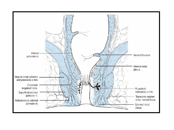
Anatomy and Physiology • The rectum is approximately 12 to 15 cm in length. • Three distinct submucosal folds, the valves of Houston, extend into the rectal lumen. • At S 4, the rectosacral fascia (Waldeyer's fascia) extends forward and downward and attaches to the fascia propria at the anorectal junction. • Anteriorly, Denonvilliers' fascia separates the rectum from the prostate and seminal vesicles in male and from the vagina in female.
• The dentate or pectinate line marks the transition point between columnar rectal mucosa and squamous anoderm. • The 1 to 2 cm of mucosa just proximal to the dentate line shares histologic characteristics of columnar, cuboidal, and squamous epithelium and is referred to as the anal transition zone. • The dentate line is surrounded by longitudinal mucosal folds, known as the columns of Morgagni, into which the anal crypts empty. These crypts are the source of cryptoglandular abscesses
Arterial supply • The superior rectal artery arises from the terminal branch of the inferior mesenteric artery and supplies the upper rectum. • The middle rectal artery arises from the internal iliac artery • The inferior rectal artery arises from the internal pudendal artery, which is a branch of the internal iliac artery. • A rich network of collaterals connects the terminal arterioles of each of these arteries, thus making the rectum relatively resistant to ischemia
Venous drainage • The superior rectal vein drains into the portal system via the inferior mesenteric vein. • The middle rectal vein drains into the internal iliac vein. • The inferior rectal vein drains into the internal pudendal vein, and subsequently into the internal iliac vein. • A submucosal plexus deep to the columns of Morgagni forms the hemorrhoidal plexus and drains into all three veins
Lymphatic drainage • Parallels the vascular supply • Lymphatic channels in the upper and middle rectum drain superiorly into the inferior mesenteric lymph nodes • Lymphatic channels in the lower rectum drain both superiorly into the inferior mesenteric lymph nodes and laterally into the internal iliac lymph nodes.
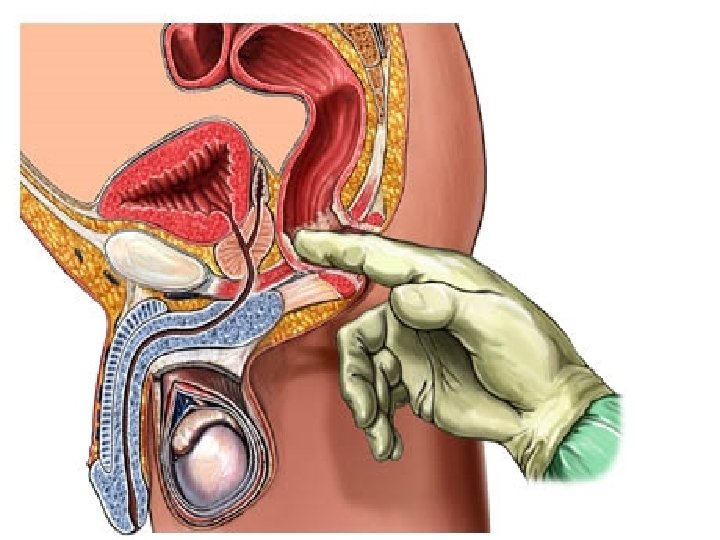
Indications for Rectal Examination • Assessment of the prostate (particularly symptoms of outflow obstruction). • When there has been rectal bleeding (prior to proctoscopy, sigmoidoscopy and colonoscopy). • Constipation. • Change of bowel habit. • Problems with urinary or faecal continence. • In exceptional circumstances to detect uterus and cervix (when vaginal examination is not possible).
Procedure • The finger is then moved through 180°, feeling the walls of the rectum. • With the finger then rotated in the 12 o'clock position, helped usually by the examiner bending knees in a half crouched position and pronating the examining wrist, the anterior wall can be palpated. • Rotation facilitates further examination of the opposing the walls of the rectum. In men, the prostate will be felt anteriorly. In women, the cervix and a retroverted uterus may be felt with the tip of the finger. • It is important to feel the walls of the rectum throughout the 360°. Small rectal wall lesions may be missed if this is not done carefully.
Examination of the Prostate Gland • Normal size is 3. 5 cms wide, protruding about 1 cm into the lumen of the rectum. • Consistency: it is normally rubbery and firm with a smooth surface and a palpable sulcus between right and left lobes. • There should not be any tenderness. • There should be no nodularity.
External Inspection • • • Skin disease. Skin tags Genital warts Anal fissures Anal fistula External haemorrhoids Rectal prolapse Skin discolouration with Crohn's disease External thrombosed piles
Internal Inspection • Simple piles (but best examined at proctoscopy) • Rectal carcinoma • Rectal polyps • Tenderness • Diseases of the prostate gland • Malignant or inflammatory conditions of the peritoneum (felt anteriorly)
Contraindications • • Imperforate Anus Unwilling patient Immunosuppressed patient Absence of anus following surgical excision Stricture Moderate to severe anal pain Prolapsed thrombosed internal hemorroids
Rectal Prolapse
• Also termed ‘rectal procidentia’ • Protrusion of the rectum beyond the anus • 6: 1 female to male predominance • Peak incidence is in the 6 th -7 th decades of life
Risk factors: • Chronic constipation • Chronic diarrhea • Mental retardation • Female sex
Anatomic abnormalities seen in patients with rectal prolapse • Deep rectovaginal or rectovesical pouch • Lax pelvic floor musculature • Failure of normal relaxation of the external sphincter • Redundant sigmoids • Pudendal nerve injury
Classification of rectal prolapse • Partial: prolapse of rectal mucosa only • Complete: prolapse with all layers • Grade 1: occult prolapse • Grade 2: prolapse to but not through anus • Grade 3: any protrusion through anus
Presentation Primary complaint is their rectum coming out May mistake it as haemorrhoids • Tenesmus • Bleeding • Mucus discharge • Constipation • Fecal incontinence • Sensation of incomplete evacuation
Complications of prolapse • Ulceration • Strangulation • Urinary and fecal incontinence • Spontaneous rupture with evisceration
Evaluation • Rectal prolapse can be incarcerated and represent a surgical emergency • So, rule out incarceration
• History – – When does it occur? Associated symptoms Pts general health and associated medical problems Association with psychiatric illness • Physical exam • Colonoscopy • Rule out additional pathology, such as a neoplasm which may be causing the prolapse
• Anorectal manometry and pudendal nerve terminal motor latency (PNTML) should be considered in patients with fecal incontinence • Patients with constipation should undergo colonic transit studies • Dynamic pelvic floor MRI • Endorectal ultrasound • Cinedefecography
Nonoperative Management • High fiber diet • Biofeedback may be helpful for patients with internal intussusception and inappropriate pelvic floor contraction • Does not play a significant role in the treatment of rectal prolapse
Surgery Abdominal approach • The first step is mobilization of the rectum • Involves dissection between the mesorectum and the presacral fascia • Mobilization is taken down to the level of the levators • Anterior mobilization should be taken to the level of the vagina or seminal vesicles
Rectopexy Ripstein procedure • First described in 1952 • After mobilization of the rectum is undertaken, a piece of prosthetic mesh is placed around the anterior wall of the rectum • Done at the level of the peritoneal reflection • low recurrence rates: 0 -9. 6% • high rate of complications: up to 52% • One of the more disastrous complications is mesh erosion into the rectum
Wells’ posterior Ivalon rectopexy • First described in 1959 • Low recurrence rates: 3. 0 -6. 0% • Morbidity rate of up to 19% • Complications: mesh erosion resulting in fistula formation
Suture rectopexy In 1959, Cutait proposed suture rectopexy without the implantation of mesh.
Suture rectopexy with resection • First described by Frykman in 1955 • Combined resection with rectopexy • Recommended for rectal prolapse patients with a long, redundant sigmoid colon • It has decreased rates of post-operative constipation • For patients with a long, redundant sigmoid and significant pre-op constipation, it is the procedure of choice • Recurrence rates of 0 -5% • Additional theoretical advantage of prevention of sigmoid volvulus • Complication rates shown to be similar to rectopexy alone
Laparoscopy • Similar recurrence rates and functional outcomes compared to similar open procedures • Longer OR times but shorter hospital stays • Cost analysis shows decreased costs due to shorter hospital stays
Anal encirclement procedure (1871) Commonly referred to as the Thiersch procedure
Mucosal sleeve resection (1900) Commonly referred to as the Delorme procedure
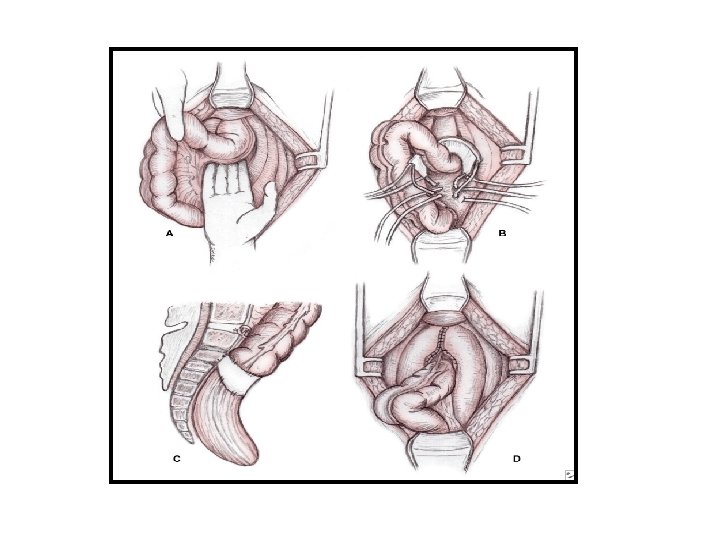
Perineal rectosigmoidectomy • differs from the Delorme procedure in that it is full thickness
Conclusion • Rectal prolapse is a complicated disease process due to a combination of factors • Thorough pre-operative workup is required to determine the appropriate procedure