
DEMO V Colon Rectum Anal Canal Ali Jasim


- Slides: 30
DEMO – V Colon, Rectum & Anal Canal Ali Jasim Alhashli Year III – Unit V (GI & Renal Systems)
STATION – 1 (Rectum & Anal canal) • Rectum: -It is continuous proximally with the sigmoid colon (rectosigmoid junction at the level of S 3 vertebra) & distally with the anal canal (anorectal flexure: forming a sharp 80 angle in a posterior direction). *Note: anorectal flexure is important for fecal continence. This flexure is also maintained by the tonus of the puborectalis muscle. During defecation, the relaxation of puborectalis muscle results in straightening of the anorectal junction. -In the rectum, there are three internal folding known as transverse rectal folds: two on the left and one on the right side. - The peritoneum covers: * Anterior and lateral surfaces of the superior third of the rectum. * Only the anterior surface of the middle third of the rectum. * No surface of the inferior third of the rectum. - Relations of the rectum: * Males: fundus of the urinary bladder – terminal parts of the ureters - vas deference – seminal glands & prostate. * Females: vagina.
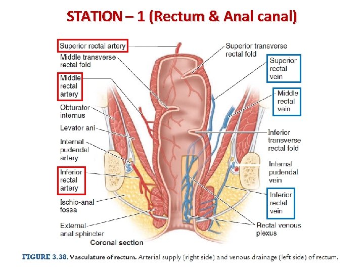
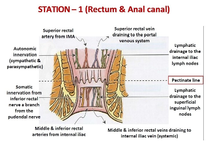
STATION – 1 (Rectum & Anal canal) • Vasculature of the rectum: - Arterial supply: *Superior rectal artery: a branch from inferior mesenteric artery. *Middle rectal artery: a branch from inferior vesicle artery (in males) & from uterine artery (in females). * Inferior rectal artery: a branch from internal pudendal artery. - Venous drainage: * Superior rectal vein drains into the portal venous system. * Middle & inferior rectal veins drain into the systemic system (to the IVC). • Lymphatic drainage: - Superior half of the rectum: *Pass to pararectal lymph nodes and then to the inferior mesenteric lymph nodes. - Inferior half of the rectum: * Pass through sacral lymph nodes and then to internal iliac lymph nodes.
STATION – 1 (Rectum & Anal canal)
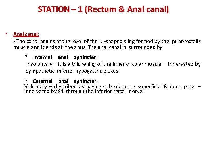
STATION – 1 (Rectum & Anal canal) • Anal canal: - The canal begins at the level of the U-shaped sling formed by the puborectalis muscle and it ends at the anus. The anal canal is surrounded by: * Internal anal sphincter: Involuntary – it is a thickening of the inner circular muscle – innervated by sympathetic inferior hypogastric plexus. * External anal sphincter: Voluntary – described as having subcutaneous superficial & deep parts – innervated by S 4 through the inferior rectal nerve.
STATION – 1 (Rectum & Anal canal)
STATION – 1 (Rectum & Anal canal)
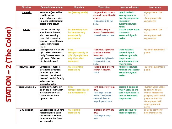
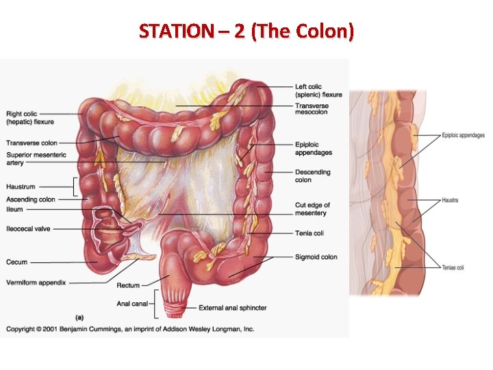
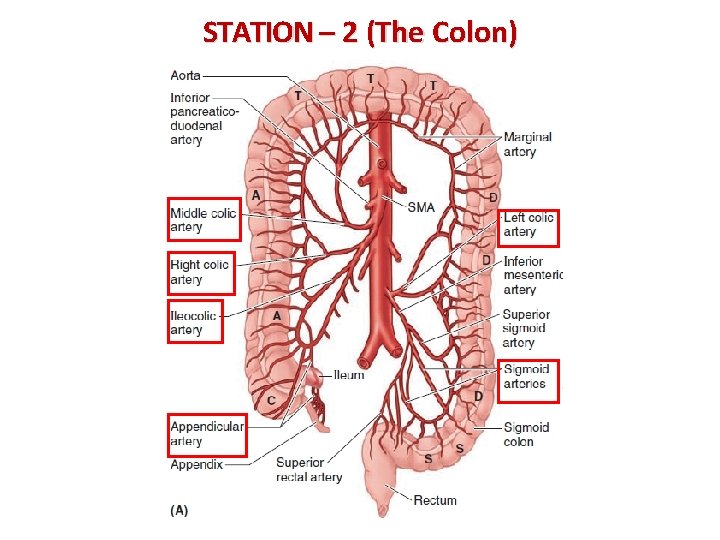
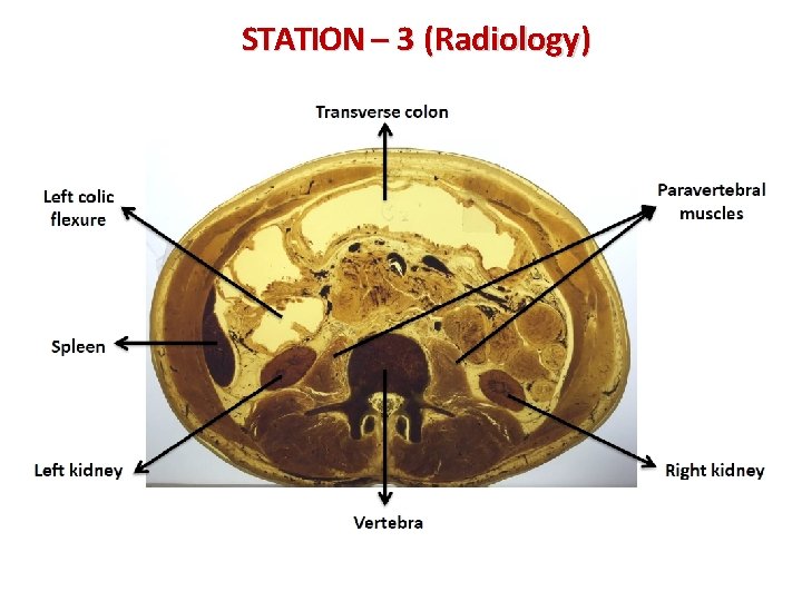
STATION – 2 (The Colon) Structure General characteristics Appendix Vermiform (worm-like) blind intestinal diverticulum extending from the posteromedial aspect of the cecum Cecum Ascending colon Transverse colon Descending colon Sigmoid colon Mesentery Vasculature Lymphatic drainage Innervation Yes (mesoappendix) • Appendicular artery: abranch from iliocolic artery. • Iliocolic vein to the SMV. • Sympathetic: T 10 T 12 • Parasympathetic: vagus nerve. First part of the large intestine-continuous with the ascending colon- blind intestinal pouch in the right lower quadrant (right iliac fossa). Passing superiorly on the right side of abdominal cavity from cecum to the right lobe of the liver (right colic flexure). No mesentery but it is almost entirely enveloped by peritoneum. • Iliocolic artery a branch from the SMA. • Iliocolic vein to SMV. Lymph nodes in mesoappendix to iliocolic lymph nodes to superior mesenteric lymph nodes. Iliocolic lymph nodes to superior mesenteric lymph nodes. It lies retroperitoneally. In 25% of people there might be a short mesentary. Yes (transverse mesocolon) From epicolic & paracolic lymph nodes to right colic lymph nodes to superior mesenteric lymph nodes. Middle colic lymph nodes to the superior mesenteric lymph nodes. • Superior mesenteric plexus Largest-most mobilecrosses the abdomen from the right colic flexure to the left colic flexure * bends inferiorly to become the descending colon. Extending from the left colic flexure into the left iliac fossa where it is continuous with the sigmoid colon. • Iliocolic & right colic arteries branches from SMA. • Iliocolic & right colic veins draining to SMV. • Middle colic artery a branch from SMA. • SMV It lies retroperitoneally and has no mesentery. • Left colic artery from IMA. • Drainage through IMV to the splenic vein and then to hepatic portal vein Epicolic & paracolic lymph nodes to left colic lymph nodes to inferior mesenteric lymph nodes. S-shaped loop linking the descending colon and the rectum. It extends from the left iliac fossa to S 3 vertebra Yes (sigmoid mesocolon) • Sigmoid artery from IMA. • Drainage through IMV Same as above (for descending colon) • Sympathetic: lumbar splanchnic nerves, inferior mesenteric ganglion, periarterial plexus on the IMA. • Parasympathetic: pelvic splanchnic nerve. Same as above (for descending colon) • Sympathetic: T 10 T 12 • Parasympathetic: vagus nerve. • Superior mesenteric plexus
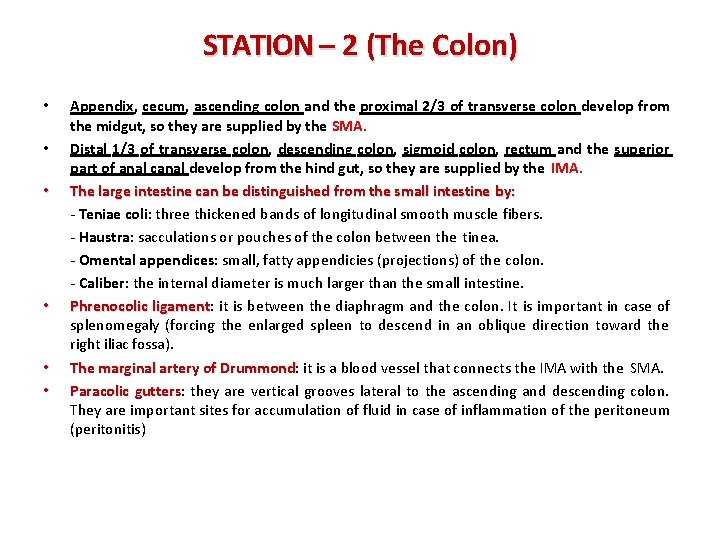
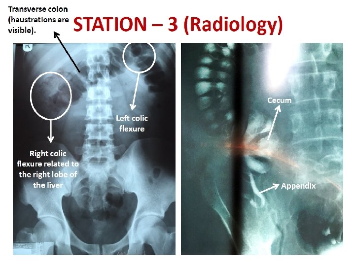
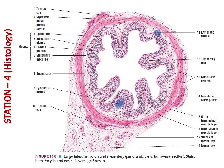
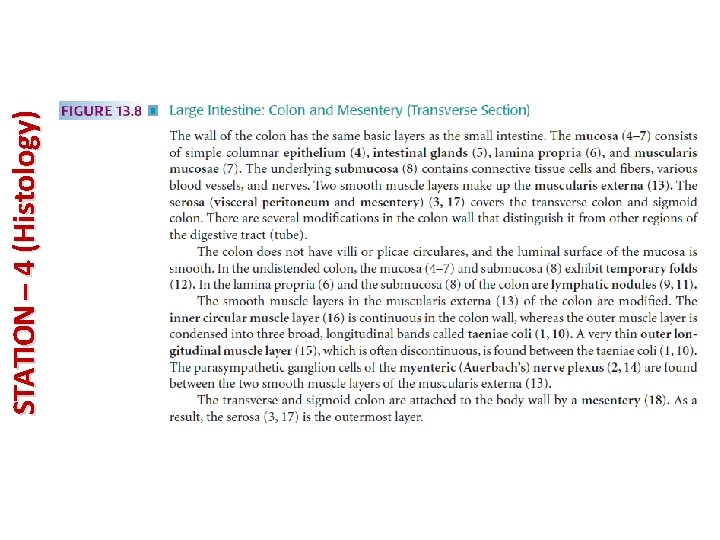
STATION – 2 (The Colon) • • • Appendix, cecum, ascending colon and the proximal 2/3 of transverse colon develop from the midgut, so they are supplied by the SMA. Distal 1/3 of transverse colon, descending colon, sigmoid colon, rectum and the superior part of anal canal develop from the hind gut, so they are supplied by the IMA. The large intestine can be distinguished from the small intestine by: - Teniae coli: three thickened bands of longitudinal smooth muscle fibers. - Haustra: sacculations or pouches of the colon between the tinea. - Omental appendices: small, fatty appendicies (projections) of the colon. - Caliber: the internal diameter is much larger than the small intestine. Phrenocolic ligament: ligament it is between the diaphragm and the colon. It is important in case of splenomegaly (forcing the enlarged spleen to descend in an oblique direction toward the right iliac fossa). The marginal artery of Drummond: Drummond it is a blood vessel that connects the IMA with the SMA. Paracolic gutters: gutters they are vertical grooves lateral to the ascending and descending colon. They are important sites for accumulation of fluid in case of inflammation of the peritoneum (peritonitis)
STATION – 2 (The Colon)
STATION – 2 (The Colon)
STATION – 3 (Radiology)
STATION – 3 (Radiology)
STATION – 3 (Radiology)
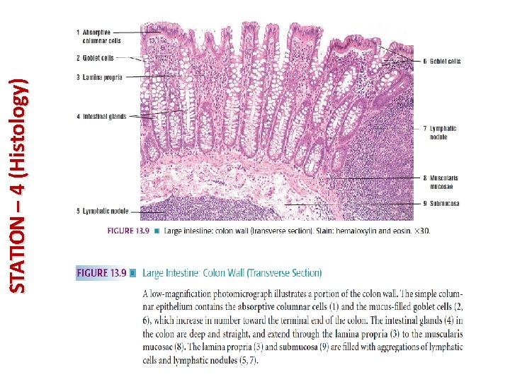
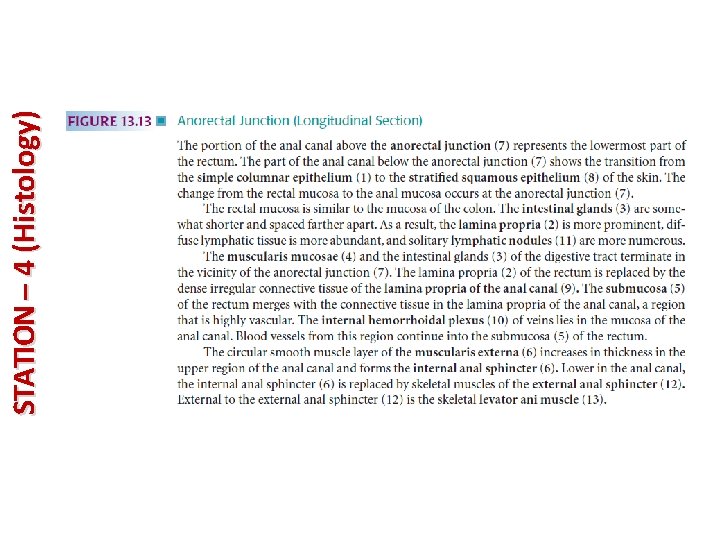
STATION – 4 (Histology)
STATION – 4 (Histology)
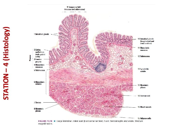
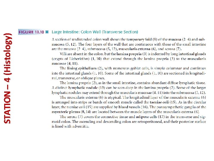
STATION – 4 (Histology)
STATION – 4 (Histology)
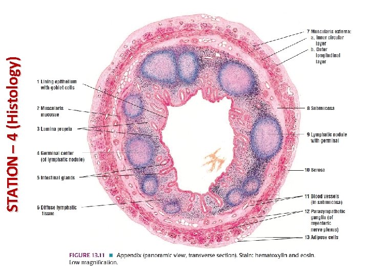
STATION – 4 (Histology)
STATION – 4 (Histology)
STATION – 4 (Histology)
STATION – 4 (Histology)
STATION – 4 (Histology)
STATION – 4 (Histology)
STATION – 4 (Histology)
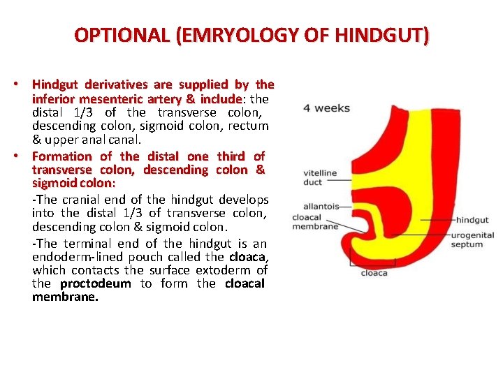
OPTIONAL (EMRYOLOGY OF HINDGUT) • Hindgut derivatives are supplied by the inferior mesenteric artery & include: include the distal 1/3 of the transverse colon, descending colon, sigmoid colon, rectum & upper anal canal. • Formation of the distal one third of transverse colon, descending colon & sigmoid colon: -The cranial end of the hindgut develops into the distal 1/3 of transverse colon, descending colon & sigmoid colon. -The terminal end of the hindgut is an endoderm-lined pouch called the cloaca, which contacts the surface extoderm of the proctodeum to form the cloacal membrane.
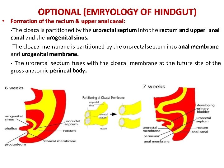
OPTIONAL (EMRYOLOGY OF HINDGUT) • Formation of the rectum & upper anal canal: -The cloaca is partitioned by the urorectal septum into the rectum and upper anal canal and the urogenital sinus. -The cloacal membrane is partitioned by the urorectalseptum into anal membrane and urogenital membrane. - The urorectal septum fuses with the cloacal membrane at the future site of the gross anatomic perineal body.
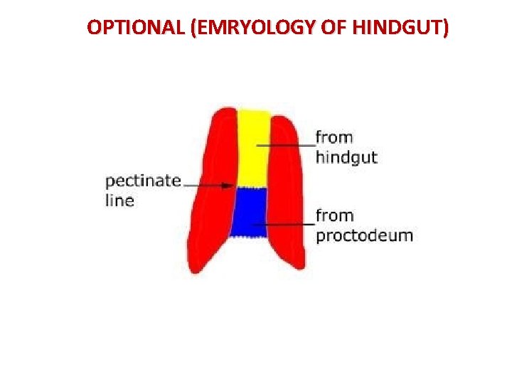
OPTIONAL (EMRYOLOGY OF HINDGUT)
GOOD LUCK!