Ascites Presented by Medicine Unit 1 Ascites Derived

Ascites Presented by Medicine Unit 1

Ascites �Derived from the Greek word “askos”, meaning bag or sac. � Defined as the accumulation of fluid in the peritoneal cavity. �It is a common clinical finding, with many extraperitoneal and peritoneal causes , but most common from liver cirrhosis.

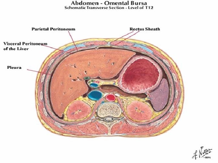
Peritoneal cavity � It is a potential space between the parietal peritoneum and visceral peritoneum, the two membranes separate the organs in the abdominal cavity from the abdominal wall. � Derived from the coelomic cavity of the embryo. � Largest serosal sac in the body and secretes approximately 50 ml of fluid per day.
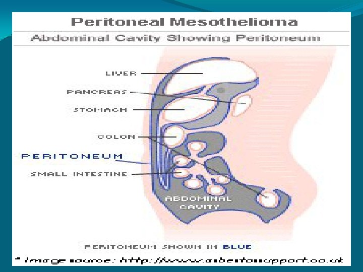

Peritoneal fluid � It is a normal, lubricating fluid found in the peritoneal cavity. �The fluid is mostly water with electrolytes, antibodies, white blood cells, albumin, glucose and other biochemicals. � Reduce the friction between the abdominal organs as they move around during digestion.

EPIDEMIOLOGY � Sex Healthy men have little or no intraperitoneal fluid, but women may normally have as much as 20 m. L, depending on the phase of their menstrual cycle. � Mortality/Morbidity Ambulatory patients with an episode of cirrhotic ascites have a 3 -year mortality rate of 50%. The development of refractory ascites carries a poor prognosis, with a 1 -year survival rate of less than 50%.

CAUSES OF ASCITES WITH ANASARCA � congestive cardiac failure � nephrotic syndrome � hypoproteinemia with severe anemia {nutritional} � pericardial effusion � Constrictive pericarditis � Myxoedema � Filariasis � Protein losing enteropathy � Epidemic dropsy
 Peritonitis – �acute")
ASCITES WITHOUT ANASARCA • Cirrhosis of liver (may lead to anasarca) Peritonitis – �acute pyogenic peritonitis �Tuberculous peritonitis �Malignant peritonitis �Sponteneous bacterial peritonistis • Portal vein thrombosis • Meig’s syndrome • Pancreatic ascites (e. g from acute pancreatitis 0 • Hepatic vein thrombosis ( Budd chiari syndrome) • Rupture of hollow viscus within abdomen • Lymphoma or leukemias • Chylous ascites • Haemoperitoneum following trauma • Rare – Vasculitis, Peritoneal dialysis, IVC obstruction

STAGING OF ASCITES �Ascites may be semi-quantified using the following system: �Stage 1+ is detectable only after careful examination. �Stage 2+ is easily detectable but of relatively small volume. �Stage 3+ is obvious, but not tense, ascites. �Stage 4+ is tense ascites.

: � Portal HTN secondary to chronic liver diseases (")
Most Common causes(90% of cases): � Portal HTN secondary to chronic liver diseases ( cirrhosis) • Intra-abdominal malignancy • Congestive Heart Failure • Mycobacterium tuberculosis

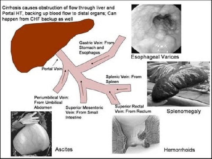
Portal Hypertension �It is a high blood pressure in the portal vein and its tributaries(portal venous system). �It is defined as a portal pressure gradient (the difference in pressure between the portal vein and the hepatic veins) of 5 mm Hg or greater.

Causes of portal hypertension � Intrahepatic causes: liver cirrhosis and hepatic fibrosis (e. g. due to Wilson's disease, hemochromatosis, or congenital fibrosis). � Prehepatic causes : portal vein thrombosis or congenital atresia. � Posthepatic obstruction occur at any level between liver and right heart, including hepatic vein thrombosis, IVC congenital malformation, and constrictive pericarditis.

Cirrhosis most common causes of cirrhosis: • Alcoholic liver disease or alcoholic hepatitis • viral hepatitis (B or C) • fatty liver disease
 � Infections")
Other causes of ascites Diseased peritoneum (SAAG < 1. 1 g/d. L) � Infections � � Bacterial peritonitis Tuberculous peritonitis Fungal peritonitis Human immunodeficiency virus (HIV)-associated peritonitis � Malignant conditions � � Peritoneal carcinomatosis Primary mesothelioma Pseudomyxoma peritonei Hepatocellular carcinoma � Other rare conditions � � Familial Mediterranean fever Vasculitis Granulomatous peritonitis Eosinophilic peritonitis
![Other causes of ascites Normal peritoneum � Portal hypertension (serum-ascites albumin gradient [SAAG] >1.](http://slidetodoc.com/presentation_image_h/e4f155fe64eeb4b1d7e32823b7ddfabd/image-19.jpg "Other causes of ascites Normal peritoneum � Portal hypertension (serum-ascites albumin gradient [SAAG] >1.")
Other causes of ascites Normal peritoneum � Portal hypertension (serum-ascites albumin gradient [SAAG] >1. 1 g/d. L) � Hepatic congestion, congestive heart failure, constrictive pericarditis, tricuspid insufficiency, Budd-Chiari syndrome � Liver disease, cirrhosis, alcoholic hepatitis, fulminant hepatic failure, massive hepatic metastases � Hypoalbuminemia (SAAG < 1. 1 g/d. L) � Nephrotic syndrome � Protein-losing enteropathy � Severe malnutrition with anasarca � Miscellaneous conditions (SAAG < 1. 1 g/d. L) � Chylous ascites � Pancreatic ascites � Bile ascites � Nephrogenic ascites � Urine ascites � Ovarian disease

Pathophysiology 1. Increased hydrostatic pressure 2. Decreased colloid osmotic pressure 3. Increase permeability of peritoneal capillaries 4. Leakage of fluid into the peritoneal cavity � Miscellaneous causes • • • Myxedema Ovarian disease (Meig’s syndrome) Chronic hemodialysis

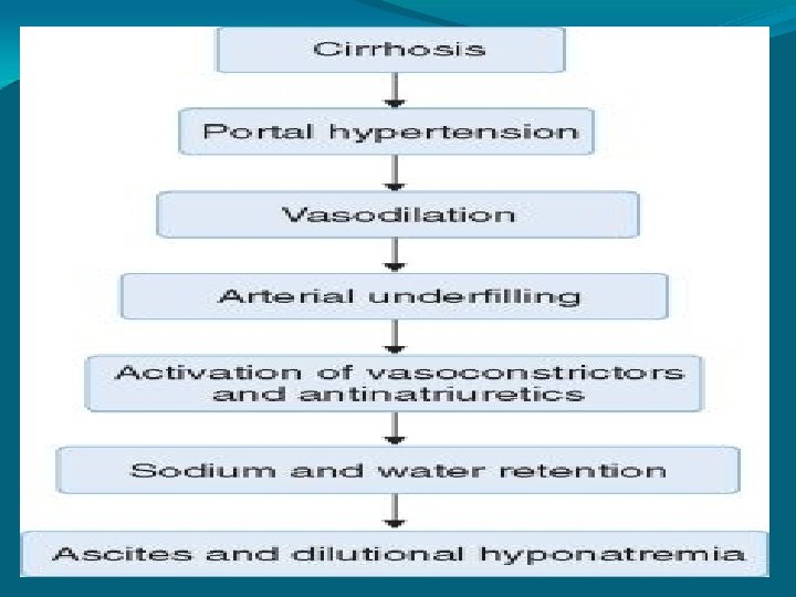
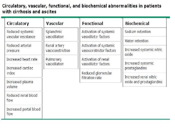
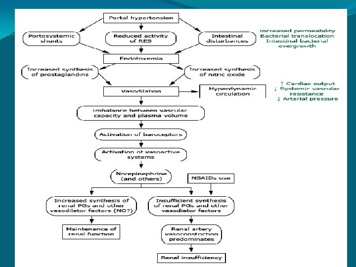
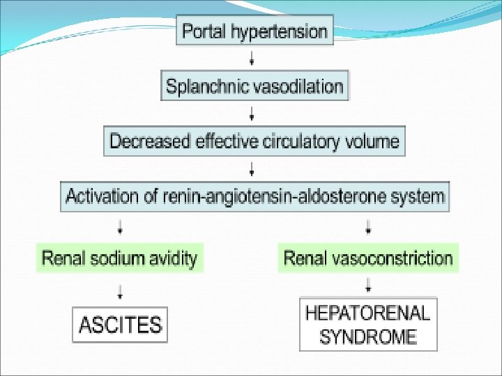
Pathophysiology � � The accumulation of ascitic fluid represents a state of totalbody sodium and water excess, but the event that initiates the unbalance is unclear. Three theories of ascites formation have been proposed: under filling, overflow, and peripheral arterial vasodilation. The under filling theory suggests that the primary abnormality is inappropriate sequestration of fluid within the splanchnic vascular bed due to portal hypertension and a consequent decrease in effective circulating blood volume. This activates the plasma renin, aldosterone, and sympathetic nervous system, resulting in renal sodium and water retention.

Pathophysiology � � The overflow theory suggests that the primary abnormality is inappropriate renal retention of sodium and water in the absence of volume depletion. This theory was developed in accordance with the observation that patients with cirrhosis have intravascular hypervolemia rather than hypovolemia. The most recent theory, the peripheral arterial vasodilation hypothesis, includes components of both of the other theories. It suggests that portal hypertension leads to vasodilation, which causes decreased effective arterial blood volume. As the natural history of the disease progresses, neurohumoral excitation increases, more renal sodium is retained, and plasma volume expands. This leads to overflow of fluid into the peritoneal cavity. The vasodilation theory proposes that underfilling is operative early and overflow is operative late in the natural history of cirrhosis.

�Although the sequence of events that occurs between the development of portal hypertension and renal sodium retention is not entirely clear, portal hypertension apparently leads to an increase in nitric oxide levels. Nitric oxide mediates splanchnic and peripheral vasodilation. Hepatic artery nitric oxide synthase activity is greater in patients with ascites than in those without ascites. �Regardless of the initiating event, a number of factors contribute to the accumulation of fluid in the abdominal cavity: �Elevated levels of epinephrine and norepinephrine are well -documented factors. �Hypoalbuminemia and reduced plasma oncotic pressure favor the extravasation of fluid from the plasma to the peritoneal fluid, and, thus, ascites is infrequent in patients with cirrhosis unless both portal hypertension and hypoalbuminemia are present.


Diagnosis: 1 -history Pts should be questioned about: • Liver diseases • Risk factors for Hepatitis C ( needle sharing, tattoos, cocaine, heroin use and emigration from Egypt or Southeast Asia) • Risk factors for Hepatitis B (needle sharing, tattoos, acupuncture, and emigration from China, Korea, Taiwan, or Southeast Asia).
")
• • Pts with obesity, diabetes, hyperlipidemia and Nonalcoholic steatohepatitis ( NASH ) should be ruled out. Pts with ascites who lack risk factors for cirrhosis should be questioned about cancer, heart failure, TB, dialysis, and pancreatitis. Operative injury to the ureter or bladder can lead to leakage of urine into peritoneal cavity. HIV pts may have infections lead to ascites.
: Mild ascites")
Diagnosis 2 -Clinical Features • A- Asymptomatic (fluid <100 - 400 ml): Mild ascites • B- symptomatic (fluid >400 ml): Increased abdominal girth, presence of abdominal pain or discomfort, early satiety, pedal edema, weight gain and respiratory distress depending on the amount of fluid accumulated in the abdomen.

� SYMPTOMS- � Progressive swelling of abdomen with gain in weight. � Aching pain all over the abdomen do stretching; bloated feeling of abdomen. � Dyspnoea & even orthopnea (in large collection of fluid). � Swelling of lower limb ( due to ccf, nephrotic syndrome, functional ivc obstruction). � Paraesthesia in the distribution of lateral femoral cuteneous nerve (meralgia paraesthetica). � Oligouria � Dyspepsia, heartburn d/t GERD

� SIGNSGENERAL SURVEYo the patient may be in propped up position due to dyspnoea. o Bipedal pitting edema. o Neck veins may engorged (due to hypervolaemia ) o Obvious swelling of abdomen.

ABDOMEN� � � � � Swelling of abdomen & there is fullness in flanks. The skin looks shiny. Diverication of recti; there may be presence of abdominal (umbilical ) herniation or abdominal striae ( striae albicantes). Prominent veins in flanks Umbillicus – flushed or everted with transverse slit. Fluid thrill present. Shifting dullness present. Measurements – Maximum girth of abdomen is at the level of abdomen. Spino-umbillical distance ( line joining anterior superior iliac spine & umbillicus ) is more or less equal on both sides. The distance between xiphisternum & umbillicus is more then the distance between umbillicus & symphysis pubis.

RESPIRATORY SYSTEM – � � Basal collapse- diminished movements , impaired note on percussion , diminished vesicular breath sound & crepitations may be heared in the lower part of chest on both sides. Hydrothorax (commonly right sided ; due to defects in diaphragm ) may be bilateral. CVS� � � Apex beat – deviated upwards & outwards. There may be diffuse pulsation over precordium. Soft systolic murmer in pulmonary area may be present ( due to associated severe anemia).

NERVOUS SYSTEM: �Nothing particular there may be features of hepatic encephalopathy. GENITO URINARY SYSTEM: �scrotal edema or hydrocele may be evident as secondary effects of ascites or as a clue to etiological (e. g. nephrotic syndrome) diagnosis of ascites.
 �Tympany at the top of")
Physical examination findings: �Umbilicus Eversion (often with umbilical herniation) �Tympany at the top of the abdomen �Fluid wave �Peripheral edema �Shifting dullness (> 500 ml fluid) �Bulging flanks (>500 ml fluid)

WHERE DO YOU GET FLUID THRILL? � � � Fluid thrill is present in the precence of fluid in the peritoneal cavity whether it is encysted or free. Ascites Ovarian cyst Hydramnio Rarely in large hydronephrosis Obesity

ESSENTIAL CRITERIA FOR POSITIVE FLUID THRILL: � Large amount of fluid (atleast 2 litre fluid is necessary to elicit fluid thrill). � Fluid should remain undertension. � Minimal fluid ( ½ to 1 litre)is required to demonstrate shifting dullness.

SHIFTING DULLNESS While the patient lies supine , the intestine will float in midline & will give tympnitic note on percussion. To follow the cardinal rules of percussion , we percuss the midline first & then the flanks- I. e. we percuss from ‘more resonant to less resonant’ area. �UNILATERAL SHIFTING DULLNESS: This is found in splenic rupture & is known as Ballance’s sign. The blood present in left flank becomes clotted (near the spleen) & does not shift to right side in right lateral position but the blood present in the right side (haemoperitoneum) is shifted to the left side.

Shifting Dullness

WHAT IS HORSESHOE SHAPED DULLNESS IN ASCITES? �In moderate ascites, the distribution of fluid in supine of patient is confined to the flanks , dependant parts & the hypogastrium. Thus , in supine position epigastrium & umbillicus are tympanitic on percussion ( due to floating of intestine) but a horse-shoe shaped dullness with concave upper border is elicited due to dull flanks & dull hypogastrium. � Percuss the abdomen in various directions from the central area of tympanicity (i. e. the umbillical region) to outwards & demonstrate the horseshoe shaped dullness.

Bulging Flanks and Umbilical Hernia
 �FAECES-")
DIFFERENTIAL DIAGNOSIS �FAT- OBESITY( INVERTD UMBILICUS WITH ABSENT FLUID THRILL AND SHIFTING DULLNESS) �FAECES- MEGACOLON OR LOW GUT OBSTRUCTION( SYMMETRICAL ENLARGEMENT WITH VISIBLE PERISTALSIS) �FOETUS- PREGNANCY (FOETAL PARTS ARE PALPABLE; CENTRAL DULLNESS) �FLATUS- GASEOUS DISTENTION(FLANKS ARE NOT BULGRD; TYMPANIC NOTE ALL OVER ABDOMEN) �FLUID- OVARIAN CYST, ACSITIS �FULL BLADDER (FLANKS ARE TYMPANIC. PALPATION CAUSE PAINOR DISCONFORT AND DESIRE TO MICTURITION; USALLY ROUNDED CYSTIC SWELLING IN HYPOGASTRIUM WHICH IS DULL ON PERCUSSION SHIFTING DULLNESS ABSENT)

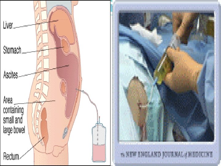
Diagnosis 3 -paracentesis �It is a diagnostic procedure to establish the etiology of new-onset ascites or to rule out spontaneous bacterial peritonitis in patients with preexisting ascites. �Large volume paracentesis is performed in hemodynamically stable patients with tense or refractory ascites to alleviate discomfort or respiratory compromise. �For diagnostic purposes, a small amount (20 cc) may be enough for adequate testing.

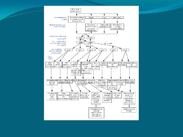
Ascitic fluid analysis Cell count: • A white blood cell count is the most important. • A neutrophil > 250 cells/mm 3 spontaneous bacterial peritonitis • An elevated lymphocyte tuberculosis or peritoneal carcinomatosis • Gram stain and culture: for bacteria and acid fast bacilli

�Red cell count > 50. 000/microliter hemorrhagic ascites, which usually is due to malignancy, tuberculosis or trauma.

Serum-Ascites Albumin Gradient �Best single test for classifying ascites into portal hypertensive and non-portal hypertensive causes. �Calculated by: Serum albumin – Ascites albumin= SAAG >1. 1 g/d. L= Portal HTN SAAG < 1. 1 g/d. L= Non-Portal hypertensive cause
 � If the serum albumin minus ascitic fluid")
WHAT IS SERUM- ASCITES ALBUMIN GRADIENT(SAAG) � If the serum albumin minus ascitic fluid albumin is equal to or greater than 1. 1 g/dl, the patient has portal hypertension. other than cirrhosis of liver this is a feature of ccf, buddchiari syndrome, portal vein thrombsis, myxoedema. � If the gradient is less than 1. 1 g/dl, usually it suggests malignancy acstis, tuberculous asctis pancreatic asctis or nephrotic syndrome * saag is based on oncotic – hydrostatic balance and directly correlates with protal pressure.

SAAG >1. 1 1. Liver Disease 2. Hepatic Congestion 3. CHF 4. Tricuspid Insufficiency 5. Massive Hepatic Metastasis SAAG <1. 1 1. Peritoneal carcinomatosis 2. Peritoneal Infection (TB, Fungal, CMV) 3. Nephrotic syndrome 4. Pancreatic ascites

• Total protein: Helpful in diagnosing spontaneous bacterial peritonitis Pts with a value<1 g/dl protein and glucose of <50 mg/dl have high risk of SBP • Cytology: for malignant cells • Amylase: to exclude pancreatic ascites

Ascitic Fluid Appearance: The gross appearance of the ascitic fluid can be helpful in the differential diagnosis. Turbid or cloudy: infected fluid. Milky: Triglyceride concentration of greater than 200 mg/dl (often greater than 1000 mg/dl), malignancy is usually MC cause, but cirrhosis may present with chylous fluid. Pink or Bloody: Pink fluid usually traumatic tap. Frankly bloody may occur in hepatocellular carcinoma, or other malignancy related ascites. Brown: Deeply jaundiced pts may present with brown ascitic fluid, which may represent gallbladder rupture or perforated duodenal ulcer.

WHAT’S CHYLOUS ASCITES � CHYLOUS- turbid, creamy or milky peritoneal fluid due to presence of lymph. it shows sudan iii staining fat globules and contains a large amount of chylomicrons. ethre dissolves the turbidity of chylous ascitis. � CHYLIFORM ASCTES- turbid fluid due to large number of leucocytes, degenerated cells or tumour cells. addition of alkali clears the turbidity. � PSUEDOCHYLOUS ASCITES- turbid fluid dye to increased amount of lecitithin, globulin and calcium phosphate in the ascitic fluid. CHYLOUS ASCITES CAUSES: � Tuberculosis, Filariasis, Trauma To The Abdomen, Intra-abdominal Malignancy, Nephrotic Syndrome, Congenital Lymphangectasisa Or Obstruction To The Thoracic Duct(eg. Lymphoma)

Diagnosis 4 -imaging Studies A- Chest and Plain Abdominal Films • Elevation of the diaphram (usually with >500 ml of fluid) • Abdominal haziness • Bulging Flanks • Poor definition of intra abdominal organs -

• Medial displacement of the cecum and ascending colon. • Hellmer's sign: the lateral liver angle is displaced medially from the thoracoabdominal wall in a patient with a large extraperitoneal fluid collection extending into the flank (Pathologic processes in both the intra- and extraperitoneal spaces).

bilateral pleural effusions in a patient with ascites

loss of any definition of the edge of the spleen or liver and displacement of the bowel loops out of the pelvis and bulging flanks

Pelvic Ascites

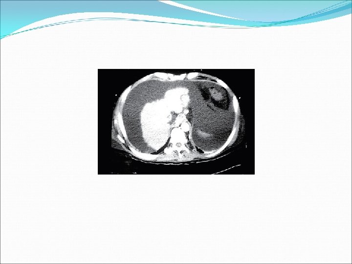
Imaging Studies B- CT scan • • • Well visualized Fluid may be visualized in the: Right perihepatic space Posterior subhepatic space (Morison pouch) Pouch of Douglas

Large ascites displacing bowel posteriorly

Perihepatic ascites
![[CT] Small amount of ascitic fluid in the pouch of Douglas and surrounding the](http://slidetodoc.com/presentation_image_h/e4f155fe64eeb4b1d7e32823b7ddfabd/image-62.jpg "[CT] Small amount of ascitic fluid in the pouch of Douglas and surrounding the")
[CT] Small amount of ascitic fluid in the pouch of Douglas and surrounding the adjacent small bowel loops

Abdominal CT, showing Morison's pouch as the dark margin surrounding the right kidney (at lower left corner of image).

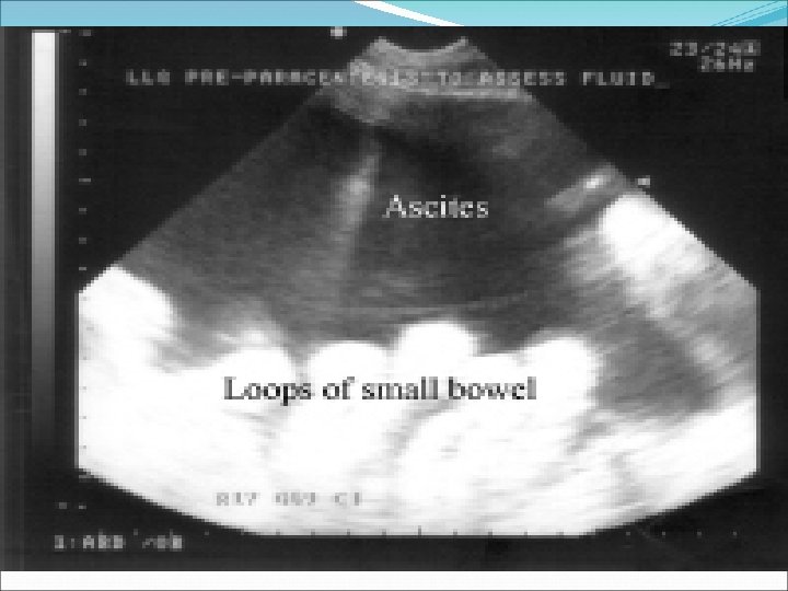
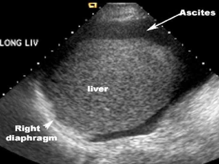
Imaging Studies C- Ultrasound • Easiest and most sensitive technique for detection of ascitic fluid. • Volume as small as 5 -10 ml can be seen.
 between the liver and right")
. Morison's pouch with abnormal fluid collection (red arrows) between the liver and right kidney

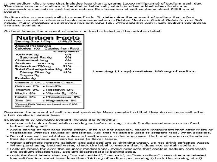
Management of Ascites • • • The goal is to prevent Na loading and increase renal excretion of Na and H 2 O and produce a net re-absorption of fluid from the ascites back into the circulating volume. Dietary Na restriction Diet of 2 g sodium per day Fluid Restriction: Only done when serum Na is <128 mmol/L Check Labs Ck serum electrolytes and creatinine every other day. Weigh the patient and measure urinary output daily.

Management of Ascites Diuretic therapy: • Spironolactone: diuretic of choice (25 -200 mg PO daily or bid) • Lasix: (20 -80 mg/d PO/IV/IM) • Zaroxolyn: (works on Edema of CHF) (5 -20 mg/dose PO q 24 hr) • Mannitol: (0. 5 -2 g/kg IV over 30 -60 min, repeat q 68 hrs) • Amilioride: 5 -20 mg/d PO

Management of Ascites Large Volume Paracentesis • To relieve symptomatic tense ascites and peripheral edema. • Up to 20 L can be removed over 4 -6 hr. • Removal of 5 L or more of ascitic fluid during a single session.
 Severe bowel distention Previous abdominal surgery (if")
Paracentesis Contraindications: • • Acute abdomen (absolute) Severe bowel distention Previous abdominal surgery (if necessary perform open procedure) Pregnancy (if necessary perform after first trimester using an open technique above the umbilicus) Distended bladder that cannot be relieved by foley catheder Infection at site of insertion (cellulitis or abscess) Thrombocytopenia (relative) Coagulopathy (relative)

Paracentesis Complications: • • • Bladder perforation Small or large bowel perforation Stomach perforation Laceration of major vessels ( mesenteric, iliac, aorta) Laceration of catheter or guide wire and loss in peritoneal cavity (requires laparotomy) Abdominal wall hematoma Incisional hernia Wound infection Wound dehiscence

Management of Ascites Transjugular Intrahepatic Portasystemic Shunt: The TIPS procedure is an interventional radiologic technique that reduces portal pressure and may be the most effective treatment for diuretic resistant ascites. Risks: �Hepatic Encephalopathy (30% of pts) �Thrombosis and shunt stenosis.

TIPS Procedure side to side portacaval shunt, usually placed through the right internal jugular vein. A needle is placed through the IJV into the hepatic vein.

hunt: : • Developed to return ascitic fluid from the peritoneal cavity directly to the systemic circulation. • Consists of an intraabdominal tube connected through a valve to silicone tube that transverses the subcutaneous tissue up to the neck and enters one of the jugular veins. • This leads to diuresis and mobilization of ascites.

Risks of Peritoneovenous shunt: �DIC �Infection �Variceal bleeding �Small bowel obstruction �Shunt occlusion �Death Due to these risk this procedure is rarely used. Peritoneovenous shunts are therapeutic but do not improve survival rates in patients with cirrhosis and ascites.

Management of Ascites liver transplantation: • Tx of choice • Corrects portal hypertension • Changes the natural course of progressive liver failure due to cirrhosis • Not all pts are candidates for transplant, and those who are may wait for years for a donor • Many die from complications of ascites while waiting for transplant donor
 recommendations for the management of")
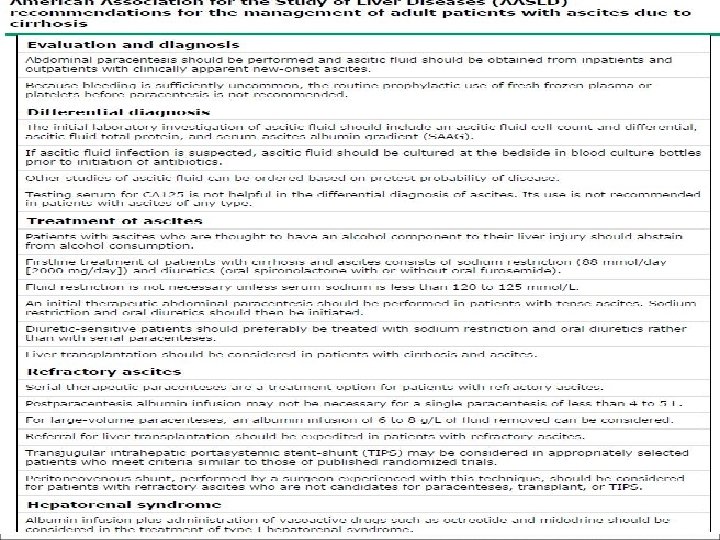
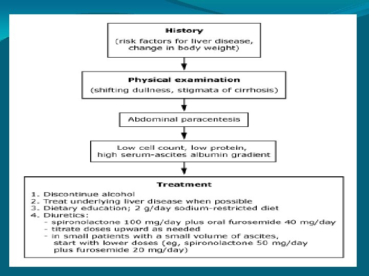
American Association for the Study of Liver Diseases (AASLD) recommendations for the management of adult patients with ascites due to cirrhosis � Evaluation and diagnosis � Abdominal paracentesis should be performed and ascitic fluid should be obtained from inpatients and outpatients with clinically apparent new-onset ascites. � Because bleeding is sufficiently uncommon, the routine prophylactic use of fresh frozen plasma or platelets before paracentesis is not recommended. � Differential diagnosis � The initial laboratory investigation of ascitic fluid should include an ascitic fluid cell count and differential, ascitic fluid total protein, and serum-ascites albumin gradient (SAAG). If ascitic fluid infection is suspected, ascitic fluid should be cultured at the bedside in blood culture bottles prior to initiation of antibiotics. Other studies of ascitic fluid can be ordered based on pretest probability of disease. Testing serum for CA 125 is not helpful in the differential diagnosis of ascites. Its use is not recommended in patients with ascites of any type.

�Treatment of ascites � Patients with ascites who are thought to have an alcohol component to their liver injury should abstain from alcohol consumption. Firstline treatment of patients with cirrhosis and ascites consists of sodium restriction (88 mmol/day [2000 mg/day]) and diuretics (oral spironolactone with or without oral furosemide). Fluid restriction is not necessary unless serum sodium is less than 120 to 125 mmol/L. An initial therapeutic abdominal paracentesis should be performed in patients with tense ascites. Sodium restriction and oral diuretics should then be initiated. Diuretic-sensitive patients should preferably be treated with sodium restriction and oral diuretics rather than with serial paracenteses. Liver transplantation should be considered in patients with cirrhosis and ascites. �Refractory ascites � Serial therapeutic paracenteses are a treatment option for patients with refractory ascites. Postparacentesis albumin infusion may not be necessary for a single paracentesis of less than 4 to 5 L. For large-volume paracenteses, an albumin infusion of 6 to 8 g/L of fluid removed can be considered. Referral for liver transplantation should be expedited in patients with refractory ascites. Transjugular intrahepatic portasystemic stent-shunt (TIPS) may be considered in appropriately selected patients who meet criteria similar to those of published randomized trials. Peritoneovenous shunt, performed by a surgeon experienced with this technique, should be considered for patients with refractory ascites who are not candidates for paracenteses, transplant, or TIPS.

Complications from Ascites 1. • • Refractory Ascites: Fluid overload that is unresponsive to Na-restricted diet and high dose anti-diuretic treatment. Usually in the setting of chronic or acute liver diseases with associated portal hypertension.
")
POSSIBLE CAUSES OF REFRACTORY ASCITIS: �NON COMPLIANCE (NOT FOLLOWING THE ADEQUATE RESTRICTION OF SODIUM) �FUNCTIONAL RENAL FAILURE IN CIRRHOSIS OF LIVER �INFECTION �SPONTANEOUS BACTERIAL PERITONITIS �HEPATOMA �G. I. BLEEDING �NSAIDS (DEPRESS THE DIURETIC EFFECT OF FRUSEMIDE AND SPIRONOLACTONE) �PORTAL OR HEPATIC VEIN THROMBOSIS �SUPERIMPOSED CARDIAC OR RENAL FAILURE

Treatment of Refractory Ascites: Liver transplantation is treatment of choice. If unsuitable, treatment with: �Serial paracentesis � TIPS � Peritoneovenous shunt

Complications of Ascites 2. Hepatorenal syndrome: Life-threatening medical condition that consists of rapid deterioration in kidney function in individuals with cirrhosis or fulminant liver failure. HRS is usually fatal unless a liver transplant is performed, although various treatments, such as dialysis, can prevent advancement of the condition. It is a common complication of cirrhosis, occurring in 18% of cirrhotics within one year of their diagnosis, and in 39% of cirrhotics within five years of their diagnosis.

Type 1 HRS: • Doubling of initial serum creatinine level to >205 mg/dl or a 50% cause decreasing in 24 -hour creatinine clearance to <20 ml/min in < 2 weeks. • Mortality is >90% without liver transplantation.

Type 2 HRS: • RF has a slower progressive course. • Occur in the setting of chronic or acute liver disease with portal hypertension. • Low GFR (with creatinine >1. 5 mg/dl) • No evidence of shock, bacterial infection, or treatment with nephrotoxic agents + absence of GI fluid losses or renal fluid losses. • No improvement in renal function following diuretic withdrawal. • Proteinuria <500 mg/dl and no US evidence of renal disease or obstructive uropathy.

Treatment of hepatorenal syndrome: • Supportive • Liver transplantation: Tx of Choice. • It corrects both liver and kidney disease. • Is associated with up to 60% survival rate in 3 years. • Shortage of donor organs leads to a high rate of death in these patients.

Complications of Ascites 3 - Spontaneous Bacterial Peritonits � 20% of patients with cirrhotic ascites � Diagnosed with neutrophil count of >250/mm 3 � Gram – neg organisms in 60% of cases (E. coli and Klebsiella pneumoniae ) � Gram + organisms 25% of cases (Strep species )

� Symptoms: Abdominal pain, fever, development of hepatic encephalopathy, diarrhea, hypothermia and shock. � Ascitic Protein level<1 g/dl is a risk for Spontaneous Bacterial Peritonits. � Treatment: Cefotaxime sodium

PROGNOSIS The prognosis for patients with ascites due to liver disease depends on the underlying disorder, the degree of reversibility of a given disease process, and the response to treatment.

Thank you
- Slides: 95