Approach to cough DR DUAA HIASAT introduction Despite


 White. (B) Yellow. (C) Green. (D) Rusty red")

 induces anxiety in many patients because of its association")


-“The Common Cold” Caused by")


 Also called “Post-nasal drip syndrome” (PNDS) Mechanism: secretions from")
 Signs (may be absent): inflamed nasal mucosa, secretions in")


")

 Classic symptoms: heartburn, sour ﺣﺎﻣﺾ taste in mouth Cough may")
 Diagnostic/Therapeutic trial: gastric acid suppression with proton pump inhibitor (e.")
 Eosinophilic airway inflammation WITHOUT variable airflow obstruction or airway hyperresponsiveness")


- Slides: 49
Approach to cough DR. DUAA HIASAT
introduction Despite advances in modern medical technology, a thorough clinical history and examination are fundamental to respiratory medicine. For many common respiratory disorders a detailed history, careful examination, chest radiograph and simple tests of ventilatory function are sufficient to make the diagnosis.
COUGH is the most common symptom of respiratory disease. Cough is usually an involuntary reflex but may be a voluntary act. The function of cough is to remove secretions or particles from the pharynx and airways. Involuntary cough is a reflex action initiated by stimulation of sensory receptors from the pharynx to the alveoli. After a rapid increase in intrathoracic pressure caused by contraction of respiratory muscles against a closed glottis, the glottis opens with an explosive release of air into the upper airway.
Description of cough Duration : Acute Cough = < 3 Weeks Duration Subacute Cough = 3 – 8 Weeks Duration Chronic Cough = > 8 Weeks Duration
Sound A feeble non-explosive 'bovine' cough with hoarseness may occur with respiratory muscle weakness but is more usually associated with lung cancer invading the left recurrent laryngeal nerve with resultant paralysis of the left vocal cord. Patients with severe airflow obstruction (asthma or COPD) often have prolonged wheezy coughing. The cough of laryngeal inflammation, infection and tumour tends to be harsh, barking or painful and may be associated with hoarseness and stridor ( )ﺻﺮﻳﺮ
cont Sound of the cough q dry centrally painful and non-productive cough is a feature of tracheitis and pneumonia moist ( ) ﺭﻃﺐ cough usually indicates secretions in the upper and larger airways and occurs in bronchial infection and bronchiectasis. ( disease state defined by localized, irreversible dilation of part of the brochial tree caused by destruction of the muscle and elastic tissue )
Timing : - nocurnal = asthma - daytime = GERD or chronic sinus disease - during and after swallowing liquids = neuromuscular disase of the oropharynx Nature : - dry - productive
SPUTUM PRODUCTION Expectorated respiratory secretions are known as sputum or phlegm and need to be specifically asked about. Patients may find it difficult to discuss sputum production because of a natural reluctance ﻣﻤﺎﻧﻌﺔ , and it may be regularly swallowed. There are four main types of sputum
Types of sputum type Appearance causes Serous Clear, watery Acute pulmonary oedema Frothy, pink ( ﻭﺭﺩﻱ ، )ﻣﺰﺑﺪ Alveolar cell cancer Clear, grey Chronic bronchitis/COPD White, viscid ﻟﺰﺝ Asthma purulent Yellow, green Bronchopulmonary infection: pneumonia bronchiectasis cystic fibrosis lung abscess Rusty ﺻﺪﺉ Rusty, golden yellow Pneumococcal pneumonia Mucoid ﻣﺨﺎﻃﻲ
Amount Ask how many teaspoons of sputum are coughed up each day. . Regular coughing up of large volumes of purulent sputum influenced by posture is characteristic of bronchiectasis. The sudden production of large amounts of purulent sputum on a single occasion suggests the rupture of a lung abscess or empyema into the bronchial tree. Large volumes of watery sputum with a pink tinge in an acutely breathless patient suggests pulmonary oedema, whereas large volumes of watery sputum for weeks (bronchorrhoea) is a symptom of alveolar cell cancer An empyema (from Greek: ἐμπύημα) is a collection of pus within a naturally existing anatomical cavity, such as the lung pleura. It must be differentiated from an abscess, which is a collection of pus in a newly formed cavity
Colour The colour of sputum is helpful. Clear or 'mucoid' sputum is produced by patients with COPD without active infection. Yellowish sputum is found in acute lower respiratory tract infection (live neutrophils) and also in asthma (eosinophils). Green sputum (dead neutrophils) indicates chronic infection as in exacerbations of COPD, bronchiectasis, etc. Purulent sputum is usually green because of the presence of lysed neutrophils and their breakdown products, specifically the green-pigmented enzyme verdoperoxidase. The first sputum produced in the morning by a patient with COPD may be green because of nocturnal stagnation of neutrophils. In the early stages of pneumococcal pneumonia sputum may be a characteristic rusty red colour as pneumonic inflammation passes through the red hepatization phase. In coal miners with pneumoconiosis the rupture of necrotic areas of pulmonary fibrosis can result in the expectoration of black sputum (melanoptysis
Different colours of sputum. (A) White. (B) Yellow. (C) Green. (D) Rusty red
Taste or smell 'Foul' or 'vile' tasting or smelling sputum suggests anaerobic bacterial infection and can occur in bronchiectasis, lung abscess and empyema. In some patients with bronchiectasis a change of sputum taste indicates an infective exacerbation
HAEMOPTYSIS Haemoptysis (coughing up blood) induces anxiety in many patients because of its association with lung cancer. It is important to determine whether the blood has been coughed up from the respiratory tract, been vomited from the upper gastrointestinal tract or has suddenly appeared in the mouth without coughing, suggesting a nasopharyngeal origin. Haemoptysis is an important symptom and should always be investigated
Amount and appearance - Patients often describe whether the haemoptysis is a small or large amount of pure blood. Streaking ﻣﺴﺤﻪ of clear sputum with blood or the presence of blood clots in the sputum for more than a week is suggestive of lung cancer. Haemoptysis with purulent sputum suggests an infective cause such as bronchiectasis. Diffuse staining of sputum with blood (pink froth) can occur in acute pulmonary oedema. Coughing up large amounts of pure blood is fortunately rare but potentially life-threatening; the most frequent causes are bronchiectasis, tuberculosis, and lung cancer. Less frequent causes include pulmonary infarction, lung abscess, mycetoma, cystic fibrosis, aorto-bronchial fistula and Wegener's granulomatosis ü ü Wegener's granulomatosis is an incurable form of vasculitis (inflammation of blood vessels) that affects the nose , lungs , kidneys and other organs Mycetoma is a chronic subcutaneous infection caused by actinomycetes or fungi
Duration and frequency Haemoptysis occurring intermittently for a few years, usually in association with a respiratory tract infection occurs in bronchiectasis. Daily haemoptysis for a week or more is a common symptom of lung cancer, other causes include tuberculosis and lung abscess. Single episodes of haemoptysis may need immediate investigation if they are very large or associated with symptoms, e. g. pleuritic chest pain and breathlessness suggesting pulmonary thromboembolism and infarction.
Causes of haemoptysis q Tumour : Lung cancer Infection : Tuberculosis Lung abscess , Mycetoma. Cystic fibrosis Vascular: Pulmonary infarction Arteriovenous malformation Trauma : Inhaled foreign Chest trauma Iatrogenic: bronchoscopic biopsy transthoracic lung biopsy bronchoscopic diathermy Cardiac : Mitral valve disease Acute left ventricular failure
THE HISTORY Past history Eczema, hayfever Allergic tendency relevant to asthma Childhood asthma In the past asthma was commonly termed wheezy bronchitis Pneumonia, pleurisy Recognized cause of bronchiectasis Recurrent episodes may be a manifestation of bronchiectasis Tuberculosis Reactivation if not previously treated effectively Respiratory failure may complicate thoracoplasty Connective tissue disorders, e. g. rheumatoid arthritis , Lung diseases are recognized complications, e. g. pulmonary fibrosis, effusions, bronchiectasis Recent travel, immobility = Pulmonary thromboembolism Neuromuscular disorders = Respiratory failure , Aspiration
q Drug history : Cough Angiotensin-converting enzyme inhibitors Family history : Cystic fibrosis and alpha-1 -antitrypsin deficiency have recessive inheritance. There is an inherited predisposition for atopic asthma, and a family history of asthma, eczema and hayfever is common. Take care with 'asthma' in parents or grandparents who were smokers because it may have been misdiagnosed COPD. Diseases such as COPD, lung cancer and tuberculosis often 'run' in families. Although subtle genetic susceptibilities are possible it is more likely that a family history of COPD and lung cancer reflects the increased likelihood of children smoking when parents smoke. A family history of tuberculosis can represent significant past exposure that may reactivate later in life. In patients with asbestos-related ﺍﻟﺤﺮﻳﺮ ﺍﻟﺼﺨﺮﻱ disease without obvious occupational exposure, parental occupation may reveal that significant childhood exposure occurred either because of proximity to the parental workplace or as a result of asbestos-contaminated work clothes being brought home for cleaning
Social history Take a full social history as described in. and obtain a detailed smoking history. Cigarette smoking is the most important cause of COPD and lung cancer. A smoking history should include the ages when smoking commenced and was given up (if applicable) and average tobacco consumption over the years in terms of cigarettes per day or ounces of tobacco per week. Patients often underestimate the magnitude of their habit. Calculate pack year consumption: smoking one pack of 20 cigarettes a day for a year is equivalent to 'one pack year'. Patients with COPD usually have a consumption of greater than '20 pack years' , Ask about exposure to pets because of their association with asthma (dogs, cats, rodents, horses), allergic alveolitis (birds).
Occupational history : A detailed occupational history is particularly important for respiratory disease. A history of significant exposure to a recognized hazard may aid diagnosis, have implications for current employment, be the basis of compensation and lead to the prevention of further disease in work colleagues. The changing nature of industry has led to a decrease in exposure to inorganic dusts such as silica, coal dust and asbestos. Nowadays more people are exposed to chemicals, moulds, enzymes, vegetable/plant dusts, animal proteins and drug manufacture, some of which can induce asthma or allergic alveolitis. Patients with occupational lung disease need a detailed occupational history, knowledge of recognized hazardous exposures and consideration of as yet unrecognized hazards
Lung disease Exposure Occupation Pulmonary fibrosis Asbestos construction workers, plumbers ﻋﻤﺎﻝ ﺍﻟﺒﻨﺎﺀ ﻭﺍﻟﺴﺒﺎﻛﻴﻦ Quartz (silica) Miners, quarry workers Stone masons ﻋﻤﺎﻝ ﺍﻟﻤﻨﺎﺟﻢ ﻭﻋﻤﺎﻝ ﺍﻟﻤﺤﺎﺟﺮ Beryllium Nuclear, aerospace industries ﺍﻟﺼﻨﺎﻋﺎﺕ ﺍﻟﻔﻀﺎﺋﻴﻪ COPD/emphysema Coal ﺍﻟﻔﺤﻢ Coal miners ﻋﻤﺎﻝ ﺍﻟﻤﻨﺎﺟﻢ Malignancy Asbestos Shipyard/construction workers, plumbers Asthma Animals Vets, laboratory workers Grains, flour Farmers, bakers, millers ﻭﺍﻟﺨﺒﺎﺯﻳﻦ ، ﺍﻟﻤﺰﺍﺭﻋﻴﻦ ﻭﺃﺼﺤﺎﺏ ﺍﻟﻤﻄﺎﺣﻦ Hardwood dusts ﻏﺒﺎﺭ ﺍﻟﺨﺸﺐ ﺍﻟﺼﻠﺐ Joiners, carpenters ﺍﻟﻨﺠﺎﺭﻳﻦ
EXAMINATION OF THE RESPIRATORY SYSTEM ü ü GENERAL EXAMINATION Examination of the respiratory system is incomplete without a simultaneous general assessment. Observe patients as you first meet them, looking particularly for breathlessness, weight loss, mental state, etc. Respiratory rate Use of accessory muscles Cyanosis
Hands Discoloration of the fingers and nails
Peripheral cyanosis Tremor : The commonest tremor in patients with respiratory disorders is a fine finger tremor similar to that in hyperthyroidism caused by excessive use of beta-agonist or theophylline bronchodilator drugs Jugular venous pressure (JVP)
EXAMINATION OF THE THORAX Inspection Abnormalities in the shape of the chest Palpation percussion Auscultation
Acute Cough in Adults Cough lasting less than 3 weeks Key questions: 1. Is it life-threatening? 2. Are antibiotics needed?
Acute Cough History, Examination, Investigations Life-threatening Dx Infectious Pneumonia, severe exacerbation of asthma or COPD, PE, Heart Failure, other serious disease URTI LRTI Non-life-threatening Dx Exacerbation of pre-existing condition Asthma Bronchiectasis Environmental or Occupational UACS COPD Figure 1: The acute cough algorithm for the management of patients aged ≥ 15 years with cough lasting < 3 weeks. For diagnosis and treatment recommendations refer to the section indicated in the algorithm. PE = pulmonary embolism; Dx = diagnosis; Rx = treatment. For other abbreviations, see handout.
Acute Cough: Life-Threatening Congestive heart failure Pneumonia Asthma Exac. COPD Exac. Pulmonary Embolism Other
Acute Cough: Non-Life-Threatening Upper respiratory tract infection (URTI or URI)-“The Common Cold” Caused by viruses, e. g. rhinoviruses Nasal congestion, drainage Post-nasal drainage irritates larynx Inflammatory mediators increase sensitivity of sensory afferents Antibiotics are NOT indicated Decongestants, cough suppressants of questionable value
Acute Cough: Non-Life-Threatening Lower respiratory tract infection-- “Acute Bronchitis” Cough, with or without phlegm Most bronchitis in otherwise healthy adults is caused by viruses (rhinovirus, adenovirus, RSV) If it’s likely viral in origin, do not prescribe antibiotics Bacterial causes to consider: Mycoplasma pneumoniae, chlamydophila pneumoniae Bordetella pertussis (whooping cough) Make sure it’s not pneumonia
Acute Cough: Non-Life-Threatening Exacerbation of pre-existing condition COPD: always consider bacterial infection Asthma: try to identify the underlying cause (exposure, viral URTI, viral LRTI, other) Bronchiectasis: always consider bacterial infection (gram negative rods, staph. aureus, organisms resistant to antibiotics) Upper airway cough syndrome (UACS) Environmental or occupational exposure: allergens, irritants
Subacute Cough in Adults Cough lasting 3 -8 weeks Key questions: 1. Is it post-infectious? 2. If post-infectious, are antibiotics needed?
Subacute Cough History and Physical Exam Post-infectious Pneumonia and other serious diseases Pertussis Bronchitis Non-postinfectious Workup same as chronic cough New onset or exacerbation of preexisting condition UACS Asthma GERD Bronchitis NAEB AECB Figure 2: Subacute cough algorithm for the management of patients aged ≥ 15 years with cough lasting 3 to 8 weeks. For diagnosis and treatment recommendations refer to section indicated in algorithm. AECB = acute exacerbation of chronic bronchitis. For other abbreviations, please see syllabus.
Subacute Cough: Pneumonia
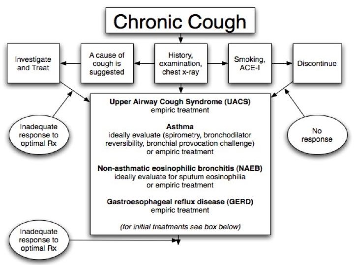
Chronic Cough in Adults Cough lasting longer than 8 weeks Top 4 in immunocompetent patient with normal CXR: Upper airway cough syndrome Asthma Gastroesophageal reflux disease Non-asthmatic eosinophilic bronchitis Cough may have more than one cause-- a diagnostic challenge!
Upper Airway Cough Syndrome (UACS) Also called “Post-nasal drip syndrome” (PNDS) Mechanism: secretions from nose/sinuses stimulate upper airway cough receptors; inflammation increases receptor sensitivity Classic symptoms: “tickle” ﺩﻏﺪﻏﻪ in throat; throat clearing, hoarseness, nasal congestion Cough may be the only symptom in ~ 20%
Upper Airway Cough Syndrome (UACS) Signs (may be absent): inflamed nasal mucosa, secretions in posterior oropharynx Consider underlying causes: allergies, chronic sinusitis, overuse of alpha-agonist nasal sprays Diagnostic/Therapeutic trial: 1 st generation antihistamine/decongestant combination medication for 2 weeks
Asthma Mechanism: inflammatory mediators, mucus, bronchoconstriction stimulate cough receptors Classic symptoms: intermittent wheeze Cough may be the only symptom in 7 -57% patients (depends on study)-- “Cough-variant asthma” Signs (often absent): expiratory wheezing on chest exam
Asthma Diagnostic tests: Spirometry, before and after bronchodilator: partially reversible airflow obstruction Methacholine inhalation challenge: positive • Diagnostic/ Therapeutic trial: inhaled corticosteroid + bronchodilator for ≥ 8 weeks
Gastroesophageal Reflux Disease (GERD)
A Vicious Cycle COUGH REFLUX INCREASED ABDOMINAL PRESSURE
Gastroesophageal Reflux Disease (GERD) Classic symptoms: heartburn, sour ﺣﺎﻣﺾ taste in mouth Cough may be only symptom in 75% patients with chronic cough Diagnostic tests: 24 -hour esophageal p. H probe (best) Esophagram
Gastroesophageal Reflux Disease (GERD) Diagnostic/Therapeutic trial: gastric acid suppression with proton pump inhibitor (e. g. omeprazole) for ≥ 2 months, combined with diet and lifestyle modification
Non-Asthmatic Eosinophilic Bronchitis (NAEB) Eosinophilic airway inflammation WITHOUT variable airflow obstruction or airway hyperresponsiveness • Diagnostic tests: - Spirometry: normal - Methacholine challenge: - normal - Induced sputum: >3% eosinophils • Diagnostic/Therapeutic trial: inhaled corticosteroid for ≥ 4 weeks
Continued on next slide…
Chronic Cough, continued: