Sputum Examination is a thick fluid produced in


 1. Before collecting sputum the mouth should")


- Slides: 13
Sputum Examination is a thick fluid produced in the lungs and in the adjacent airways
Possible Pathogens • BACTERIA – Gram Positive – Gram Negative – Mycobacterium tuberclosis • FUNGI & ACTINOMYCETES • PARASITES
Collection of Sputum Sample (1 st day) 1. Before collecting sputum the mouth should be pre-rinsed and this removes contaminants from oral cavity. 2. Give the patient a clean dry wide necked leak-proof container and request him or her to cough deeply to produce a sputum specimen. 3. For best results early morning freshly expectorated sputum specimen should be collected. 4. Label the container, and complete the request form. 5. When Pneumonia or Bronchopneumonia is suspected, deliver the sputum to the laboratory with a little delay as soon as possible because organisms such as S. pneumoniae & H. influenzae require culturing as soon as possible. Specimen for the isolation for S. Pneumoniae & H. Influenzae must never be refrigerated.
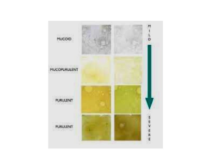
Appearance • Purulent: Green Looking, mostly pus • Mucopurulent: Green Looking with pus & mucus. • Mucoid: Mostly mucus • Mucoslivary:
Microscopic • After macroscopic examination transfer material to a clean slides. Smears made on clear slides should be air dried, fixed over flame and stained. • Different stains used: • 1: - Gram’s stain • 2: - Ziehl-Neelsen stain for AFB • der oil immersion
Smear the sputum Stained by Gentian violet stain Keep for 1 min. Bacteria get violet color Pour Gram’s Iodine For 1 min. Wash with Alcohol Wash with water and dry Mount un
Zeil-Neelson Smear to detect AFM Chance of detecting AFB in sputum smears are significantly increased if the organisms are first concentrated by centrifugation sodium hypochlorite(Bleach) is recommended for liquefying the sputum because it kills M. Tuberculosis
Na. OC 1 centrifugation technique to concentrate AFB 1: Transfer 1 -2 ml of sputum to a screw cap universal bottle. 2: Add an equal volume of concentrated Na. OC 1 solution and mix well. 3: Leave at room temperature for 10 -15 min shaking at interval to break down the mucus in the sputum. 4: Add about 8 ml of distilled water mix well. 5: Centrifuge at 3000 g for 15 min. 6: Using a glass pasture pipette to remove and discard the supernatant fluid. Mix the sediment. Transfer a drop of well mixed sediment to a clean glass slide. Make a thin smear & Allow to air dry.
Smear the sputum Fixed by Heating Pour carbol fuchin and heat it from below Keep for 5 min. Wash with water Decolorize with 20%H 2 SO 4 Wash with Malachite green/methyene blue for 1 min. Wash & dry Mount under oil immersion
Specimen Culture To obtain as pure a culture as possible of a respiratory pathogens it is necessary to reduce the number of commensals inoculated. Ways of reducing commensal number include washing the sputum free from sliva or liquefying and diluting it. The technique using saline washed sputum described. The dilution technique requires a liquid agent such as dithiothreitol (Sputolysin. Sputasol) which is expensive and unstable. Each specimen received for culture should be plated on agar.
Different agars 1: Blood agar 2: Chocolate agar 3: Mac. Conkey’s agar 4: Thioglycollate broth
1: Wash a purulent part of sputum in about 5 ml of sterile physiological saline. 2: Inoculate the washed sputum on plate of: Blood agar and Chocolate agar 3: Add an aptochin disc to the blood plate within the area of 2 nd spread. This will help to identify S. pneumoniae. 4: Incubate the blood agar plate aerobically and the chocolate agar plate in a carbon dioxide enriched atmosphere. Examine and report the cultures Blood agar and chocolate agar cultures: Look especially for a significant growth of: streptococcus Pneumoniae Haemophilus influenzae Stephlococcus aureus