VARICOSE VEINS DEFINITION When a vein becomes dilated

VARICOSE VEINS

DEFINITION: When a vein becomes dilated, elongated and tortuous, the vein is said to be “VARICOSE” SITES: The common sites of varicosity are: 1. Superficial venous system of the lower limb – either the Long or short saphenous or both. 2. Oesophageal varix 3. Varicosity of the haemorrhoidal veins (Piles) 4. Varicosity of the spermatic veins (Varicocele)

VENOUS DRAINAGE OF THE LOWER LIMB It can be conveniently described under the three heads 1. Deep veins 2. Superficial veins 3. Perforating or communicating veins

VENOUS DRAINAGE OF LOWER LIMB
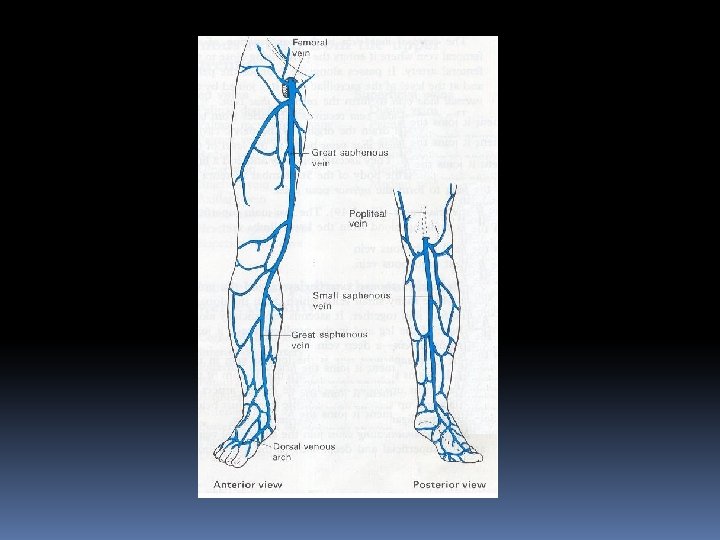

Deep veins: The following comprises the deep veins: 1. Posterior tibial vein and its tributaries 2. The peronial vein 3. Anterior tibial vein 4. Popliteal vein 5. Femoral vein

DEEP VEINS

Characteristic features of deep veins: 1. There are numerous valves which direct the flow of blood upward and prevent downward flow. 2. Venous plexes of soleus muscle (which are devoid of valves) and perforating veins empty into the posterior tibial and peroneal vein. The entry may be at the same site.
: These veins lie in subcutaneous fat between the skin and the")
Superficial Veins (SV): These veins lie in subcutaneous fat between the skin and the deep fascia. SV comprises of : 1. Long Saphenous vein. It begins in the medial marginal vein of the foot and ends in the femoral vein about 3 cm below the inguinal ligament. It contains 10 to 20 valves.


LONG SAPHENOUS VEIN VARICOSITY
: 1. As there is communication between the long and short saphenous")
Surgical importance (LSV): 1. As there is communication between the long and short saphenous veins varicosity may spread from one system to the other. 2. At the time of operation for varicose vein, when a ligature is advised to the S. F. Junction, the veins draining into the long saphenous vein near the saphenous opening must be ligated and cut otherwise these small veins will become varicosed due to incompetent S. F. junction. 3. In case of varicosity of the long saphenous vein, the small veins from the sole of the foot and the ankle which drain into this venous system through the medial marginal veins become dilated and gives rise to swelling of ankle, know as “ANKLE FLARE”

Ankle flare with mild ulceration

2. Short Saphenous vein: This vein begins in the lateral marginal vein of the foot and ends in popliteal vein 3 to 7. 5 cms above the level of the knee joint. It possess 7 to 13 valves.

Perforators: These veins communicate between the superficial and deep veins. There are valves in these veins which allow the blood flow from superficial to deep veins. If these valves become incompetent the veins become high pressure leaks during muscular contraction and this transmission of high pressure in the deep veins to the superficial veins results in dilatation of the superficial veins producing varicose veins.

Perforators types: A. Indirect perforators: These are numerous small vessels which starts from the superficial venous system, pierce the deep fascia and communicate with a vessel in an underlying muscle. B. Direct perforators – These veins directly connect the superficial and deep veins. These are. . . In the thigh – between the long saphenous and femoral vein (Dodd) In the leg - (Lower leg perforators – Cockett) - Gastrocnemius perforator - Boyd

PERFORATORS OF LOWER LIMB

SURGICAL PATHOLOGY: Normally blood flow from the superficial veins to deep system through the competent perforators. But if this mechanism breaks down, either due to destruction of the valves of the deep veins (following dvt) or of the perforators or of the superficial venous system, the blood becomes stagnated in the superficial veins which leads to varicosity.


AETIOLOGY: Morphological factor – Varicose veins of the lower limbs are the penalty the man has to pay for its erect posture. The veins have to drain against gravity. The superficial veins have loose fatty tissue to support them and thus suffer from varicosity. There are three types of varicose veins based on aetiology. 1. Primary varicose veins – due to defect in the valves 2. Secondary varicose veins – due to venous obstruction (Pregnancy, pelvic tumours, dvt, hormonal causes, retroperitoneal lymphadenopathy) 3. Congenital varicose veins – Congenital arterio venous fistula or cavernous (venous) haemangioma.

Predisposing factors: A. Prolonged standing B. Obesity C. Pregnancy D. Old age E. Athlets

OBESITY

ATHELETS

Clinical features: Majority of the patients come with dilated veins in the leg. They are minimal to start with and at the end of they are sufficiently large because of venous engorgement. Symptoms: 1. Dragging pain in the leg or dull ache is due to heaviness 2. Night cramps occur due to change in the diameter of veins. 3. Sudden pain in the calf region with fever and oedema of the ankle region suggests DVT. 4. Patients can present with ulceration, eczema, dermatitis and bleeding. 5. A generalised swelling of the leg may be present which is due to DVT

Signs: Dilated veins are present in the medial aspect of the leg and the knee some times they are visible in the thigh also. Single dilated varix at the SF junction called “SAPHENA VARIX” A group of veins near the medial malleolus is ANKLE FLARE A localised, dilated segment of the vein, if present is an indication of blow out. It signifies underlying perforator.

SAPHENA VARIX

Ankle flare with mild ulceration

BLOW OUT

Clinical classification of chronic lower extremity venous disease: Grade 0: No visible or palpable signs of venous disease. Grade 1 : Telangiectases, reticular veins or malleolar flare. Grade 2 : Varicose veins. Grade 3 : Edema without skin changes. Grade 4 : Skin changes ascribed to venous disease (Pigmentation, eczema or lipodermatosclerosis) Grade 5 : Skin changes as defined above with healed ulceration. Grade 6 : Skin changes as defined above with active ulceration.

Physical Examination: Local Examination: A. Inspection of : 1. Varicose veins 2. Swelling of the leg 3. Skin of the limb 4. Impulse on coughing
 Cough impulse or Morrissey’s test –")
B. Palpation : Tests for varicose veins: (1) Cough impulse or Morrissey’s test – Saphenofemoral incompetence. (2) Brodie - Trendelenburg – A. Saphenofemoral incompetence. B. Perforator in competence. (3) Multiple tourniquet test – Site for perforator in competence. (4) Schwartz test – Superficial column of blood. (5) Modified Perthes test – Deep vein thrombosis. (6) Fegan’s Test – To locate the perforators in the deep fascia. 7. Pratt’s test – To know the position of the leg perforators.

C. Percussion: If the most prominent parts of the varicose veins are tapped, an impulse can be felt by the finger at the saphenous opening. This is known as SCHWARTZ TEST D. Auscultation: The importance of auscultation is limited to the arteriovenous fistula, where a continuous machinery murmur may be heard.
: Are only enlarged if there be venous ulcer. F.")
E. Regional lymph nodes (Inguinal): Are only enlarged if there be venous ulcer. F. Other limb : Should be examined for presence of varicose veins. GENERAL EXAMINATION: A. Examination of the abdomen B. Vaginal and rectal examinations C. Peripheral arterial pulses D. Sensations & Movements
 Doppler ultrasound is the most important minimum level investigation to be done")
INVESTIGATIONS: (1) Doppler ultrasound is the most important minimum level investigation to be done before treatment a patient with venous disease. (2) Duplex ultrasound Imaging: In this investigation high resolution B – Mode ultrasound imaging and Doppler ultrasound are used. (3) Various Special investigations: Ascending phlebography, thermography, radioisotope scanning, radioactive fibrinogen studies and ultrasonics to know the condition of the deep vein, position of the thrombus and position of the incompetent perforators.

Complications of varicose veins: 1. Haemorrage: It may occur from minor trauma to the dilated vein. The bleeding is profuse due to high pressure. Simple elevation of leg does a lot to stop such bleeding. 2. Thrombo Phlebitis: This means inflammation of the superficial veins. In this case the veins become red and feel like tender cords in the subcutaneous tissue. The skin is inflamed and pyrexia is usually present. It is treated by bed rest, elevation, crepe bandage, antibiotic and anti-inflammatory drugs.

PHLEBITIS

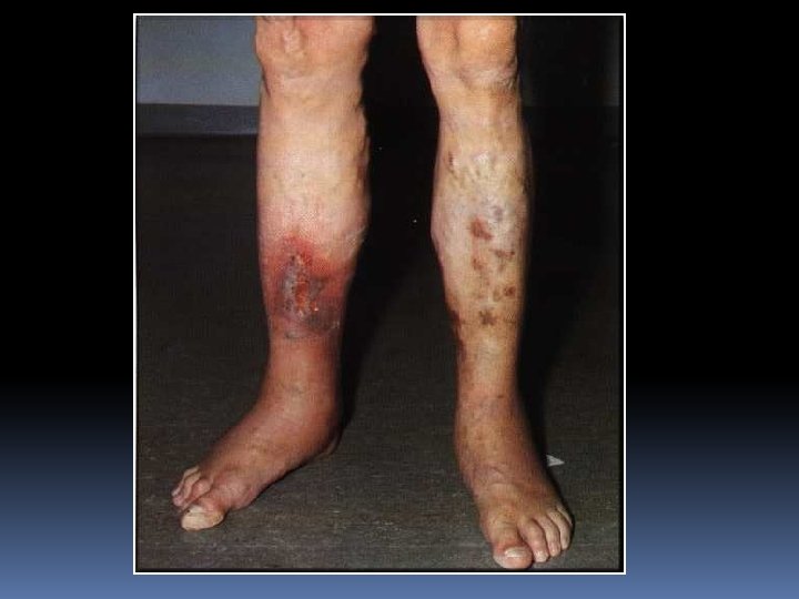
3. Ulceration : This is more due to deep vein thrombosis rather than varicose veins alone. These ulcers are mostly found on or near the medial malleolus because of plenty of perforating veins present there. After thrombosis has been recanalised the valves of the deep veins are irreparably damaged. The deoxygenenated blood stagnates in the lower part of the leg, so the superficial tissue present there loss its vitality to certain extent and a gravitational ulcer follows. On long standing, Marjolin’s ulcer may develop. Treatment of varicose ulcer by Bisgard’s method – Conservative line

ULCERS

MARJOLINS ULCER

4. Eczema and dermatitis: It occurs due to extravasation and break down of R. B. C. in the lower leg. Break down of RBC produce haemosiderin and biliviridin. Biliviridin causes itching which precipitates venous ulcer. Treatment is application of ointment containing zinc oxide and coal tar twice daily.

5. Lipodermatosclerosis: This means the skin becomes thickened fibrosed and pigmented. This is due to high venous pressure which causes fibrin accumulation around the capillary and it also activates white cells. 6. Calcification of vein: 7. Pigmentation: Haemosiderin deposits produced breakdown causes black pigmentation. from RBC

LIPODERMATOSCLEROSIS

LIPODERMATOSCLEROSIS

8. Periostitis : In case of long standing ulcer over medial surface of tibia. 9. Equinus deformity: This results from longstanding ulcer. The patient finds that walking on toes relieves pain so he continues to do so and ultimately the Achilles tendon becomes shorter to cause this.

SHIN SPLINTS DUE TO PERIOSTITIS

EQUINUS DEFORMITY

TREATMENT OF VARICOSE VEINS: Varicose veins can be treated by three methods: 1. Conservative line of treatment Elastic crepe bandage and elevation of leg forms the fundamental steps in treatment of varicose veins. 2. Injection line of treatment By injection sclerotherapy: the most commonly used sclerosant used is sodium tetradecyl sulphate Ultrasound guided foam sclerotherapy: this has become an alternative to the blind sclerotherapy practiced in the past. Polidocanol is used rather than sodium tetradecyl sulphate.

3. Surgery – Types of operations a. Ligation: Trendelenburg’s operation b. Ligation with stripping C. Others: 1. Subfascial endoscopic perforator surgery 2. Multiple Cosmetic Phebectomy 3. Subfascial ligation of Cockett and Dodd 4. Radical surgery approach

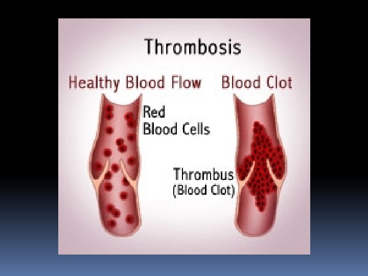
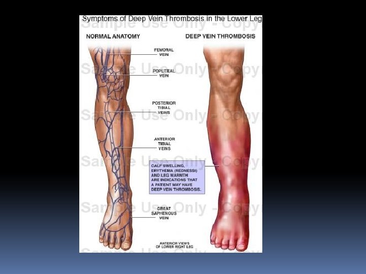
VENOUS THROMBOSIS Venous thrombosis is a very common surgical problem, which has great influence on the morbidity and mortality of surgical patients. Types: 1. Superficial vein thrombosis (Thrombophlebitis) 2. Deep vein thrombosis (Phlebothrombosis)
 It is thrombosis of the deep venous system. It can")
DEEP VEIN THROMBOSIS (DVT) It is thrombosis of the deep venous system. It can be acute or recurrent, occlusive or non-occlusive, free or fixed thrombus, propagative or non-propagative. Sites: 1. Pelvic veins – Common 2. Leg veins – Common in femoral and popliteal veins (Common on left side) 3. Upper limb veins – Not uncommon (Axillary vein thrombosis).

Predisposing factors for venous thrombosis: 1. Stasis 2. Hypercoagulability of the blood 3. Injury to the vessel wall The above three factors constitutes Virchow’s triad

STASIS

HYPERCOAGULABILITY

VESSEL WALL INJURY

Causes for DVT: 1. Genetic a. Mutation in factor-V b. Antithrombin deficiency c. Protein C and S deficiency 2. Acquired – Major injuries, following operation, visceral cancer, tobacco smoking, diabetes, congestive heart failure, shock, polycythemie vera, pregnancy, infection, varicose veins, obesity, using of contraceptive pills and long period of sitting or bed rest. 3. Spontaneous – due to defective fibronolysis mechanism.
 causing")
Phlegmasia alba dolens: It is DVT of femoral vein (deep femoral vein commonly) causing painful congestion and oedema of leg, with lymphangitis, which further increases the oedema and worsens the situation (White leg) Phlegmasia cerulea dolens: It is extensive DVT of iliac and pelvic veins causing blue leg with either venous gangrene or areas of infarction

PHLEGMASIA CERULEA DOLENS

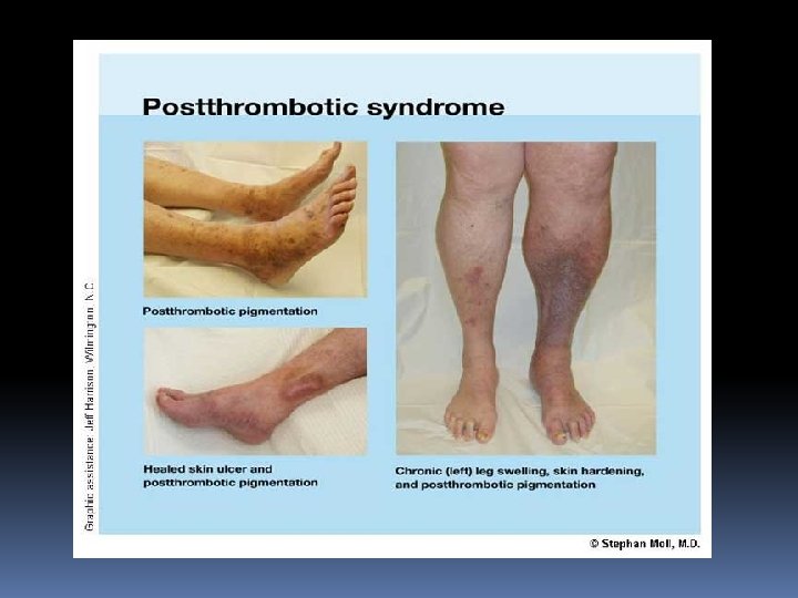
Fate of thrombus: 1. Propagation of the thrombus leading to obstruction of the lumen of the involved vessel. 2. Embolisation 3. Dissolution due to endogenous fibrinolytic activity 4. Organization or recanalisation leading to secondary changes to the venous wall and manifest as Chronic Venous Ischemia.
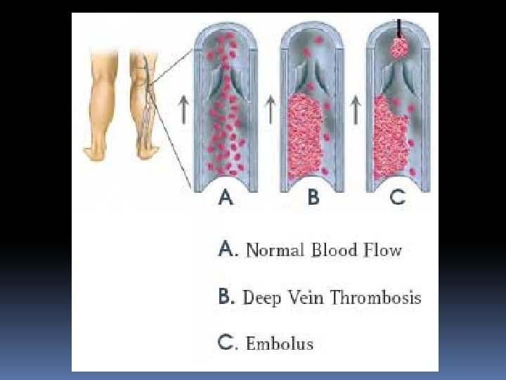

FATE OF THROMBUS - EMBOLISM

Clinical Features: Fever is the earliest and common symptom. Pain and swelling in the calf and thigh, commonly associated with fever. Positive Homan’s sign: Passive forceful dorsiflexion of the foot with extended knee will cause tenderness in the calf. Positive Homan’s sign is confirmative sign of DVT; but absence of Homan’s sign is not a reliable indicator of absence of DVT. Mose’s sign: Gentle squeezing of lower part of the calf from side-to-side is painful. Gentleness is very important otherwise it may dislodge a thrombus to form an embolus.

Neuhof’s sign: Thickening and deep tenderness is elicited while palpating deep in calf muscles. Linton’s test: After applying proximal tourniquet: with elevation after walk; superficial veins are still prominent. Most often, DVT is asymptomatic (60%) and presents suddenly with features of pulmonary embolism like chest pain, breathlessness and haemoptysis.

INVESTIGATIONS: Venous Doppler, Duplex scanning, Venogram, Radioactive I 125 fibrinogen study, haemogram with platelet count, MR venography. Treatment: 1. Thrombolytic therapy - Strepto kinase or urokinase. These drugs are given either systemically or catheter defected intra-clot infusion. 2. Venous thrombectomy in life threatening thrombosis. 3. Inferior Vena Cava(IVC) filters - Reduces the rate of pulmonary embolism.

Prophylaxis: Thromboprophylaxis is achieved by administering either conventional heparin or LMWH. Prevention is the key word in the management of DVT rather than treatment. Intermittent pneumatic compression of the cough through the operation, maintains the blood flow in lower limbs.

- Slides: 69